IMR Press / JMCM / Volume 4 / Issue 1 / DOI: 10.31083/j.jmcm.2021.01.208
Open Access Review
Antimicrobial peptides-An alternative candidates to antibiotics against Staphylococcus aureus and its antibiotic-resistant strains
Show Less
1 Department of Chemistry and Biochemistry, Mendel University in Brno, Zemedelska 1, CZ-613 00 Brno, Czech Republic
2 Central European Institute of Technology, Brno University of Technology, Purkynova 123, CZ-612 00 Brno, Czech Republic
*Correspondence: vojtech.adam@mendelu.cz (Vojtech Adam)
J. Mol. Clin. Med. 2021, 4(1), 1–17; https://doi.org/10.31083/j.jmcm.2021.01.208
Submitted: 24 October 2020 | Revised: 17 November 2020 | Accepted: 8 December 2020 | Published: 20 March 2021
Copyright: © 2021 The Authors. Published by IMR Press.
This is an open access article under the CC BY 4.0 license (https://creativecommons.org/licenses/by/4.0/).
Abstract

Staphylococcus aureus and its antibiotic-resistant strains are the cause of soft tissue infections representing some severe life-threatening infections. These situations have caused great concern for its treatment worldwide. Thus, the need to introduce new antibiotics or an alternative to antibiotics markedly increasing. Antimicrobial peptides (AMPs) have been shown to have various properties and uses in the biological system since their discovery. This review is based on the increasing concern for S. aureus, its resistant strains, the associated infections, pathogenicity, and the mechanism of resistance to antibiotics. Lastly, the overall significance of AMPs against S. aureus showed that they can be ideal candidates as an alternative to antibiotics with high potential for future therapeutics.

Keywords
Antimicrobial peptides
Antibiotics
Antibiotic-resistance
Staphylococcus aureus
1. Introduction

Bacterial infection by antibiotic-resistant bacteria has been a serious global public health concern in the last decades. Usually, antibiotics exhibit good activity against bacterial infections and all of them come along with side effects. However, the emergence of antibiotic-resistant strains with time has further worsened the condition with increasing pressure on the clinical setting [1]. These have increased the morbidity and mortality rate and prolonged hospital stay [2, 3, 4]. These antibiotic-resistant strains are the leading cause of hospital and community-acquired infections. The overuse, misuse of antibiotics, and an incomplete course of antibiotics are among the main reasons for antibiotic resistance [5, 6, 7]. Further, the ability of the bacteria to evolve and gain resistance to antibiotics via various modalities like reduced permeability, efflux via pumps, alterations of the target enzyme, horizontal gene transfer, and ability to produce enzymes which degrade the antibiotic, has made the situation more critical [8]. Thus, this scenario has pushed the researcher and clinical settings to develop new antibiotics or generate some new alternatives to antibiotics.

The bacterial strains that are normally acquired even inside the intensive care units (ICU) are Acinetobacter baumannii (19.5%), Enterococcus spp. (10.6%), Staphylococcus aureus (10.7%), Pseudomonas aeruginosa (15.6%), Stenotrophomonas maltophilia (11.5%), and Klebsiella pneumoniae (9.7%) [9] and the infections caused by these antibiotic-resistant bacteria are hard to cure. According to the World Health Organization (WHO) report of 2017 regarding antibiotic resistance, they listed resistant bacteria based on three priority levels as critical, medium, and high. Where S. aureus, vancomycin-resistant S. aureus (VRSA) and methicillin-resistant S. aureus (MRSA) were listed as a high priority [1]. However, in 2019, the Centre for Disease Control and Prevention (CDC) listed bacteria and fungi as urgent, serious, and concerning. According to their report, MRSA is listed as a serious threat [10]. Moreover, the current uprising of multi-drug resistant strains has made the situation even more critical which calls for modalities to overcome bacterial antibiotic resistance [12, 13]. Thus the interest to develop new antibiotics with proper use is highly increasing. In this respect, the development of an alternative to antibiotics is inevitable. The application of nanoparticles, combined therapy (conjugate formations), plant extracts, algal metabolites, and antimicrobial peptides are some of the common alternatives.

Antimicrobial peptides (AMPs) are amphipathic and their natural occurrence provides innate immunity via non-specific and effective defensive activity against invading pathogens. Further, their ability to enter bacteria, mostly soluble in water or aqueous buffer or organic solvent, possibility to modify and their ability to form a secondary structure like beta sheets and helices, has made them a promising candidate alternative to antibiotics [4, 14, 15]. AMPs showed prominent activity against fungi and bacteria with decreased resistance compared to antibiotics. AMPs also display anticancer activity. They have limitations including stability, non-specific interaction, high production costs, lack of information regarding their mechanism of action, and sometimes high toxicity [16]. Despite these drawbacks, AMPs are also clinically tested and approved for their appearance in the world of medicines.

2. Staphylococcus aureus and its resistant strains

The Staphylococcus aureus and its related resistant taxonomic classes can sustain harsh conditions like human skin. Fig. 1 gives a clear view of the timeline for the emergence of S. aureus resistant strains. They are opportunistic pathogens which can cause opportunistic infections. They are the cause of both community and healthcare-associated bloodstream infections. They cause infections via various production of secreted and multiple cell surface virulence factors. Further, S. aureus endovascular pathogenesis is the result of the cumulative effects of different virulence factors [17]. The presence of protein A (Spa) on the S. aureus surface acts as a virulence factor that prevents the immune system from performing phagocytosis [18, 19]. Moreover, they are associated with soft tissue infections like surgical site infections, folliculitis, cutaneous abscesses, boils, carbuncles, impetigo, scalded skin syndrome, purulent cellulitis, and furuncles. They are also responsible for life-threatening infections like sepsis, bacteremia, pneumonia, toxic shock syndrome, osteomyelitis, pseudomembranous enteritis, and endocarditis [20]. S. aureus can colonize in the region of lesion of atopic dermatitis patients as well as patients with chronic infections (up to 93% approximately) like chronic leg ulcers. However, S. aureus also plays a role in sepsis in burn patients leading to death [21].
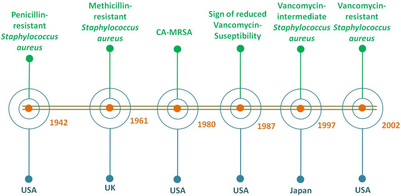
Fig. 1.

The timeline based on the first reports of the emergence of S. aureus resistant strains since 1900s.

2.1 Methicillin-Resistant Staphylococcus aureus

MRSA, a significant nosocomial pathogen with versatile nature associated with postsurgical wound infections in hospitals. The first case was in the United Kingdom in the year 1961 [22]. Reports by the WHO showed that out of most cases of MRSA infection, 64% of people die in comparison to people with non-MRSA infections [23]. They may be transmitted via physical contact but rarely by air. The infection by MRSA has increased morbidity, hospital stays, mortality, and overall cost. Reports showed that S. aureus contains at least 11 major types (I-XI) of the staphylococcal cassette chromosome mec (SCCmec) mobile elements that are responsible for the emergence of MRSA [24]. Therefore, the overall frequencies of the healthcare-associated (HA) and community-associated (CA) MRSA infection and their ability to gain antibiotic resistance have increased [25]. Furthermore, the presence of a hypervariable region in the spa gene (Xr) and SSCmec elements often used for MRSA typing [24].

The first case of a CA-MRSA infection was reported in the USA in the year 1980. Moreover, significant epidemiological changes in the CA-MRSA had been observed in the past decade [26]. Infections caused by CA-MRSA are usually skin infections but rarely necrotising pneumonia with a high tendency to infect young individuals living in the community. Patients with necrotizing pneumonia often seem to have influenza-like sickness or influenza virus infections with the symptoms of multiple cavity lung infiltrates leads to a mortality rate of more than 50% [27]. They also cause severe infections like Waterhouse-Friderichsen syndrome, necrotising fasciitis, and sepsis. CA-MRSA has a distinct pattern of resistance containing SCCmec IV or V or VI with resistance toward fewer categories of antibiotics [28, 29, 30]. Reports also showed that most CA-MRSA produces a bi-component cytolytic toxin for neutrophils encoded by lukSF-PV genes [31, 32].

The isolates of CA-MRSA mostly contain a phage-encoded Panton-Valentine leukocidin (PVL). PVL is a toxin that is associated with severe infections like necrotising pneumonia and also skin infections [33]. It causes necrosis of epithelial cells and human leukocytes lysis. They also produce an α-hemolysin, pore-forming toxin, that destroys a wide range of host cells [34]. Further, these CA-MRSA’s are also gaining resistance towards other antibiotics, and the problem to treat them is also becoming urgent clinical issues.

However, other dangerous isolates are the HA-MRSA that can survive in the hospital environment due to their multidrug resistance ability. The HA-MRSA can colonize in patients who have been admitted to the hospital and underwent medical treatments therein or were recently discharged from the hospital. Even more than six months, the carrier state can persist in these patients. Thus, the patient infections caused by MRSA within the healthcare system or in hospitals are known as healthcare-associated MRSA infections [35]. Albeit, the bacterial peptides group named the phenol soluble modulins (PSM) which causes inflammation, skin infection, and neutrophil cytolysis, is found mainly in CA-MRAS. PSM showed lower expression in HA-MRSA isolates [36]. HA-MRSAs are resistant to a greater number of antibiotic categories due to the presence of SCCmec I, II, and III. These elements facilitate the survival of these pathogens in the healthcare system [37]. Further, the horizontal gene transfer of SSCmec III among S. aureus is not feasible due to the larger size of the mobile element. Thus, the possible spread of HA-MRSA in the community is through healthcare workers being the carriers which come in contact with the community [38, 39, 40]. Thus, more precaution measures are needed to be taken by the healthcare workers.

2.2 Vancomycin resistant Staphylococcus aureus

Antibiotic resistance in S. aureus against vancomycin being the last resort antibiotic against infections caused by gram-positive bacteria is also a major concern [4]. According to the Clinical and Laboratory Standard Institute depending on the susceptibility to vancomycin, S. aureus is classified into 3 groups including VRSA with MIC 16 μg/mL, vancomycin-susceptible S. aureus (VSSA) with MIC 2 μg/mL, and vancomycin-intermediate S. aureus (VISA) with MIC 4-8 μg/mL [41].

The VISA was discovered in 1997 in Japan. However, retrospective studies regarding vancomycin susceptibility were observed around 1987 in the USA. They are partially resistant to vancomycin due to polygenic factors. It includes a series of mutations in genes which encode molecules that are related to cell envelope biosynthesis, cell wall thickness increment, altered surface protein profile, changes in growth characteristics, and other factors [42, 43, 44, 45]. Though it is accepted that VISA occurred due to the accumulation of a series of gene mutations, only a few are important like genes encoding two-component regulatory systems (GraSR, WalKR, and VraSR) [46, 47, 48, 49, 50, 51]. However, the detailed molecular mechanism of VISA resistance is yet needed to be explored by researchers. The VISA infections are related to persistent infections, hospitalization, prolonged vancomycin treatment or failure, and lack in clinical settings. Moreover, the heterogeneous VISA (hVISA) is the S. aureus population with vancomycin MIC ranges from 2 to 4 μg/mL. It has been hypothesized that when these hVISA’s are constantly under prolonged treatment with glycopeptides, they acquire homogenous resistance to vancomycin [52, 53].

Vancomycin-resistant enterococci (VRE) were identified in Europe which was found endemic in ICUs soon after its discovery [54, 55]. The resistance mediated by transposon Tn1546 present in a conjugative plasmid caused an increased risk in the spread of vancomycin resistance to universally susceptible microorganisms including S. aureus [56, 57, 58, 59]. Further, it was confirmed that in a mixed infected mouse, the van elements were transferred to MRSA from Enterococcus faecalis but it did not spread hence causing a lesser concern [60]. However, there are cases of co-colonization and co-infection of VRE and MRSA, which are common in patients with prolonged hospital stay as well as stay in ICU [61, 62]. The first VRSA isolate was reported in 2002 from Michigan, USA [63, 64]. In numerous cases, isolates of both VRE and VRSA were obtained together from the same patients [65]. It has also been reported that vancomycin acts as a selective pressure for its resistance imparted by vanA gene clusters. The majority of hospital-associated infections caused by VRSA isolates belong to clonal complex 5 (CC5) lineages [66, 67]. Therefore, the misuse of vancomycin for treatment has to stop. Whereas, the development of alternatives is increasingly required.

3. Mechanism of resistance and possible threats

The resistance towards different antibiotics in S. aureus has emerged due to various mechanisms such as acquiring mobile genetic elements via horizontal gene transfer, increasing the expression of efflux pumps and mutations altering target binding sites [68]. Fig. 2 provides the overall mechanisms of antibiotic resistance in S. aureus.
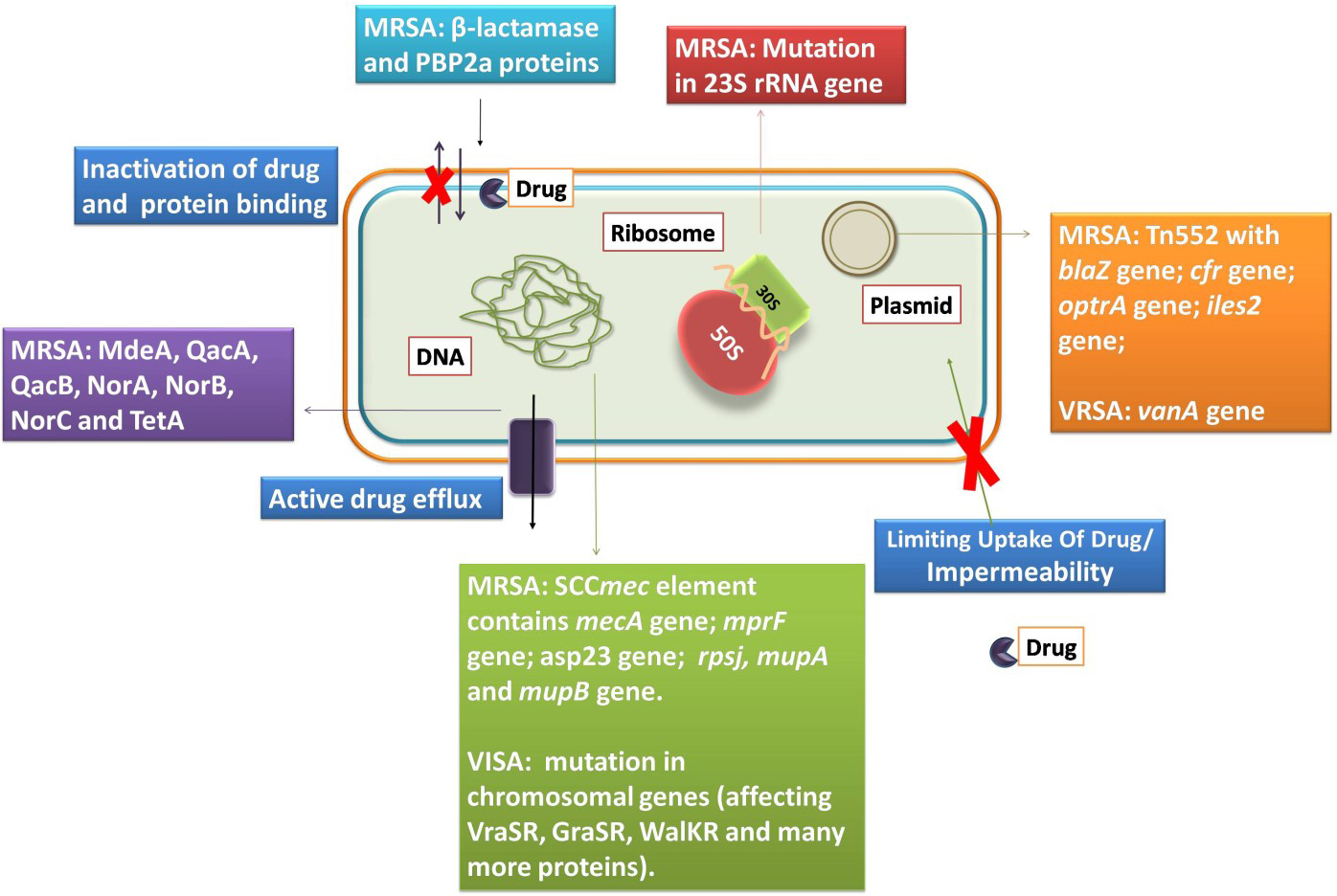
Fig. 2.

The S. aureus and its mechanisms of resistance to antibiotics.

3.1 Mechanisms of antibiotic resistance in MRSA

The first reported penicillin-resistant S. aureus was described in 1942, in the USA [69]. The production of β-lactamase caused penicillin-resistance in S. aureus. Typically, BlaZ, a serine β-lactamase which forms an acyl-enzyme intermediate is produced in S. aureus. It is essentially the same enzyme as the transpeptidase of PBP2 with the difference in their kinetics of deacylation [70]. The transposon Tn552 or similar elements carry the blaZ gene. This gene is either integrated into the chromosome or located in the plasmid like pI524 [68]. The expression of blaZ is inducible as it is controlled by the BlaR sensor and BlaI repressor proteins [70, 71]. This lipoprotein enzyme is partly localized on the outer cytoplasmic membrane to protect PBP2s (penicillin-binding protein 2). However, some is also released to the surrounding vicinity which inhibits the activity of penicillin.

MRSA strains contain a large mobile genetic element named SCCmec containing the mecA gene responsible for resistance. Thus, resistance to methicillin and oxacillin is mediated via the acquisition of this mecA gene (encodes PBP2a). PBP2a is not interacting with drugs due to the presence of the active site serine (i.e., of the transpeptidase) in a deep pocket [72, 73]. According to reports, CA-MRSA carries a small SCCmec with higher virulence but lower resistance to multiple antibiotics [74, 75]. The mecA gene is expressed upon exposure to a drug that is under the regulation of MecIR regulatory protein [72]. However, in the presence of BlaI and BlaR, the activity of mecA gene expression was seen to be repressed. Thus, the presence of Mec and Bla regulator can cause a variation in the PBP2a expression.

Further, the non-synonymous or nonsense mutation in mecA can usually cause a change in the amino acid sequence of PBP2a resulting in ceftaroline resistance [76, 77]. It was observed that when passaged in ceftaroline the COL strain showed a mutation in PBP2, PBP4, and gdpP with a high level of resistance towards it [79]. It was later reported the wild type MRSA also showed ceftaroline resistance [78]. Hence, prolonged treatment with ceftaroline can lead to the emergence of resistance to this antibiotic.

MRSA infections like endocarditis and bacteremia were treated with daptomycin, an alternative to vancomycin which was approved in 2003 in the USA. However, certain. aureus strains showed some changes like enhanced membrane fluidity, lower daptomycin surface binding, changes in the cytoplasmic membrane, increase net positive charge, and reduced susceptibility to daptomycin-induced depolarization [80]. When the gene mprF encoding for the lysyl-phosphatidyl glycerol synthetase got mutated, it increased the outer membrane positive charge and reduced the susceptibility to daptomycin [81]. The inactivation of the asp23 gene causes decrease in daptomycin susceptibility [82]. Thus, these alterations in S. aureus strains facilitated the acquisition of resistance to daptomycin.

Furthermore, MRSA also acquired resistance to oxazolidinone via a mutation in the 23S rRNA, to chloramphenicol-florfenicol resistance (cfr) gene, and optrA gene (influx or efflux of drugs) [83, 84]. Aminoglycoside modifying enzymes like bidomain AAC(6)le-APH(2)la acetyltransferase and phosphotransferase, APH(3)IIIa phosphotransferase, and ANT(4)Ia nucleotidyltransferase, typically induce resistance to aminoglycosides in MRSA [85]. The presence of two efflux pumps TetA(K) and TetA(L) along with TetO/M determinants present in Tn916 and Tn1545 conjugative transposons located in chromosome, confers tetracycline resistance in MRSA. Further, the mutation in rpsJ (ribosomal protein S10) gene also lead to tetracycline resistance in MRSA [68, 86]. Resistance to fluoroquinolones is mediated by mutation in the quinolone resistance determining region (topoisomerase mutants) and the presence of antibiotic efflux pumps (MdeA, QacA, QacB, NorA, NorB, and NorC) in S. aureus [87, 88, 89, 90]. MRSA is also resistant to pleuromutilins by target modification (mutation in ribosomal protein or cfr gene), efflux pump, and ribosomal protection (vga genes) [91, 92]. Moreover, mupirocin resistance in MRSA was conferred by ileS-2, mupA, and mupB genes [93, 94]. Thus, it is of paramount importance and utmost need to develop new antibiotics or alternatives to treat MRSA infections that are resistant to almost all classes of antibiotics.

3.2 Mechanisms of antibiotic resistance in VRSA and VISA

Vancomycin was antibiotic of choice for the treatment of MRSA in the late 1980s. However, vancomycin resistance emerged in the same year in VRE. Later, it was followed by reports of vancomycin resistance in S. aureus.

The mechanism of action of vancomycin is to bind the D-Ala-D-Ala dipeptide of lipid II. This binding prevents the transpeptidation and transglycosylation in gram-positive bacteria [95]. In S. aureus, the cell wall is underneath the polysaccharide capsule layer that allows host-pathogen interactions and helps to maintain cell integrity [96]. It consists of N-acetylmuramic acid cross-linked to N-acetylglucosamine by stem pentapeptides (UDP-Mur-NAc-L-Ala-D-iso-Gln-L-Lys-D-Ala-D-Ala) and glycine bridges. Each precursor is synthesized in the cytoplasm of the cells and transported to the cell wall division septum for assembly [97]. Vancomycin resistance in VRSA is due to the presence of the vanA operon. It is present in the Tn3 family of the mobile transposable element named Tn1546. The latter is a part of both conjugative and non-conjugative plasmids originally present in VRE [98]. These conjugative plasmids or transposons were transferred to S. aureus from VRE by horizontal gene transfer thereby establishing VRSA.

The vanA operon-mediated resistance depends on two main events. The first is the synthesis of the D-Ala-D-Lactate peptidoglycan precursor that does not bind to vancomycin. The second, the hydrolysis of the dipeptide precursor (D-Ala-D-Ala) that binds to vancomycin [99]. The vanA operon is composed of a minimum of 7 genes (vanRSHAXYZ). It includes two promoters for each ORF (open reading frames). Whereas the regulatory apparatus is encoded with the help of vanS (membrane-bound histidine kinase) as well as vanR (cytoplasmic transcriptional regulator) representing the two-component system that regulates the transcriptional activation of all vanHAXYZ genes [100]. The VanS (sensor kinase) is anchored to the cytoplasmic membrane with the help of two transmembrane segments. This anchoring helps to predict the sensory domain. It also binds to vancomycin with the help of its ligand-recognition domain and ATP-dependent autophosphorylation (i.e., present on the highly conserved region of histidine residues) [101]. The transfer of the phosphoryl group to the VanR (the response regulator) facilitates its dimerization, increasing its DNA binding affinity, and leads to transcriptional activation of both promoters in the vanA gene cluster. Besides the regulatory genes, the vanA gene cluster also includes the vanH gene which encodes for the dehydrogenase that reduces pyruvate to D-Lactate; vanX encodes for a D,D-dipeptidase that hydrolyzes the dipeptide D-Ala-D-Ala, preventing its incorporation in LIPID II precursors; whereas, vanA which encodes for the ligase is responsible for the synthesis of the dipeptide D-Ala-D-Lac dipeptide. The vanY gene encodes for D,D-carboxypeptidase which helps to eliminate the natural peptidoglycan precursors. The role of the vanZ gene in antibiotic resistance is not known yet but it is usually referred to have an accessory function [102]. Thus, the replacement of D-Ala-D-Ala by D-Ala-D-Lactate helps to prevent vancomycin binding and confers resistance to this antibiotic.

In the case of VISA, isolates gain resistance by obtaining multiple mutations in chromosomal genes due to treatment failure by vancomycin or during prolonged antibiotic treatment. This affects cell homeostasis and cell wall biosynthesis like reduction in crosslinking providing false D-Ala-D-Ala for binding and increase thickness [52, 103]. Due to these mutations, the hVISA and VISA are gaining increased resistance. The VISA strain Mu50 showed a mutation in MsrR which is responsible for the addition of secondary wall teichoic acid and wall polymer capsular polysaccharide to peptidoglycan. Another mutation affects the VraSR, a two-component signal transduction system (TCSTS). This mutation not only caused the constitutive expression of the VraR (response regulator) but also increased the expression of more than 40 genes which are related to cell wall biosynthesis and its stimulation. Thus, these mutations give rise to hVISA features of this strain.

Similarly, mutations in graR (response regulator of GraSR TCSTS) and RNA polymerase B subunit led to the VISA phenotype. This characteristic VISA phenotype emerges upon exposure to vancomycin. Furthermore, the mutation in sle1 and fdh2 reduced autolytic activity, creating a strain with indistinguishable Mu50 phenotype [104]. In strain Mu3, mutations in rpoB, walK, rpoC, cmk, and other genes were the key reason for the emergence of VISA strains [105]. Similarly, in the ST239 VISA strain a mutation in WalKR (a TCSTS) was crucial for some VISA strains [106]. Due to mutations in these TCSTS, dtl, and rpoc, the VISA strain also displayed resistance to daptomycin as they are also the pathway that overlaps with daptomycin resistance in other strains [107]. Thus, the mutation due to prolonged and failed treatment as well as gene acquisition are the reason for the appearance of these resistant strains. Thus, the burning need for new antibiotics or alternatives to antibiotics is inevitable.

4. Antimicrobial peptides against Staphylococcus aureus

Alexander Fleming first discovered the lysozyme, an enzyme which displayed antimicrobial activity which was obtained from the human nasal mucus and tears in1922 [108]. Lysozymes are antimicrobial enzymes with AMPs like properties that are widely present in animals. However, in 1939, the first reported antimicrobial peptides named Gramicidin A, B, and C, was used for clinical purposes [109]. AMPs usually consist of 10-50 amino acid residues with characteristics like net positive charge, presence of secondary structure, and amphipathic nature. They are part of the host defense of almost all living organisms. They not only help in killing microbes but also control the host physiological functions [110]. In the past decade, the number of studies increasing our understanding of the antibacterial activity of AMPs has considerably increased. We have used parameters like antimicrobial peptides against Staphylococcus aureus, antibacterial, and antimicrobial to place a search in the Web of Science portal. The results showed the total number of articles was 1467 from the beginning of 2000. There is a steady increase in interest in this field of AMPs until 2020, as shown in Fig. 3. It is well known that a wide variety of AMPs are present in each species [111], and it is beyond the scope of any review to give an overview of all of these peptides. Thus, a complete directory of the AMPs is not possible. As such, we also did not intend to do so in this review. However, information about the S. aureus, associated infections, and mechanisms of resistance to highlight the key targets and threats associated is presented herein. Further, the key functions of AMPs and their recent activity are demonstrated that elevate their importance and level of consideration for their study, development, and clinical value.
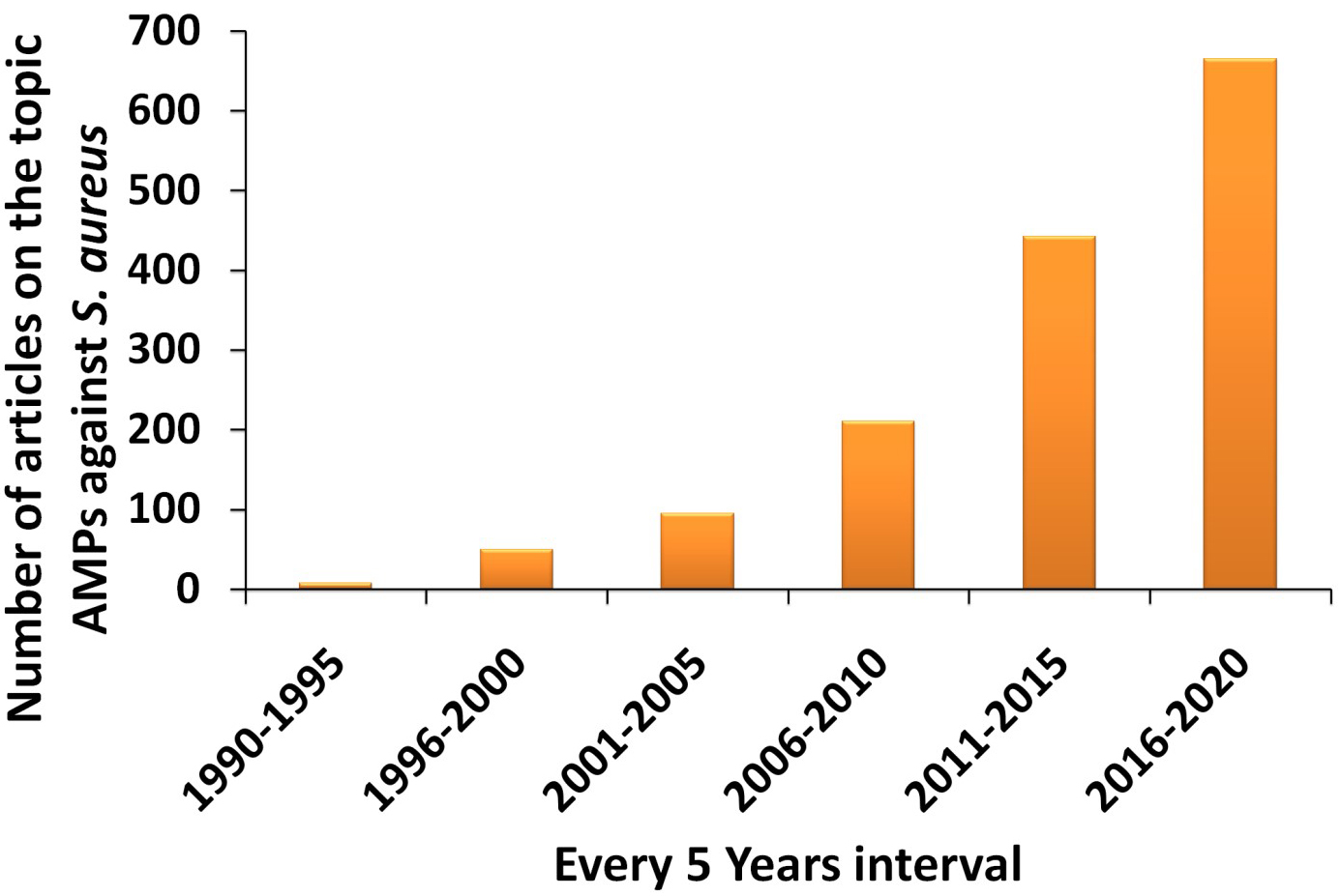
Fig. 3.

Statistical chart showing the increase number of articles on AMPs against S. aureus from year 2000 until 2020.

4.1 AMPs and host innate immune system

Initially, the innate immune system provides the first line of defense against pathogenic invasion and colonization. It consists of physical and chemical defense attributes. The genes encode for host defense peptides as part of the host biological defense; these peptides are also known as antimicrobial peptides [112, 113]. Studies consistently revealed that in response to infections, cationic peptides (like defensins) were produced by phagocytic cells [114]. A human liver-specific peptide is known as hepcidin. Hepcidin is a cysteine-rich peptide that makes its similar to defensins. They have an iron-regulatory activity as well as antimicrobial activity [115]. Reports showed that AMPs from higher organisms have broad-spectrum activities against microorganisms [116]. The expression of genes that encode for the AMPs in mammals is not tissue-specific but occurs throughout the body. Although, reports showed that a combination of host defense molecules is produced at a single site by the expression of multiple AMP due to the coordinated transcriptional regulation of AMP genes [117, 118].

Cathelicidins and defensins are the most abundant human AMPs. The α-defensin family consists of two peptides (HD-5((Human α-defensin-5) and HD-6) found within the small intestine (in paneth cells) and four peptides (HNP (Human Neutrophil Peptide) 1-4) in neutrophils. However, the active forms of HNP1-4 peptides can be part of the oxygen-independent antibacterial mechanism of neutrophils. Moreover, the β-defensins are found throughout the body (in endothelial cells) at various sites. The human β-defensin 1 (hBD1) is expressed constitutively at a low level. Moreover, the hBD2-4 depend on inducing factors including cytokines and microbes [112, 113, 119]. Cathelicidins are cationic AMPs that are also part of innate immunity exhibiting a broad-spectrum antimicrobial activity [120]. The expression of LL-37, a cathelicidin peptide, also depends on induced factors like cytokines and bacteria. They have antibacterial activity and play a role in the host innate immune system [121, 122]. The mCRAMP a murine ortholog of LL-37, coordinates and modulates the host innate immune system in in vivo studies against bacterial invasion and helps in survival but shows weak inhibitory effects against bacteria in vitro [123, 124].

4.2 AMPs as immune regulators

The importance of the host-derived peptides is not only restricted to antimicrobial activity during microbial invasion or phagocytosis. However, they can also cross-talk with adaptive and innate immunity. It was reported that α-defensin has numerous roles in maintaining intact mucosal barriers, innate immunity, and adaptive immunity. They act as a chemotactic agent that modulate the cytokines response of human lymphocytes and monocytes [125]. Further study using anti-infective peptides like Innate Defense Regulator-1 (IDR-1), in response to microbial products (lipopolysaccharide), showed a reduction in inflammation in vivo. It is due to the lower production of the pro-inflammatory cytokines. Thus, the anti-infective peptide promotes the recruitment of monocytes and enhances monocyte chemokines level [126]. The AMPs also have a plethora of activities like immune-modulatory activity (induction or inhibition of pro-inflammatory cytokines, chemotaxis of various leukocytes, enhanced wound healing, anti-inflammatory properties, and chemokine productions), regulate epithelial cell differentiation, anti-angiogenesis or angiogenesis, vasculogenesis, anti-obesity, and modulation of host cell gene expression or mast cell degranulation [127, 128, 129, 130, 131, 132, 133].

4.3 Experimental approach and clinical trials

Numerous approaches like computational design approaches, site-directed mutagenesis, template-assisted methodologies, mechanism-based strategies, and synthetic libraries are in use to design AMPs. Although the strategies are different conceptually, the aim is identical. All these strategies attempt to develop de novo designed synthetic peptides or modify the naturally occurring peptides for improved antibacterial activity [134]. Furthermore, to understand AMPs activity at different pH values, solubility, structure, composition, molecular weight, charge, amphiphilicity, and other parameters, different biophysical and biochemical methods are used [135, 136]. According to the secondary structures, peptides are divided into four groups named as β-sheets peptides, α-helical peptides, extended peptides and loops containing peptides [137, 138]. Further, to understand the 3D-structure of the AMP, the peptide sequence is used for molecular dynamics simulation [4, 15]. The MD simulation helps to predict the possible structure of the peptide in the given time period in an artificial environment created in silico.

Following the characterization, the biological activity of the peptides needs to be evaluated. The antibacterial activities of the peptides against the bacterial strains are evaluated by determining the minimal inhibitory concentrations, and minimum bactericidal concentration, growth curve analysis, disk diffusion assay, live dead cell assay and other assays [140, 141]. However, after obtaining the information about the activity, an in depth understanding of the mechanism of action (which can vary between peptides) is required.

The actual mechanism of action of most AMPs is still not known in detail. The most general mechanism of action is the cytoplasmic membrane pore formation that includes a carpet-like model, barrel-stave model, and toroidal model. The carpet-like model is based on the AMPs concentration. When the AMPs concentration is high, they become accumulated on the cell surface and dissolve in the cell membrane [142]. In the barrel-stave model, the formation of the membrane-spanning pores occurs due to the direct interaction of AMPs into the target membrane [143]. Lastly, in the toroidal model, a curvature is induced in the membrane due to membrane-spanning pores by AMPs with intercalated lipids [144]. Additionally, other mechanisms by which peptides act are charged lipid clustering, electroporation, membrane thinning or thickening, inhibition of protein synthesis machinery, non-bilayer intermediates, modulation of anion carriers, inhibition of cell wall component biosynthesis, nucleic acid targeting, and non-lytic membrane depolarization [145, 146, 147, 148, 149, 150, 151]. The interactions of most cationic AMPs are with the bacterial cell surface. It occurs due to the negatively charged components embedded in lipid bilayers [152, 153]. Thus, the mechanism of action and activity of AMPs is influenced by amino acids chain length, overall charge of the peptide, structural conformation, amino acid composition, hydrophobicity, and amphipathicity [139]. These mechanisms are studied using different biophysical techniques like circular dichroism, FTIR spectroscopy, NMR, and other techniques and by performing various biological assays like cellular leakage assay, cell dyes, time-course of killing assay, and other assays [4, 15, 140, 141]. Lastly, the toxicity of the AMPs is determined to understand its safety for better treatments using different assays like the MTT assay, hemolytic assay, microscopic analysis, in vivo model study, serum stability, and other relevant toxicity assays [4, 15, 141, 154].

Therefore, to study the newly developed or designed AMPs against S. aureus and its resistant strains, the abovementioned experimental approaches, as illustrated in Fig. 4, must be performed with or without further amendments to understand the structure and biological activities of these AMPs and their possible biological applications.
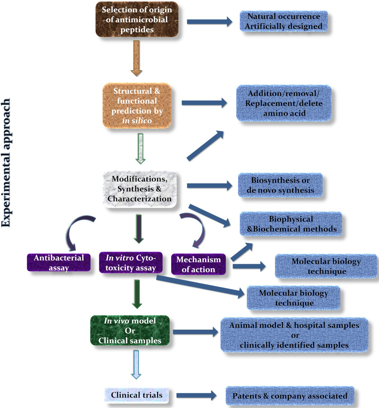
Fig. 4.

Experimental strategy to study AMPs.

The infections caused by the S. aureus strain (i.e. not the resistant strains) can be treated by vancomycin, the last resort antibiotic against gram-positive bacteria. However, vancomycin can cause severe renal toxicity when used at extremely high concentrations. The toxicity also depends on the length of the treatments [157, 158]. Therefore, the infection by these resistant strains is becoming more difficult to treat. Moreover, the situation of community-acquired infections is steadily increasing. Thus, this situation had an impact on the clinical settings. Hence, new antibiotics or alternatives like AMPs are becoming highly necessary, however they must undergo clinical trials to explore their clinical activity. In our recent study, we designed the PBDM (Probiotic Bacteriocin Derived and Modified) peptides from a bacteriocin (m2163) present in Lactobacillus casei and synthesized it. Further, in silico, in vitro, and in vivo studies were performed with these peptides. These studies showed that the peptides were effective against MRSA, VRSA, and wild type S. aureusin vitro [15]. Thus, the shorter length forms of AMPs with modified sequences can be derived from other larger peptides displaying good antibacterial activity. Moreover, the list of peptides depicted in Table 1 shows the growing importance of the AMPs and its variations against S. aureus strains in 2020.

Table 1.Summary of antimicrobial peptides and their variable activities against S. aureus in 2020.
Antimicrobial peptides Origin S. aureus and its variants MIC value Ref.
Enterocin EF35 Enterococcus faecalis ECF35 Staphylococcus aureus < 125 µg/mL [164]
Enterocin TJUQ1 Enterococcus faecium TJUQ1 S. aureus 46.50 ± 1.81 µg/mL [165]
CMP-Van-Lipo (Collagen mimetic peptide tethered vancomycin liposomes) Synthetic Methicillin Resistant S. aureus van concentration (4 to 10 µg/mL) [166]
HF-18 Hagfish intestinal peptide Drug resistant S. aureus 4 µg/mL [167]
HSER-CQDs Retinoic acid receptor responder protein 2 and Carbon quantum dots Vancomycin Resistant S. aureus 25 µg/mL [4]
Fr.A2 and Fr.B1 Camel milk and cow milk S. aureus 130000 µg/mL [168]
Backbone cyclized KR-12 dimers Mammalian defense peptide LL-37 S. aureus 1.25 µM [169]
Short cationic dialkyl lipopeptides (C10)2-KKKK-NH2and (C12)2-KKKK-NH2 Synthetic Methicillin susceptible S. aureus (MSSA), MRSA 4000-16000µg/mL and 8000-32000 µg/mL [170]
TC26 Cyprinus carpio (C-terminal of tissue factor pathway inhibitor 1) S. aureus 10 µM [171]
Alafosfalin; Phosphonopeptides S. aureus, MRSA and MSSA 8 µg/mL; [172]
di-alanyl fosfalin; 16 µg/mL;
β-chloro-L-alanyl-β-chloro-L-alanine; 2-4 µg/mL;
Fosfomycin 16 µg/mL
Melittin (MEL); Bee venom; S. aureus 5 µM; [173]
MEL in presence of pyrrolidinium- based ionic liquids Synthetic 2-200 µM
Fermentaion of Kenaf seed protein produces bioactive peptides Kenaf seed protein S. aureus 4000 µg/mL [174]
Myticusin-beta Mytilus coruscus S. aureus NA [175]
AMP-CBP-FAM Synthetic S. aureus 0.25 µg/mL [176]
Melittin; Apis mellifera MRSA, entertoxin producer S. aureus SEC and SED 7.2 µg/mL, 0.7 µg/mL and 3.6 µg/mL; [177]
Apitoxin 6.7 µg/mL, 7.2 µg/mL and 5.4 µg/mL
S7 (the most effective peptide) Triplet-tryptophan-pivot peptides; Synthetic S. aureus and MRSA 4 and 2 µM [178]
Bacteriocins (Bac22 and Bac4463) capped silver nanoparticle Lactobacillus strains; Synthetic S. aureus 2 and 8 µg/mL [179]
Teixobactin Eleftheria terrae Susceptible S. aureus and MRSA 1 µg/mL [180]
Indole-triazole-peptide conjugate (compound 9 series) Synthetic S. aureus 10-16 µg/mL [181]
Acetylated and non-acetylated QAK Antheraea mylitta; Synthetic S. aureus 7.5 and 60 µg/mL [182]
Intestinalin Clostridium intestinale (LysC); Synthetic S. aureus 6 µg/mL [183]
GAM019 (G19) and GIBIM-P5S9K (G17) peptide encapsulated on poly-lactic-glycolic-acid (PLGA) Synthetic MRSA 0.7 and 0.2 µM [184]
Analog of BSI-9 (compound 9-13;15) BSI-9; Synthetic S. aureus 1-8 µg/mL [185]
TC19 Human thrombocidin-1-derived peptide S. aureus Lethal concentration (3.75 µM in PT buffer; 7.5-15 µM in PBS buffer; 15-30 µM in 50% plasma) [186]
A1-A6; B1-B6 Synthetic and modified version of A1 and B1 MRSA 1-8 µg/mL; 0.5-8 µg/mL [187]
Melittin Purchased from Mimotopes Peptide company (Mulgrave, Australia) MRSA and MSSA 0.5-8 µg/mL; 0.5-2 µg/mL [188, 189]
Short self assembling cationic antimicrobial peptide mimetic library based on 3,5 diaminobenzoic acid scaffold (C2, C4, C6 and C7) Synthetic S. aureus 27, 15.6, 29, and 3.9 µg/mL [190]
RCP (Polypeptide enrich extract) Skin of Rana chensinensis S. aureus 157.8 µg/mL [191]
ID13; DLP4 DLP4 (an insect defensin) S. aureus 4 µg/mL; 16 µg/mL [192]
Pep-cur complex Octaarginine (P) and cumin (C) S. aureus C= 3.0 ± 0.2 nmol [193]
P = 5.2 ± 0.1 nmol
PBDM1 and PBDM2 Lactobacillus casei ATCC 334 (bacteriocin m2163) S. aureus, MRSA and VRSA 10-15 µg/mL; 10 µg/mL [15]
OVTp12 Ovotransferrin hydrolysate (egg) S. aureus 32 µg/mL [194]
C12-KTKCKfKLKC-NH2 and C14-KTKCKfKLKC-NH2 lipopeptide Synthetic S. aureus and MRSA 3.90-31.25 µg/mL [195]
Compound 14 Amphiphilic sofalcone derivatives 1-17; Synthetic S. aureus 1.56 µg/mL [196]
Adepamycin Adenanthera pavonina seeds (sequence of plant trypsin inhibitor) S. aureus 1.8 µM [197]
caP4 Curcuma pseudomontana L. (Zingiberaceae) S. aureus 8 µg/mL [198]
Bac8c; Bovine (improved from Bac2A) S. aureus and MRSA 2 and 8 µg/mL; [199]
Lipoic acid (LA) Bac8c 1 and 4 µg/mL
Polydopamine/hydroxyapatite/nisin (PDA/HAP/Nisin) Synthetic S. aureus 30 µg/mL [200]
Natural peptide; S3K; G2K-S3K Didymocentrus krausi (MK049518) S. aureus 12.5 µg/mL; [154]
3.13-6.25 µg/mL;
3.13-6.25 µg/mL
P6.2 Synthetic S. aureus 12.72 µM [201]
ent A- col E1 Enterocin A and Colicin E1; Synthetic S. aureus 10 µg/mL [202]

The innate immune system associated AMPs are good candidates for the development of the alternatives to antibiotics. However, they exert some cytotoxicity towards normal host cells that limits their inclusion as therapeutics. Thus, to overcome this hurdle, increased AMPs efficacy, stability, and reduced toxicity, strategies like structural analogs or modifications as well as combinations with conventional antibiotics or nanoparticles can be used as shown in Fig. 5 [155]. Zharkova et al., reported in their study that antibiotics with intercellular targets (e.g., rifampicin and gentamicin) and AMPs which are highly membrane-active (e.g., hBD-3 and protegrin 1) showed synergistic effects due to increased bioavailability [159]. Jelinkova et al., showed that the combination of Hecate peptide with vancomycin improved the efficacy of the components after forming the conjugate and reduced the overall toxicity. It allowed the treatment of VRSA, MRSA, and the non-resistant S. aureus [141]. In another study, we used the combination strategy by combining the HSER peptide (derived from retinoic acid receptor responder protein 2) with carbon quantum dots (CQDs) and tested this combination against resistant strains of S. aureus and E. coli. The results revealed that the HSER-CQDs were more effective at a lower concentration in comparison to the individual components. The HSER-CQDs was found to be non-toxic and hemocompatible [4]. Therefore, these combinatorial strategies help to overcome resistance, reduce dosage, enhance the selectivity of the compound, and reduce side effects [160, 161, 162]. AMPs can also be used for targeted delivery or can be encapsulated within some nano-capsule to reduce their toxicity, proper release, and increase stability [156]. Thus, AMPs are molecules which can be modified in various ways to improve their activity for therapeutic purposes based on clinical demands.
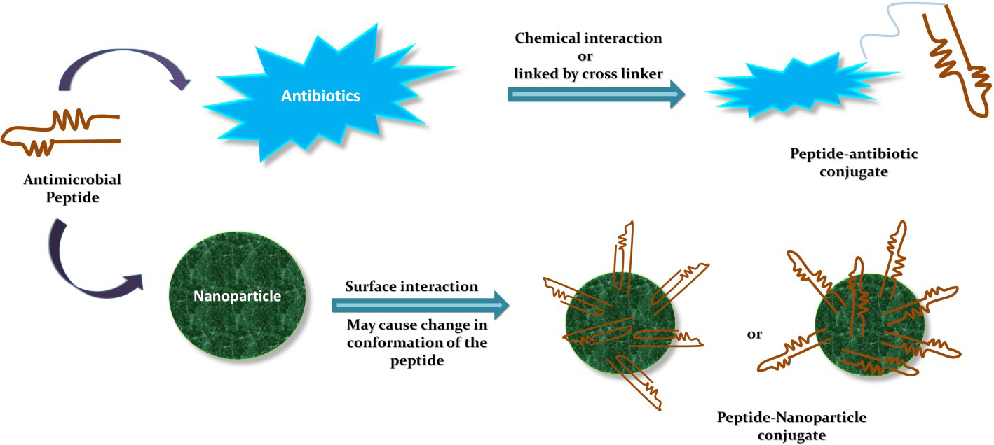
Fig. 5.

Combinatorial strategies using AMPs.

4.4 AMPs against S. aureus and the present status of AMPs

Moreover, the studies on in vivo infection models helped to understand the efficacy of the AMPs for treating infections in the mammalian systems. At the beginning of this section, we discuss PBDM peptides. An in vivo study with VRSA infected BALB/c mice model showed that these PBDM peptides were able to treat VRSA infection and cure them without visible side effects [15]. Another study by Fan Yu, et al., showed a new concept of entrapping bacteria instead of killing by using an HDMP (human defensin-6-mimic peptide). HDMP inhibits bacterial invasion by trapping bacteria using its self-assembled fibrous network. This mechanism was apparent with both S. aureus- and MRSA-infected BALB/c mouse model. However, HDMP is active only against gram-positive bacteria but not against gram-negative bacteria [163]. Thus, these in vivo studies helped to understand the activity of this peptide in the live system. These in vivo studies will help to determine the future of these peptides whether it will be heading towards the clinical studies or towards the need to generate modified peptides or to use these peptides as conjugates or entrap them in a capsule to reduce their toxicity and targeted delivery.

Although a great deal of research is focused on AMPs, only a very small number of AMPs is in clinical use when compared to conventional antibiotics. Some of the peptides that are undergoing clinical trials are Pexiganan (MRSA,) LTX-109 (against nasal colonies of MRSA/MSSA), MU1140 (MRSA), NP432 (MRSA), and AP138 (MRSA implant infections). Most of them are undergoing preclinical evaluation excluding Pexiganan which is undergoing Phase III clinical testing whereas LTX-109 is under clinical phase I/II. Even if the whole process of clinical trials takes more than 8 years (Phase I 1.5 years; Phase II 2 years; Phase III 3 years; introduction to the market 1.5 years; Phase IIIb/IV- for further clinical use and re-confirmations of its use) to eventually bring a drug into the market for proper medical use [203]. Therefore, it is necessary to launch a greater number of clinical trials to bring out the potential of AMPs in the world of therapeutics against the ever growing bacterial infections especially with antibiotic resistant bacteria.

5. Concluding remarks

The infections caused by the MRSA and VRSA strains have become a serious health issue due to their resistance to antibiotics. Thus, the treatments against these strains are prolonged and/or can end up in a failure. The discussion about their mechanisms of antibiotic resistance will not only help the researchers to target specific genes and proteins for the development of new therapeutics but it will also help them to further investigate the unsolved tasks for a better understanding of the pathogenesis.

Though many efforts are aimed at producing new antibiotics, the choice of an alternative to antibiotics can also be an interesting approach. There are reports of resistance towards AMPs which can lead to the development of future resistance to host AMPs. However, this can be overcome by synthetically modified peptides [156, 204]. Apart from this, the limitation of its high cost of production, degradation by proteases, toxicity, and their unknown pharmacokinetics can also be a problem [205]. However, these can be solved by the use of relatively shorter peptides that will reduce the overall production costs. The use of different drug delivery systems can reduce their toxicity, maintain proper release, and improve stability. They can also be used in combination with antibiotics for better activity and can overcome resistance. Thus, combination strategies for therapy will become the modality of choice to overcome antibiotic resistance. Despite their limitation, the growing interest in AMPs as an alternative to antibiotics against S. aureus will improve its status in the future for developing new therapeutics. It will be an interesting subject of research to explore these AMPs as an alternative to antibiotics not only against the S. aureus but also against other pathogens.

Author contributions

M. conceived and designed the framework of the articles, wrote the paper, prepared the figures and thoroughly checked the paper; V.A. supervised the work and checked the paper for detail correction.

Acknowledgment

Thanks to all the peer reviewers and editors for their opinions and suggestions.

Funding

This work was financially supported by ERDF “Multidisciplinary research to increase application potential of nanomaterials in agricultural practice” (No. CZ.02.1.01/0.0/0.0/16_025/0007314) and CEITEC 2020 (LQ1601) with financial support from the Ministry of Education, Youth and Sports of the Czech Republic under the National Sustainability Programme II is gratefully acknowledged.

Conflict of interest

The authors declare no conflict of interest.

References
[1]
Tacconelli E, Carrara E, Savoldi A, Harbarth S, Mendelson M, Monnet DL, et al. Discovery, research, and development of new antibiotics: the WHO priority list of antibiotic-resistant bacteria and tuberculosis. The Lancet Infectious Diseases. 2018; 18: 318-327.
[2]
Agyepong N, Govinden U, Owusu-Ofori A, Essack SY. Multidrug-resistant gram-negative bacterial infections in a teaching hospital in Ghana. Antimicrobial Resistance and Infection Control. 2018; 7: 37.
[3]
Singh N, Manchanda V. Control of multidrug-resistant Gram-negative bacteria in low- and middle-income countries—high impact interventions without much resources. Clinical Microbiology and Infection. 2017; 23: 216-218.
[4]
Mazumdar A, Haddad Y, Milosavljevic V, Michalkova H, Guran R, Bhowmick S, et al. Peptide-carbon quantum dots conjugate, derived from human retinoic acid receptor responder protein 2, against antibiotic-resistant gram positive and gram negative pathogenic bacteria. Nanomaterials. 2020; 10: 325.
[5]
Ventola CL. The antibiotic resistance crisis: part 1: causes and threats. Pharmacy and therapeutics.2015; 40: 277.
[6]
Hanretty AM, Gallagher JC. Shortened courses of antibiotics for bacterial infections: a systematic review of randomized controlled trials. Pharmacotherapy. 2018; 38: 674-687.
[7]
Robinson TP, Bu DP, Carrique-Mas J, Fèvre EM, Gilbert M, Grace D, et al. Antibiotic resistance is the quintessential one Health issue. Transactions of the Royal Society of Tropical Medicine and Hygiene. 2016; 110: 377-380.
[8]
Jelinkova P, Mazumdar A, Sur VP, Kociova S, Dolezelikova K, Jimenez AMJ, et al. Nanoparticle-drug conjugates treating bacterial infections. Journal of Controlled Release. 2019; 307: 166-185.
[9]
Tan R, Liu J, Li M, Huang J, Sun J, Qu H. Epidemiology and antimicrobial resistance among commonly encountered bacteria associated with infections and colonization in intensive care units in a university-affiliated hospital in Shanghai. Journal of Microbiology, Immunology, and Infection. 2014; 47: 87-94.
[10]
Centers for Disease Control and Prevention Antibiotic resistance threats in the united states, 2019. 2019. Available at: http://dx.doi.org/10.15620/cdc:82532 (Accessed: 13 November 2019).
[11]
Davies J, Davies D. Origins and evolution of antibiotic resistance. Microbiology and Molecular Biology Reviews. 2010; 74: 417-433.
[12]
McNeece G, Naughton V, Woodward MJ, Dooley JSG, Naughton PJ. Array based detection of antibiotic resistance genes in Gram negative bacteria isolated from retail poultry meat in the UK and Ireland. International Journal of Food Microbiology. 2014; 179: 24-32.
[13]
Juknius T, Tamulevičius T, Gražulevičiūtė I, Klimienė I, Matusevičius AP, Tamulevičius S. In-situ measurements of bacteria resistance to antimicrobial agents employing leaky mode sub-wavelength diffraction grating. Sensors and Actuators B: Chemical. 2014; 204: 799-806.
[14]
Wang S, Wang Q, Zeng X, Ye Q, Huang S, Yu H, et al. Use of the antimicrobial peptide sublancin with combined antibacterial and immunomodulatory activities to protect against methicillin-resistant Staphylococcus aureus infection in mice. Journal of Agricultural and Food Chemistry. 2017; 65: 8595-8605.
[15]
Mazumdar A, Haddad Y, Sur VP, Milosavljevic V, Bhowmick S, Michalkova H, et al. Characterization and in vitro analysis of probiotic-derived peptides against multi drug resistance bacterial infections. Frontiers in Microbiology. 2020; 11: 1963.
[16]
Brunetti J, Falciani C, Roscia G, Pollini S, Bindi S, Scali S, et al. In vitro and in vivo efficacy, toxicity, bio-distribution and resistance selection of a novel antibacterial drug candidate. Scientific Reports. 2016; 6: 26077.
[17]
Pérez-Montarelo D, Viedma E, Murcia M, Muñoz-Gallego I, Larrosa N, Brañas P, et al. Pathogenic characteristics of Staphylococcus aureus endovascular infection isolates from different clonal complexes. Frontiers in Microbiology. 2017; 8: 917.
[18]
Votintseva AA, Fung R, Miller RR, Knox K, Godwin H, Wyllie DH, et al. Prevalence of Staphylococcus aureus protein a (spa) mutants in the community and hospitals in Oxfordshire. BMC Microbiology. 2014; 14: 63.
[19]
Liu Y, Wang H, Du N, Shen E, Chen H, Niu J, et al. Molecular evidence for spread of two major methicillin-resistant Staphylococcus aureus clones with a unique geographic distribution in Chinese hospitals. Antimicrobial Agents and Chemotherapy. 2009; 53: 512-518.
[20]
McGuinness WA, Malachowa N, DeLeo FR. Vancomycin Resistance in Staphylococcus aureus. The Yale Journal of Biology and Medicine. 2017; 90: 269-281.
[21]
Thomer L, Schneewind O, Missiakas D. Pathogenesis of Staphylococcus aureus bloodstream infections. Annual Review of Pathology. 2016; 11: 343-364.
[22]
Eriksen KR. “Celbenin”-resistant staphylococci. Ugeskr Laeger. 1961; 123: 384-386.
[23]
WHO. Antimicrobial resistance. 2020. Available at: https://www.who.int/news-room/fact-sheets/detail/antimicrobial-resistance (Accessed: 31 July 2020).
[24]
Fasihi Y, Kiaei S, Kalantar-Neyestanaki D. Characterization of SCCmec and spa types of methicillin-resistant Staphylococcus aureus isolates from health-care and community-acquired infections in Kerman, Iran. Journal of Epidemiology and Global Health. 2017; 7: 263-267.
[25]
Gurung RR, Maharjan P, Chhetri GG. Antibiotic resistance pattern of Staphylococcus aureus with reference to MRSA isolates from pediatric patients. Future Science OA. 2020; 6: FSO464.
[26]
P R V, M J. A comparative analysis of community acquired and hospital acquired methicillin resistant Staphylococcus aureus. Journal of Clinical and Diagnostic Research. 2013; 7: 1339-1342.
[27]
Centers for Disease Control and Prevention (CDC). Severe methicillin-resistant Staphylococcus aureus community-acquired pneumonia associated with influenza-Louisiana and Georgia, December 2006-January 2007. Morbidity and mortality weekly report. 2007; 56: 325.
[28]
Tsuji BT, Rybak MJ, Cheung CM, Amjad M, Kaatz GW. Community- and health care-associated methicillin-resistant Staphylococcus aureus: a comparison of molecular epidemiology and antimicrobial activities of various agents. Diagnostic Microbiology and Infectious Disease. 2007; 58: 41-47.
[29]
Ma XX, Ito T, Tiensasitorn C, Jamklang M, Chongtrakool P, Boyle-Vavra S, et al. Novel type of staphylococcal cassette chromosome mec identified in community-acquired methicillin-resistant Staphylococcus aureus strains. Antimicrobial Agents and Chemotherapy. 2002; 46: 1147-1152.
[30]
Ito T, Ma XX, Takeuchi F, Okuma K, Yuzawa H, Hiramatsu K. Novel type V staphylococcal cassette chromosome mec driven by a novel cassette chromosome recombinase, ccrC. Antimicrobial Agents and Chemotherapy. 2004; 48: 2637-2651.
[31]
Baggett HC, Hennessy TW, Rudolph K, Bruden D, Reasonover A, Parkinson A, et al. Community-onset methicillin-resistant Staphylococcus aureus associated with antibiotic use and the cytotoxin Panton-Valentine leukocidin during a furunculosis outbreak in rural Alaska. the Journal of Infectious Diseases. 2004; 189: 1565-1573.
[32]
Otto M. A MRSA-terious enemy among us: end of the PVL controversy? Nature Medicine. 2011; 17: 169-170.
[33]
Hoppe P, Holzhauer S, Lala B, Bührer C, Gratopp A, Hanitsch LG, et al. Severe infections of Panton-Valentine leukocidin positive Staphylococcus aureus in children. Medicine. 2019; 98: e17185.
[34]
Bhakdi S, Tranum-Jensen J. Alpha-toxin of Staphylococcus aureus. Microbiological Reviews. 1991; 55: 733-751.
[35]
Friedman ND, Kaye KS, Stout JE, McGarry SA, Trivette SL, Briggs JP, et al. Health care–associated bloodstream infections in adults: a reason to change the accepted definition of community-acquired infections. Annals of Internal Medicine. 2002; 137: 791-797.
[36]
Wang R, Braughton KR, Kretschmer D, Bach TL, Queck SY, Li M, et al. Identification of novel cytolytic peptides as key virulence determinants for community-associated MRSA. Nature Medicine. 2007; 13: 1510-1514.
[37]
Wang X, Li X, Liu W, Huang W, Fu Q, Li M. Molecular characteristic and virulence gene profiles of community-associated methicillin-resistant Staphylococcus aureus isolates from pediatric patients in Shanghai, China. Frontiers in Microbiology. 2016; 7: 1818.
[38]
Loomba PS, Taneja J, Mishra B. Methicillin and vancomycin resistant S. aureus in hospitalized patients. Journal of Global Infectious Diseases. 2010; 2: 275-283.
[39]
Chen Y, Liu Z, Duo L, Xiong J, Gong Y, Yang J, et al. Characterization of Staphylococcus aureus from distinct geographic locations in China: an increasing prevalence of spa-t030 and SCCmec type III. PloS ONE. 2014; 9: e96255.
[40]
Deurenberg RH, Stobberingh EE. The evolution of Staphylococcus aureus. Infection, Genetics and Evolution. 2008; 8: 747-763.
[41]
Werner G, Strommenger B, Witte W. Acquired vancomycin resistance in clinically relevant pathogens. Future Microbiology. 2008; 3: 547-562.
[42]
Boyle-Vavra S. Development of vancomycin and lysostaphin resistance in a methicillin-resistant Staphylococcus aureus isolate. Journal of Antimicrobial Chemotherapy. 2001; 48: 617-625.
[43]
Boyle-Vavra S, Challapalli M, Daum RS. Resistance to Autolysis in Vancomycin-Selected Staphylococcus aureus Isolates Precedes Vancomycin-Intermediate Resistance. Antimicrobial Agents and Chemotherapy. 2003; 47: 2036-2039.
[44]
Cui L, Ma X, Sato K, Okuma K, Tenover FC, Mamizuka EM, et al. Cell wall thickening is a common feature of vancomycin resistance in Staphylococcus aureus. Journal of Clinical Microbiology. 2003; 41: 5-14.
[45]
Muthaiyan A, Jayaswal RK, Wilkinson BJ. Intact mutS in laboratory-derived and clinical glycopeptide-intermediate Staphylococcus aureus strains. Antimicrobial Agents and Chemotherapy. 2004; 48: 623-625.
[46]
Peng H, Hu Q, Shang W, Yuan J, Zhang X, Liu H, et al. WalK(S221P), a naturally occurring mutation, confers vancomycin resistance in VISA strain XN108. the Journal of Antimicrobial Chemotherapy. 2017; 72: 1006-1013.
[47]
Hu J, Zhang X, Liu X, Chen C, Sun B. Mechanism of reduced vancomycin susceptibility conferred by walK mutation in community-acquired methicillin-resistant Staphylococcus aureus strain MW2. Antimicrobial Agents and Chemotherapy. 2015; 59: 1352-1355.
[48]
McEvoy CRE, Tsuji B, Gao W, Seemann T, Porter JL, Doig K, et al. Decreased vancomycin susceptibility in Staphylococcus aureus caused by is256 tempering of WalKR expression. Antimicrobial Agents and Chemotherapy. 2013; 57: 3240-3249.
[49]
Yoo JI, Kim JW, Kang GS, Kim HS, Yoo JS, Lee YS. Prevalence of amino acid changes in the yvqF, vraSR, graSR, and tcaRAB genes from vancomycin intermediate resistant Staphylococcus aureus. Journal of Microbiology (Seoul, Korea). 2013; 51: 160-165.
[50]
Hu Q, Peng H, Rao X. Molecular events for promotion of vancomycin resistance in vancomycin intermediate Staphylococcus aureus. Frontiers in Microbiology. 2016; 7: 1601.
[51]
Gardete S, Kim C, Hartmann BM, Mwangi M, Roux CM, Dunman PM, et al. Genetic pathway in acquisition and loss of vancomycin resistance in a methicillin resistant Staphylococcus aureus (MRSA) strain of clonal type USA300. PLoS Pathogens. 2012; 8: e1002505.
[52]
Howden BP, Davies JK, Johnson PDR, Stinear TP, Grayson ML. Reduced vancomycin susceptibility in Staphylococcus aureus, including vancomycin-intermediate and heterogeneous vancomycin-intermediate strains: resistance mechanisms, laboratory detection, and clinical implications. Clinical Microbiology Reviews. 2010; 23: 99-139.
[53]
Roch M, Clair P, Renzoni A, Reverdy M, Dauwalder O, Bes M, et al. Exposure of Staphylococcus aureus to subinhibitory concentrations of β-lactam antibiotics induces heterogeneous vancomycin-intermediate Staphylococcus aureus. Antimicrobial Agents and Chemotherapy. 2014; 58: 5306-5314.
[54]
Uttley AHC, George RC, Naidoo J, Woodford N, Johnson AP, Collins CH, et al. High-level vancomycin-resistant enterococci causing hospital infections. Epidemiology and Infection. 1989; 103: 173-181.
[55]
Uttley AH, Collins CH, Naidoo J, George RC. Vancomycin-resistant enterococci. Lancet (London, England). 1988; 1: 57-58.
[56]
Leclercq R, Derlot E, Weber M, Duval J, Courvalin P. Transferable vancomycin and teicoplanin resistance in Enterococcus faecium. Antimicrobial Agents and Chemotherapy. 1989; 33: 10-15.
[57]
Shlaes DM, Bouvet A, Devine C, Shlaes JH, al-Obeid S, Williamson R. Inducible, transferable resistance to vancomycin in Enterococcus faecalis a256. Antimicrobial Agents and Chemotherapy. 1989; 33: 198-203.
[58]
Leclercq R, Derlot E, Duval J, Courvalin P. Plasmid-mediated resistance to vancomycin and teicoplanin in Enterococcus faecium. the New England Journal of Medicine. 1988; 319: 157-161.
[59]
Simjee S, White DG, McDermott PF, Wagner DD, Zervos MJ, Donabedian SM, et al. Characterization of Tn1546 in vancomycin-resistant Enterococcus faecium isolated from canine urinary tract infections: evidence of gene exchange between human and animal enterococci. Journal of Clinical Microbiology. 2002; 40: 4659-4665.
[60]
Noble WC, Virani Z, Cree RG. Co-transfer of vancomycin and other resistance genes from Enterococcus faecalis NCTC 12201 to Staphylococcus aureus. FEMS Microbiology Letters. 1992; 72: 195-198.
[61]
Warren DK, Nitin A, Hill C, Fraser VJ, Kollef MH. Occurrence of co-colonization or co-infection with vancomycin-resistant enterococci and methicillin-resistant Staphylococcus aureus in a medical intensive care unit. Infection Control and Hospital Epidemiology. 2004; 25: 99-104.
[62]
Reyes K, Malik R, Moore C, Donabedian S, Perri M, Johnson L, et al. Evaluation of risk factors for coinfection or cocolonization with vancomycin-resistant enterococcus and methicillin-resistant Staphylococcus aureus. Journal of Clinical Microbiology. 2010; 48: 628-630.
[63]
Chang S, Sievert DM, Hageman JC, Boulton ML, Tenover FC, Downes FP, et al. Infection with vancomycin-resistant Staphylococcus aureus containing the vanA resistance gene. the New England Journal of Medicine. 2003; 348: 1342-1347.
[64]
Centers for Disease Control and Prevention (CDC). Staphylococcus aureus resistant to vancomycin-United States, 2002. Morbidity and mortality weekly report. 2002; 51: 565.
[65]
Franchi D, Climo MW, Wong AH, Edmond MB, Wenzel RP. Seeking vancomycin resistant Staphylococcus aureus among patients with vancomycin-resistant enterococci. Clinical Infectious Diseases. 1999; 29: 1566-1568.
[66]
Walters MS, Eggers P, Albrecht V, Travis T, Lonsway D, Hovan G, et al. Vancomycin-resistant Staphylococcus aureus - delaware, 2015. Morbidity and Mortality Weekly Report. 2015; 64: 1056.
[67]
Challagundla L, Reyes J, Rafiqullah I, Sordelli DO, Echaniz-Aviles G, Velazquez-Meza ME, et al. Phylogenomic classification and the evolution of clonal complex 5 methicillin-resistant Staphylococcus aureus in the western hemisphere. Frontiers in Microbiology. 2018; 9: 1901.
[68]
Jensen SO, Lyon BR. Genetics of antimicrobial resistance in Staphylococcus aureus. Future Microbiology. 2009; 4: 565-582.
[69]
Rammelkamp CH, Maxon T. Resistance of Staphylococcus aureus to the action of penicillin. Experimental Biology and Medicine. 1942; 51: 386-389.
[70]
Lowy FD. Antimicrobial resistance: the example of Staphylococcus aureus. the Journal of Clinical Investigation. 2003; 111: 1265-1273.
[71]
Zhang HZ, Hackbarth CJ, Chansky KM, Chambers HF. A proteolytic transmembrane signaling pathway and resistance to beta-lactams in staphylococci. Science. 2001; 291: 1962-1965.
[72]
Peacock SJ, Paterson GK. Mechanisms of methicillin resistance in Staphylococcus aureus. Annual Review of Biochemistry. 2015; 84: 577-601.
[73]
Lim D, Strynadka NC. Structural basis for the beta lactam resistance of PBP2a from methicillin-resistant Staphylococcus aureus. Nature structural biology. 2002; 9: 870-876.
[74]
Malachowa N, DeLeo FR. Mobile genetic elements of Staphylococcus aureus. Cellular and Molecular Life Sciences. 2010; 67: 3057-3071.
[75]
Thurlow LR, Joshi GS, Clark JR, Spontak JS, Neely CJ, Maile R, et al. Functional modularity of the arginine catabolic mobile element contributes to the success of USA300 methicillin-resistant Staphylococcus aureus. Cell Host & Microbe. 2013; 13: 100-107.
[76]
Harrison EM, Ba X, Blane B, Ellington MJ, Loeffler A, Hill RLR, et al. PBP2a substitutions linked to ceftaroline resistance in MRSA isolates from the UK. the Journal of Antimicrobial Chemotherapy. 2016; 71: 268-269.
[77]
Kelley WL, Jousselin A, Barras C, Lelong E, Renzoni A. Missense mutations in PBP2a Affecting ceftaroline susceptibility detected in epidemic hospital-acquired methicillin-resistant Staphylococcus aureus clonotypes ST228 and ST247 in Western Switzerland archived since 1998. Antimicrobial Agents and Chemotherapy. 2015; 59: 1922-1930.
[78]
Gostev V, Kalinogorskaya O, Kruglov A, Lobzin Y, Sidorenko S. Characterisation of methicillin-resistant Staphylococcus aureus with reduced susceptibility to ceftaroline collected in Russia during 2010-2014. Journal of Global Antimicrobial Resistance. 2018; 12: 21-23.
[79]
Chan LC, Basuino L, Diep B, Hamilton S, Chatterjee SS, Chambers HF. Ceftobiprole- and ceftaroline-resistant methicillin-resistant Staphylococcus aureus. Antimicrobial Agents and Chemotherapy. 2015; 59: 2960-2963.
[80]
Jones T, Yeaman MR, Sakoulas G, Yang S, Proctor RA, Sahl H, et al. Failures in clinical treatment of Staphylococcus aureus Infection with daptomycin are associated with alterations in surface charge, membrane phospholipid asymmetry, and drug binding. Antimicrobial Agents and Chemotherapy. 2008; 52: 269-278.
[81]
Ernst CM, Slavetinsky CJ, Kuhn S, Hauser JN, Nega M, Mishra NN, et al. Gain-of-function mutations in the phospholipid flippase MprF confer specific daptomycin resistance. MBio. 2018; 9: e01659-18.
[82]
Barros EM, Martin MJ, Selleck EM, Lebreton F, Sampaio JLM, Gilmore MS. Daptomycin resistance and tolerance due to loss of function in staphylococcus aureus dsp1 and asp23. Antimicrobial Agents and Chemotherapy. 2018; 63: e01542-18.
[83]
Wang Y, Lv Y, Cai J, Schwarz S, Cui L, Hu Z, et al. A novel gene, optrA, that confers transferable resistance to oxazolidinones and phenicols and its presence in Enterococcus faecalis and Enterococcus faecium of human and animal origin. The Journal of Antimicrobial Chemotherapy. 2015; 70: 2182-2190.
[84]
Livermore DM. Linezolid in vitro: mechanism and antibacterial spectrum. Journal of Antimicrobial Chemotherapy. 2003; 51: ii9-ii16.
[85]
Krause KM, Serio AW, Kane TR, Connolly LE. Aminoglycosides: an Overview. Cold Spring Harbor Perspectives in Medicine. 2016; 6: a027029.
[86]
Beabout K, Hammerstrom TG, Perez AM, Magalhães BF, Prater AG, Clements TP, et al. The ribosomal S10 protein is a general target for decreased tigecycline susceptibility. Antimicrobial Agents and Chemotherapy. 2015; 59: 5561-5566.
[87]
Hooper DC, Jacoby GA. Mechanisms of drug resistance: quinolone resistance. Annals of the New York Academy of Sciences. 2015; 1354: 12-31.
[88]
Costa SS, Falcão C, Viveiros M, Machado D, Martins M, Melo-Cristino J, et al. Exploring the contribution of efflux on the resistance to fluoroquinolones in clinical isolates of Staphylococcus aureus. BMC Microbiology. 2011; 11: 241.
[89]
Muñoz-Bellido JL, Alonzo Manzanares M, Martínez Andrés JA, Guttiérrez Zufiaurre MN, Ortiz G, Segovia Hernández M, et al. Efflux pump-mediated quinolone resistance in Staphylococcus aureus strains wild type for gyrA, gyrB, grlA, and norA. Antimicrobial Agents and Chemotherapy. 1999; 43: 354-356.
[90]
Wassenaar TM, Ussery D, Nielsen LN, Ingmer H. Review and phylogenetic analysis of qac genes that reduce susceptibility to quaternary ammonium compounds in Staphylococcus species. European Journal of Microbiology & Immunology. 2015; 5: 44-61.
[91]
Schwarz S, Shen J, Kadlec K, Wang Y, Brenner Michael G, Feßler AT, et al. Lincosamides, streptogramins, phenicols, and pleuromutilins: mode of action and mechanisms of resistance. Cold Spring Harbor Perspectives in Medicine. 2016; 6: a027037.
[92]
Novak R. Are pleuromutilin antibiotics finally fit for human use? Annals of the New York Academy of Sciences. 2012; 1241: 71-81.
[93]
Patel J, Gorwitz R, Jernigan J. Mupirocin resistance. Clinical Infectious Diseases. 2009; 49: 935-941.
[94]
Nunes EL, dos Santos KR, Mondino PJ, Bastos MDC, Giambiagi-deMarval M. Detection of ileS-2 gene encoding mupirocin resistance in methicillin-resistant Staphylococcus aureus by multiplex PCR. Diagnostic Microbiology and Infectious Disease. 1999; 34: 77-81.
[95]
Zeng D, Debabov D, Hartsell TL, Cano RJ, Adams S, Schuyler JA, et al. Approved glycopeptide antibacterial drugs: mechanism of action and resistance. Cold Spring Harbor Perspectives in Medicine. 2016; 6: a026989.
[96]
Dmitriev BA, Toukach FV, Holst O, Rietschel ET, Ehlers S. Tertiary structure of Staphylococcus aureus cell wall murein. Journal of Bacteriology. 2004; 186: 7141-7148.
[97]
Tomasz A. The staphylococcal cell wall. In Fischetti VA, Novick RP, Ferretti JJ, Portnoy DA, Rood JI (eds.) Gram-Positive Pathogens (pp. 443-455). 2nd edn. American Society of Microbiology. 2006.
[98]
Arthur M, Molinas C, Depardieu F, Courvalin P. Characterization of Tn1546, a Tn3-related transposon conferring glycopeptide resistance by synthesis of depsipeptide peptidoglycan precursors in Enterococcus faecium BM4147. Journal of Bacteriology. 1993; 175: 117-127.
[99]
Bugg TD, Wright GD, Dutka-Malen S, Arthur M, Courvalin P, Walsh CT. Molecular basis for vancomycin resistance in Enterococcus faecium BM4147: biosynthesis of a depsipeptide peptidoglycan precursor by vancomycin resistance proteins VanH and VanA. Biochemistry. 1991; 30: 10408-10415.
[100]
Hong H, Hutchings MI, Buttner MJ. Vancomycin resistance VanS/VanR two-component systems. Advances in Experimental Medicine and Biology. 2008; 631: 200-213.
[101]
Abadía Patiño L, Courvalin P, Perichon B. VanE gene cluster of vancomycin-resistant Enterococcus faecalis BM4405. Journal of Bacteriology. 2002; 184: 6457-6464.
[102]
Périchon B, Courvalin P. VanA-type vancomycin-resistant Staphylococcus aureus. Antimicrobial Agents and Chemotherapy. 2009; 53: 4580-4587.
[103]
Gardete S, Tomasz A. Mechanisms of vancomycin resistance in Staphylococcus aureus. Journal of Clinical Investigation. 2014; 124: 2836-2840.
[104]
Katayama Y, Sekine M, Hishinuma T, Aiba Y, Hiramatsu K. Complete reconstitution of the vancomycin-intermediate Staphylococcus aureus phenotype of strain Mu50 in vancomycin-susceptible S. aureus. Antimicrobial Agents and Chemotherapy. 2016; 60: 3730-3742.
[105]
Matsuo M, Cui L, Kim J, Hiramatsu K. Comprehensive identification of mutations responsible for heterogeneous vancomycin-intermediate Staphylococcus aureus (hVISA)-to-VISA conversion in laboratory-generated VISA strains derived from hVISA clinical strain Mu3. Antimicrobial Agents and Chemotherapy. 2013; 57: 5843-5853.
[106]
Howden BP, Peleg AY, Stinear TP. The evolution of vancomycin intermediate Staphylococcus aureus (VISA) and heterogenous-VISA. Infection, Genetics and Evolution. 2014; 21: 575-582.
[107]
Howden BP, McEvoy CRE, Allen DL, Chua K, Gao W, Harrison PF, et al. Evolution of multidrug resistance during Staphylococcus aureus infection involves mutation of the essential two component regulator WalKR. PLoS Pathogens. 2011; 7: e1002359.
[108]
Fleming A. On a remarkable bacteriolytic element found in tissues and secretions. Proceedings of the Royal Society of London. Series B, Containing Papers of a Biological Character. 1922; 93: 306-317.
[109]
Dubos RJ. Studies on a bactericidal agent extracted from a soil bacillus: I. preparation of the agent. Its activity in vitro. The Journal of Experimental Medicine. 1939; 70: 1-10.
[110]
Lai Y, Gallo RL. AMPed up immunity: how antimicrobial peptides have multiple roles in immune defense. Trends in Immunology. 2009; 30: 131-141.
[111]
Hancock REW, Rozek A. Role of membranes in the activities of antimicrobial cationic peptides. FEMS Microbiology Letters. 2002; 206: 143-149.
[112]
Devine DA, Hancock RE. Mammalian host defense peptides. Cambridge University Press. 2004.
[113]
Kaiser V, Diamond G. Expression of mammalian defensin genes. Journal of Leukocyte Biology. 2000; 68: 779-784.
[114]
Zeya HI, Spitznagel JK. Antibacterial and enzymic basic proteins from leukocyte lysosomes: separation and identification. Science. 1936; 142: 1085-1087.
[115]
Cuesta A, Meseguer J, Esteban MA. The antimicrobial peptide hepcidin exerts an important role in the innate immunity against bacteria in the bony fish gilthead seabream. Molecular Immunology. 2008; 45: 2333-2342.
[116]
Brogden KA. Antimicrobial peptides: pore formers or metabolic inhibitors in bacteria? Nature Reviews. Microbiology. 2005; 3: 238-250.
[117]
Laube DM, Yim S, Ryan LK, Kisich KO, Diamond G. Antimicrobial peptides in the airway. Current Topics in Microbiology and Immunology. 2006; 306: 153-182.
[118]
Russell JP, Diamond G, Tarver AP, Scanlin TF, Bevins CL. Coordinate induction of two antibiotic genes in tracheal epithelial cells exposed to the inflammatory mediators lipopolysaccharide and tumor necrosis factor alpha. Infection and Immunity. 1996; 64: 1565-1568.
[119]
O’Neil DA. Regulation of expression of β-defensins: endogenous enteric peptide antibiotics. Molecular Immunology. 2003; 40: 445-450.
[120]
Kościuczuk EM, Lisowski P, Jarczak J, Strzałkowska N, Jóźwik A, Horbańczuk J, et al. Cathelicidins: family of antimicrobial peptides. a review. Molecular Biology Reports. 2012; 39: 10957-10970.
[121]
Bals R, Wang X, Zasloff M, Wilson JM. The peptide antibiotic LL-37/hCAP-18 is expressed in epithelia of the human lung where it has broad antimicrobial activity at the airway surface. Proceedings of the National Academy of Sciences of the United States of America. 1998; 95: 9541-9546.
[122]
Hosokawa I, Hosokawa Y, Komatsuzawa H, Goncalves RB, Karimbux N, Napimoga MH, et al. Innate immune peptide LL-37 displays distinct expression pattern from beta-defensins in inflamed gingival tissue. Clinical and Experimental Immunology. 2006; 146: 218-225.
[123]
Ganz T. Defensins: antimicrobial peptides of innate immunity. Nature Reviews. Immunology. 2003; 3: 710-720.
[124]
Tjabringa GS, Ninaber DK, Drijfhout JW, Rabe KF, Hiemstra PS. Human cathelicidin LL-37 is a chemoattractant for eosinophils and neutrophils that acts via formyl-peptide receptors. International Archives of Allergy and Immunology. 2006; 140: 103-112.
[125]
Grigat J, Soruri A, Forssmann U, Riggert J, Zwirner J. Chemoattraction of macrophages, T lymphocytes, and mast cells is evolutionarily conserved within the human α-defensin family. The Journal of Immunology. 2007; 179: 3958-3965.
[126]
Scott MG, Dullaghan E, Mookherjee N, Glavas N, Waldbrook M, Thompson A, et al. An anti-infective peptide that selectively modulates the innate immune response. Nature Biotechnology. 2007; 25: 465-472.
[127]
McDermott AM. The role of antimicrobial peptides at the ocular surface. Ophthalmic Research. 2009; 41: 60-75.
[128]
Dodd GT, Mancini G, Lutz B, Luckman SM. The peptide hemopressin acts through CB1 cannabinoid receptors to reduce food intake in rats and mice. the Journal of Neuroscience. 2010; 30: 7369-7376.
[129]
Koczulla R, von Degenfeld G, Kupatt C, Krötz F, Zahler S, Gloe T, et al. An angiogenic role for the human peptide antibiotic LL-37/hCAP-18. the Journal of Clinical Investigation. 2003; 111: 1665-1672.
[130]
Tokumaru S, Sayama K, Shirakata Y, Komatsuzawa H, Ouhara K, Hanakawa Y, et al. Induction of keratinocyte migration via transactivation of the epidermal growth factor receptor by the antimicrobial peptide LL-37. Journal of Immunology. 2005; 175: 4662-4668.
[131]
Bowdish DME, Davidson DJ, Scott MG, Hancock REW. Immunomodulatory activities of small host defense peptides. Antimicrobial Agents and Chemotherapy. 2005; 49: 1727-1732.
[132]
Prasad SV, Fiedoruk K, Daniluk T, Piktel E, Bucki R. Expression and function of host defense peptides at inflammation sites. International Journal of Molecular Sciences. 2019; 21: 104.
[133]
Dürr M, Peschel A. Chemokines meet defensins: the merging concepts of chemoattractants and antimicrobial peptides in host defense. Infection and Immunity. 2002; 70: 6515-6517.
[134]
Torres MDT, Sothiselvam S, Lu TK, de la Fuente-Nunez C. Peptide design principles for antimicrobial applications. Journal of Molecular Biology. 2019; 431: 3547-3567.
[135]
Singhal N, Sharma A, Kumari S, Garg A, Rai R, Singh N, et al. Biophysical and biochemical characterization of nascent polypeptide-associated complex of picrophilus torridus and elucidation of its interacting partners. Frontiers in Microbiology. 2020; 11: 915.
[136]
Das T, Carroll J. Biophysical and biochemical characterization of peptide and protein drug product. Pharmaceutical Dosage Forms - Parenteral Medications. 2016; 98: 194-221.
[137]
Steckbeck JD, Deslouches B, Montelaro RC. Antimicrobial peptides: new drugs for bad bugs? Expert Opinion on Biological Therapy. 2014; 14: 11-14.
[138]
Nguyen LT, Haney EF, Vogel HJ. The expanding scope of antimicrobial peptide structures and their modes of action. Trends in Biotechnology. 2011; 29: 464-472.
[139]
Gennaro R, Zanetti M. Structural features and biological activities of the cathelicidin-derived antimicrobial peptides. Biopolymers. 2000; 55: 31-49.
[140]
Raheem N, Straus SK. Mechanisms of action for antimicrobial peptides with antibacterial and antibiofilm functions. Frontiers in Microbiology. 2019; 10: 2866.
[141]
Jelinkova P, Splichal Z, Jimenez AMJ, Haddad Y, Mazumdar A, Sur VP, et al. Novel vancomycin-peptide conjugate as potent antibacterial agent against vancomycin-resistant Staphylococcus aureus. Infection and Drug Resistance. 2018; 11: 1807-1817.
[142]
Pietiäinen M, François P, Hyyryläinen H, Tangomo M, Sass V, Sahl H, et al. Transcriptome analysis of the responses of Staphylococcus aureus to antimicrobial peptides and characterization of the roles of vraDE and vraSR in antimicrobial resistance. BMC Genomics. 2009; 10: 429.
[143]
Ehrenstein G, Lecar H. Electrically gated ionic channels in lipid bilayers. Quarterly Reviews of Biophysics. 1977; 10: 1-34.
[144]
Matsuzaki K, Murase O, Fujii N, Miyajima K. An antimicrobial peptide, magainin 2, induced rapid flip-flop of phospholipids coupled with pore formation and peptide translocation. Biochemistry. 1996; 35: 11361-11368.
[145]
Brogden KA. Antimicrobial peptides: pore formers or metabolic inhibitors in bacteria? Nature Reviews. Microbiology. 2005; 3: 238-250.
[146]
Lohner K. New strategies for novel antibiotics: peptides targeting bacterial cell. General Physiology and Biophysics. 2009; 28: 105-116.
[147]
Epand RM, Epand RF. Bacterial membrane lipids in the action of antimicrobial agents. Journal of Peptide Science. 2011; 17: 298-305.
[148]
Rokitskaya TI, Kolodkin NI, Kotova EA, Antonenko YN. Indolicidin action on membrane permeability: carrier mechanism versus pore formation. Biochimica Et Biophysica Acta. 2011; 1808: 91-97.
[149]
Chan DI, Prenner EJ, Vogel HJ. Tryptophan- and arginine-rich antimicrobial peptides: structures and mechanisms of action. Biochimica Et Biophysica Acta. 2006; 1758: 1184-1202.
[150]
Gifford JL, Hunter HN, Vogel HJ. Lactoferricin. Cellular and Molecular Life Sciences. 2005; 62: 2588-2598.
[151]
Haney EF, Nathoo S, Vogel HJ, Prenner EJ. Induction of non-lamellar lipid phases by antimicrobial peptides: a potential link to mode of action. Chemistry and Physics of Lipids. 2010; 163: 82-93.
[152]
Phoenix D, Dennison S, Harris F. Cationic antimicrobial peptides, in antimicrobial peptides. Wiley-VCH Verlag GmbH & Co. KGaA, Weinheim. 2013; 27652: 853.
[153]
Haney EF, Mansour SC, Hancock REW. Antimicrobial peptides: an introduction. Methods in Molecular Biology. 2017; 1548: 3-22.
[154]
Li Z, Yuan Y, Li S, Deng B, Wang Y. Antibacterial activity of a scorpion-derived peptide and its derivatives in vitro and in vivo. Toxicon. 2020; 186: 35-41.
[155]
Teixeira MC, Carbone C, Sousa MC, Espina M, Garcia ML, Sanchez-Lopez E, et al. Nanomedicines for the delivery of antimicrobial peptides (AMPs). Nanomaterials. 2020; 10: 560.
[156]
Omardien S, Brul S, Zaat SAJ. Antimicrobial activity of cationic antimicrobial peptides against gram-positives: current progress made in understanding the mode of action and the response of bacteria. Frontiers in Cell and Developmental Biology. 2016; 4: 111.
[157]
Barceló-Vidal J, Rodríguez-García E, Grau S. Extremely high levels of vancomycin can cause severe renal toxicity. Infection and Drug Resistance. 2018; 11: 1027-1030.
[158]
Zamoner W, Prado IRS, Balbi AL, Ponce D. Vancomycin dosing, monitoring and toxicity: critical review of the clinical practice. Clinical and Experimental Pharmacology & Physiology. 2019; 46: 292-301.
[159]
Zharkova MS, Orlov DS, Golubeva OY, Chakchir OB, Eliseev IE, Grinchuk TM, et al. Application of antimicrobial peptides of the innate immune system in combination with conventional antibiotics-a novel way to combat antibiotic resistance? Frontiers in cellular and infection microbiology. 2019; 9: 128.
[160]
Lorian V. Antibiotics in laboratory medicine. Lippincott Williams & Wilkins. 2005.
[161]
Turnidge J. Drug–drug combinations. Fundamentals of Antimicrobial Pharmacokinetics and Pharmacodynamics. 2014; 366: 153-198.
[162]
Chou T. Theoretical basis, experimental design, and computerized simulation of synergism and antagonism in drug combination studies. Pharmacological Reviews. 2006; 58: 621-681.
[163]
Fan Y, Li X, He P, Hu X, Zhang K, Fan J, et al. A biomimetic peptide recognizes and traps bacteria in vivo as human defensin-6. Science Advances. 2020; 6: eaaz4767.
[164]
Shastry RP, Arunrenganathan RR, Rai VR. Molecular characterization of enterocin EF35 against human pathogens and its in-silico analysis against human cancer proteins top1 and PI3K. Biocatalysis and Agricultural Biotechnology. 2020; 23: 101485.
[165]
Qiao X, Du R, Wang Y, Han Y, Zhou Z. Purification, characterization and mode of action of enterocin, a novel bacteriocin produced by Enterococcus faecium TJUQ1. International Journal of Biological Macromolecules. 2020; 144: 151-159.
[166]
Thapa RK, Kiick KL, Sullivan MO. Encapsulation of collagen mimetic peptide-tethered vancomycin liposomes in collagen-based scaffolds for infection control in wounds. Acta Biomaterialia. 2020; 103: 115-128.
[167]
Jiang M, Yang X, Wu H, Huang Y, Dou J, Zhou C, et al. An active domain HF-18 derived from hagfish intestinal peptide effectively inhibited drug-resistant bacteria in vitro/vivo. Biochemical Pharmacology. 2020; 172: 113746.
[168]
Wang R, Han Z, Ji R, Xiao Y, Si R, Guo F, et al. Antibacterial activity of trypsin-hydrolyzed camel and cow whey and their fractions. Animals. 2020; 10: 337.
[169]
Gunasekera S, Muhammad T, Strömstedt AA, Rosengren KJ, Göransson U. Backbone cyclization and dimerization of LL-37-Derived peptides enhance antimicrobial activity and proteolytic stability. Frontiers in Microbiology. 2020; 11: 168.
[170]
Greber KE, Roch M, Rosato MA, Martinez MP, Rosato AE. Efficacy of newly generated short antimicrobial cationic lipopeptides against methicillin-resistant Staphylococcus aureus (MRSA) International Journal of Antimicrobial Agents. 2020; 55: 105827.
[171]
Su Y, Wang G, Wang J, Xie B, Gu Q, Hao D, et al. TC26, a teleost TFPI-1 derived antibacterial peptide that induces degradation of bacterial nucleic acids and inhibits bacterial infection in vivo. Fish & Shellfish Immunology. 2020; 98: 508-514.
[172]
Marrs ECL, Varadi L, Bedernjak AF, Day KM, Gray M, Jones AL, et al. Phosphonopeptides revisited, in an era of increasing antimicrobial resistance. Molecules. 2020; 25: 1445.
[173]
Saraswat J, Wani FA, Dar KI, Rizvi MMA, Patel R. Noncovalent conjugates of ionic liquid with antibacterial peptide melittin: an efficient combination against bacterial cells. ACS omega, 2020. 5: 6376-6388.
[174]
Arulrajah B, Muhialdin BJ, Zarei M, Hasan H, Saari N. Lacto-fermented Kenaf (Hibiscus cannabinus L.) seed protein as a source of bioactive peptides and their applications as natural preservatives. Food Control. 2020; 110: 106969.
[175]
Oh R, Lee MJ, Kim Y, Nam B, Kong HJ, Kim J, et al. Myticusin-beta, antimicrobial peptide from the marine bivalve, Mytilus coruscus. Fish & Shellfish Immunology. 2020; 99: 342-352.
[176]
Weishaupt R, Zünd JN, Heuberger L, Zuber F, Faccio G, Robotti F, et al. Antibacterial, cytocompatible, sustainably sourced: cellulose membranes with bifunctional peptides for advanced wound dressings. Advanced Healthcare Materials. 2020; 9: 1901850.
[177]
Marques Pereira AF, Albano M, Bérgamo Alves FC, Murbach Teles Andrade BF, Furlanetto A, Mores Rall VL, et al. Influence of apitoxin and melittin from Apis mellifera bee on Staphylococcus aureus strains. Microbial Pathogenesis. 2020; 141: 104011.
[178]
Chou S, Li Q, Nina Z, Shang L, Li J, Li J, et al. Peptides with triplet-tryptophan-pivot promoted pathogenic bacteria membrane defects. Frontiers in Microbiology, 2020. 11: 537.
[179]
Sidhu PK, Nehra K. Bacteriocin‐capped silver nanoparticles for enhanced antimicrobial efficacy against food pathogens. IET Nanobiotechnology. 2020; 14: 245-252.
[180]
Hussein M, Karas JA, Schneider-Futschik EK, Chen F, Swarbrick J, Paulin OKA, et al. The killing mechanism of teixobactin against methicillin-resistant Staphylococcus aureus: an untargeted metabolomics study. Msystems. 2020; 5: e00077-20.
[181]
Suryapeta S, Papigani N, Banothu V, Dubey PK, Mukkanti K, Pal S. Synthesis, biological evaluation, and docking study of a series of 1,4‐disubstituted 1,2,3‐triazole derivatives with an indole‐triazole‐peptide conjugate. Journal of Heterocyclic Chemistry. 2020; 57: 3126-3141.
[182]
Chowdhury T, Mandal SM, Kumari R, Ghosh AK. Purification and characterization of a novel antimicrobial peptide (QAK) from the hemolymph of Antheraea mylitta. Biochemical and Biophysical Research Communications. 2020; 527: 411-417.
[183]
Plotka M, Szadkowska M, Håkansson M, Kovačič R, Al-Karadaghi S, Walse B, et al. Molecular characterization of a novel lytic enzyme LysC from clostridium intestinale URNW and its antibacterial activity mediated by positively charged N-terminal extension. International journal of molecular sciences. 2020; 21: 4894.
[184]
Gómez-Sequeda N, Ruiz J, Ortiz C, Urquiza M, Torres R. Potent and specific antibacterial activity against escherichia coli O157: H7 and methicillin resistant Staphylococcus aureus (MRSA) of G17 and G19 peptides encapsulated into Poly-Lactic-Co-Glycolic Acid (PLGA) nanoparticles. Antibiotics. 2020; 9: 384.
[185]
Thomsen TT, Mendel HC, Al-Mansour W, Oddo A, Løbner-Olesen A, Hansen PR. Analogues of a cyclic antimicrobial peptide with a flexible linker show promising activity against Pseudomonas aeruginosa and Staphylococcus aureus. Antibiotics. 2020; 9: 366.
[186]
Riool M, de Breij A, Kwakman PHS, Schonkeren-Ravensbergen E, de Boer L, Cordfunke RA, et al. Thrombocidin-1-derived antimicrobial peptide TC19 combats superficial multi-drug resistant bacterial wound infections. Biochimica Et Biophysica Acta (BBA) - Biomembranes. 2020; 1862: 183282.
[187]
Grimsey E, Collis DWP, Mikut R, Hilpert K. The effect of lipidation and glycosylation on short cationic antimicrobial peptides. Biochimica Et Biophysica Acta (BBA) - Biomembranes. 2020; 1862: 183195.
[188]
Mahmoudi H, Alikhani MY, Imani Fooladi AA. Synergistic antimicrobial activity of melittin with clindamycin on the expression of encoding exfoliative toxin in Staphylococcus aureus. Toxicon. 2020; 183: 11-19.
[189]
Hakimi Alni R, Tavasoli F, Barati A, Shahrokhi Badarbani S, Salimi Z, Babaeekhou L. Synergistic activity of melittin with mupirocin: a study against methicillin-resistant S. aureus (MRSA) and methicillin-susceptible S. aureus (MSSA) isolates. Saudi Journal of Biological Sciences. 2020; 27: 2580-2585.
[190]
Thota CK, Berger AA, Harms B, Seidel M, Böttcher C, von Berlepsch H, et al. Short self-assembling cationic antimicrobial peptide mimetics based on a 3,5-diaminobenzoic acid scaffold. Peptide Science. 2020; 112: e24130.
[191]
Wang R, Zhai S, Liang Y, Teng L, Wang D, Zhang G. Antibacterial effects of a polypeptide-enriched extract of Rana chensinensis via the regulation of energy metabolism. Molecular Biology Reports. 2020; 47: 4477-4483.
[192]
Li B, Yang N, Wang X, Hao Y, Mao R, Li Z, et al. An enhanced variant designed from DLP4 cationic peptide against Staphylococcus aureus CVCC 546. Frontiers in Microbiology. 2020; 11: 1057.
[193]
Ratrey P, Dalvi SV, Mishra A. Enhancing aqueous solubility and antibacterial activity of curcumin by complexing with cell-Penetrating octaarginine. ACS Omega. 2020; 5: 19004-19013.
[194]
Ma B, Guo Y, Fu X, Jin Y. Identification and antimicrobial mechanisms of a novel peptide derived from egg white ovotransferrin hydrolysates. LWT. 2020; 131: 109720.
[195]
Huwaitat R, Coulter SM, Porter SL, Pentlavalli S, Laverty G. Antibacterial and antibiofilm efficacy of synthetic polymyxin-mimetic lipopeptides. Peptide Science. 2020; 113: e24188.
[196]
Lin S, Chen Y, Li H, Liu J, Liu S. Design, synthesis, and evaluation of amphiphilic sofalcone derivatives as potent Gram-positive antibacterial agents. European Journal of Medicinal Chemistry. 2020; 202: 112596.
[197]
Almeida LHDO, Oliveira CFRD, Rodrigues MDS, Neto SM, Boleti APDA, Taveira GB, et al. Adepamycin: design, synthesis and biological properties of a new peptide with antimicrobial properties. Archives of Biochemistry and Biophysics. 2020; 691: 108487.
[198]
Banu SH, Kumar MC. Mechanism of Antibacterial Cationic Peptide caP4 from Curcuma pseudomontana L. (Zingiberaceae) against E. coli. International Journal of Peptide Research and Therapeutics. 2021; 27: 669-677.
[199]
Zhou W, Du Y, Li X, Yao C. Lipoic acid modified antimicrobial peptide with enhanced antimicrobial properties. Bioorganic & Medicinal Chemistry. 2020; 28: 115682.
[200]
Wang N, Yu X, Kong Q, Li Z, Li P, Ren X, et al. Nisin-loaded polydopamine/hydroxyapatite composites: Biomimetic synthesis, and in vitro bioactivity and antibacterial activity evaluations. Colloids and Surfaces a: Physicochemical and Engineering Aspects. 2020; 602: 125101.
[201]
Maturana P, Gonçalves S, Martinez M, Espeche JC, Santos NC, Semorile L, et al. Interactions of “de novo” designed peptides with bacterial membranes: Implications in the antimicrobial activity. Biochimica Et Biophysica Acta (BBA) - Biomembranes. 2020; 1862: 183443.
[202]
Fathizadeh H, Saffari M, Esmaeili D, Moniri R, Salimian M. Evaluation of antibacterial activity of enterocin A-colicin E1 fusion peptide. Iranian Journal of Basic Medical Sciences. 2020; 23: 1471-1479.
[203]
E. Greber K, Dawgul M. Antimicrobial peptides under clinical trials. Current Topics in Medicinal Chemistry. 2017; 17: 620-628.
[204]
Nijnik A, Hancock R. Host defence peptides: antimicrobial and immunomodulatory activity and potential applications for tackling antibiotic-resistant infections. Emerging Health Threats Journal. 2009; 2: 7078.
[205]
Dutta P, Das S. Mammalian antimicrobial peptides: promising therapeutic targets against infection and chronic inflammation. Current Topics in Medicinal Chemistry. 2016; 16: 99-129.
Share
Back to top