IMR Press / RCM / Volume 21 / Issue 1 / DOI: 10.31083/j.rcm.2020.01.588
Open Access Case Report
Imminent rupture of abdominal aortic aneurysm complicated by arteriovenous fistulaization and hepatorenal failure: case report and literature review
Show Less
1 Department of Critical Care Medicine, Guangdong Provincial Geriatrics Institute, Guangdong Provincial People’s Hospital, Guangdong Academy of Medical Sciences, Guangzhou 510080, P. R. China
2 Department of Emergency and Critical Care Medicine, Guangdong Provincial People’s Hospital, Guangdong Academy of Medical Sciences, Guangzhou, 510080, P. R. China
3 Department of Cardiology, Guangdong Cardiovascular Institute, Guangdong Provincial People’s Hospital, Guangdong Academy of Medical Sciences, Guangzhou 510080, P. R. China
4 Department of Echocardiography, Guangdong Cardiovascular Institute, Guangdong Provincial People’s Hospital, Guangdong Academy of Medical Sciences, Guangzhou 510080, P. R. China
*Correspondence: huang1huang2@163.com (Shouhong Wang) and qintiehe@163.com (Tiehe Qin)
These authors contributed equally.
Rev. Cardiovasc. Med. 2020, 21(1), 119–122; https://doi.org/10.31083/j.rcm.2020.01.588
Submitted: 11 November 2019 | Accepted: 14 February 2020 | Published: 30 March 2020
Copyright: © 2020 Huang et al. Published by IMR press.
This is an open access article under the CC BY-NC 4.0 license https://creativecommons.org/licenses/by-nc/4.0/.
Abstract

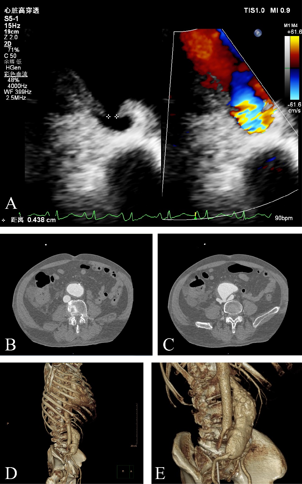
Arteriovenous fistula (AVF) is a rare complication of the abdominal aortic aneurysm (AAA) with complex clinical features. However, AVF and AAA usually cause no symptoms except when they rupture. This case study demonstrated that ultrasonography was a rapid and non-invasive method for the initial assessment of AAA and AVF. A 65-year-old man was admitted to the intensive care unit with hepatic and renal dysfunction. Physical examination revealed an abdominal vascular murmur and bilateral toe discoloration. Ultrasonic examination revealed an AAA and right common iliac artery aneurysm with an AVF located between the right common iliac artery and inferior vena cava. A computed tomography scan confirmed the sonographic findings. We propose that ultrasound should be used more commonly as part of the initial evaluation of the potential and established vascular diseases.

Keywords
Abdominal aortic aneurysm
arteriovenous fistula
hepatorenal dysfunction
ultrasound
Figures
Figure 1.
Share
Back to top