IMR Press / RCM / Volume 21 / Issue 3 / DOI: 10.31083/j.rcm.2020.03.102
Open Access Review
Cardiac sarcoidosis: diagnosis and management
Show Less
1 Pulmonary Department, Corfu General Hospital, Corfu 49100, Greece
*Correspondence: lefte_mark83@yahoo.gr (Eleftherios Markatis); icpapanikolaou@hotmail.com (Ilias C Papanikolaou)
Rev. Cardiovasc. Med. 2020, 21(3), 321–338; https://doi.org/10.31083/j.rcm.2020.03.102
Submitted: 22 May 2020 | Revised: 16 August 2020 | Accepted: 18 August 2020 | Published: 30 September 2020
(This article belongs to the Special Issue Cardiac fibrosis)
Copyright: © 2020 Markatis et al. Published by IMR Press.
This is an open access article under the CC BY 4.0 license (https://creativecommons.org/licenses/by/4.0/).
Abstract

Sarcoidosis is a chronic inflammatory disease of unknown etiology characterized by multi-organ involvement. End-organ disease consists of granulomatous inflammation, which if left untreated or not resolved spontaneously, leads to permanent fibrosis and end-organ dysfunction. Cardiac involvement and fibrosis in sarcoidosis occur in 5-10% of cases and is becoming increasingly diagnosed. This is due to increased clinical awareness among clinicians and new diagnostic modalities, since magnetic resonance imaging and positron-emission tomography are emerging as “gold standard” tools replacing endomyocardial biopsy. Despite this progress, isolated cardiac sarcoidosis is difficult to differentiate from other causes of arrhythmogenic cardiomyopathy. Cardiac fibrosis leads to congestive heart failure, arrhythmias and sudden cardiac death. Immunosuppressives (mostly corticosteroids) are used for the treatment of cardiac sarcoidosis. Implantable devices like a cardioverter-defibrillator may be warranted in order to prevent sudden cardiac death. In this article current trends in the pathophysiology, diagnosis and management of cardiac sarcoidosis will be reviewed focusing on published research and latest guidelines. Lastly, a management algorithm is proposed.

Keywords
Sarcoidosis
cardiac sarcoidosis
interstitial lung disease
fibrosis
1. Introduction

Sarcoidosis is a multisystemic granulomatous disease of unknown etiology manifesting frequently as a mild or even asymptomatic pulmonary disease (Costabel, 2001; Yamamoto et al., 1992). Despite its generally benign nature, sarcoidosis may progress to organ fibrosis and impairment with poor prognosis including death (Swigris et al., 2011; Viskum and Vestbo, 1993). The first case of heart involvement in sarcoidosis was reported by Bernstein in 1929 in a 52-year-old tailor dying from heart failure (Bernstein, 1929). Cardiac sarcoidosis (CS) is considered to be the second most common cause of death in sarcoidosis patients globally and the first among Japanese sarcoidosis patients (Iwai et al., 1994). The pathology consists of granulomatous inflammation of the pericardium, myocardium and endocardium with patchy, multifocal involvement (Roberts et al., 1977). Clinically manifest CS occurs in 3-10% of patients with sarcoidosis in America and Europe, though its prevalence from certain autopsy studies is estimated to be up to 25% (Iwai et al., 1993; Perry and Vuitch, 1995; Silverman et al., 1978). CS manifests with ventricular arrhythmia, high grade block, sudden death or symptoms of heart failure most commonly presented with a rather acute onset (Birnie et al., 2016). In certain studies, almost one third of patients with CS did not have a diagnosis of systemic sarcoidosis (Nery et al., 2014a; Tung et al., 2015). The diagnosis of CS remains a challenge, although the evolution of modern imaging modalities such as cardiac magnetic resonance imaging (CMR) and 18-fluorodeoxyglucose positron emission tomography (18F-FDG PET), as well as the application of clinical guidelines have led to increased diagnosis rates (Birnie et al., 2014; Mc Ardle et al., 2013). Recognition of patients who will need an implantable cardioverter-defibrillator is paramount. Corticosteroids are considered the standard of care for CS treatment, though there is no consensus regarding the dosage or the duration of treatment and the role of second line steroid-sparing agents (Nagai et al., 2015; Yazaki et al., 2001).

2. Epidemiology

Epidemiological studies have added to our knowledge about sarcoidosis and its phenotypes, though accurately estimating the incidence and prevalence of the disease is challenging due to its considerable heterogeneity (Hunninghake et al., 1999). Most studies suggest an annual incidence of 1-30 per 100,000 (Arkema and Cozier, 2018; Ungprasert et al., 2016a), with the highest prevalence reported in Scandinavian countries and United States (US) African-Americans, and the lowest among Asians (James and Hosoda, 1994; Morimoto et al., 2008). In most but not all studies, sarcoidosis is found more common in women (Byg et al., 2003; Deubelbeiss et al., 2010; Erdal et al., 2012; Gribbin et al., 2006; Henke et al., 1986; Hillerdal et al., 1984; Parkes et al., 1985; Rybicki et al., 1997; Thomeer et al., 2001; Ungprasert et al., 2016b). Peak onset age is between 20 and 40 years old and a second peak is seen especially in women over 50 years old (Kowalska et al., 2014; Selroos, 1969). An increase in the mean age at diagnosis has been reported the last twenty years (Foreman et al., 2006; Sawahata et al., 2015). Sarcoidosis is generally considered a benign disease with an annual rate of mortality in USA estimated between 2-4 deaths per million (Gerke, 2014; Mirsaeidi et al., 2015). African Americans experience more multiorgan involvement, and notably, African American women die at a younger age than Caucasians (Cozier et al., 2011). A lower socioeconomic status has been associated with more severe disease and new organs involved (Rabin et al., 2001; Rabin, 2004). The prevalence of CS is not exactly recognized. As mentioned above, clinically evident CS has been noted in 3% to 10% of patients, although autopsy studies reported cardiac involvement in about one-fourth of cases in the United States , in Europe and in Japan (Baughman et al., 2001; Greulich et al., 2013; James et al., 1976; Judson et al., 2012). More recent data suggest that cardiac involvement is more common in male sarcoidosis patients (Martusewicz-Boros et al., 2016a). CS incidence and hospitalizations have been rising, as a result of the increasing recognition and interest for the disease, the development of new imaging techniques, and the publication of related guidelines (Okumura et al., 2004; Patel et al., 2009; Soejima and Yada, 2009). Interestingly, the rates of in-hospital mortality between 2005 and 2014 have been found decreased in one study (Patel et al., 2018).

3. Pathogenesis and etiology

Significant research has not managed to establish a definite etiology of sarcoidosis. Genetic, environmental, lifestyle and occupational risk factors have been suggested, in order to explain the heterogeneity in disease presentation and severity among different ethnic and racial groups (Abe et al., 1987; Berlin et al., 1997; Brennan et al., 1984; Cozier et al., 2012, 2013, 2015; Iannuzzi et al., 2007; Kucera et al., 2003; Martinetti et al., 1995; McGrath et al., 2000; Newman et al., 2004). The general concept is of a gene-environment interaction, suggesting that sarcoidosis results from the exposure of genetically susceptible individuals to specific environmental agents (Chen and Moller, 2008; Verleden et al., 2001). The human leukocyte antigen (HLA) system, a group of related proteins that are encoded by the major histocompatibility (MHC) gene complex in humans, has been associated with sarcoidosis (Iannuzzi et al., 2007). The CD4+ T-cell immunological response in sarcoidosis has been linked to HLA genes, with recognized effects on disease susceptibility, chronicity and severity (Brewerton et al., 1977; Grunewald et al., 2004; Hedfors and Lindstrom, 1983; Iannuzzi et al., 2003; Ishihara et al., 1994; Lio et al., 1997; Maliarik et al., 1998; Rossman et al., 2003; Rybicki et al., 2003; Schurmann et al., 2000, 2001; Smith et al., 1981; Spagnolo and Grunewald, 2013). Cardiac involvement is most likely associated with the DQB1*0601 and DRB1*0803 alleles, while DRB1*0101 and DQB1*0501 appear protective against a predominantly pulmonary phenotype (Grubić et al., 2011; Naruse et al., 2000; Rybicki and Iannuzzi, 2007; Sato et al., 2010). Furthermore, single nucleotide polymorphisms in non-HLA genes, including CCR2, CCR5, IL1A, IL23R, TNF-α  and NOD2, FCGR, have been linked to the disease (Rivera et al., 2016). Regarding pathology, the hallmark of sarcoidosis is the formation of noncaseating granulomas characterized by the presence of epithelioid histiocytes, macrophages, giant cells and lymphocytes, mostly Th1 type (Korsten et al., 2018). A key role in the immune response is attributed to Th1-cells and increased levels of related cytokines such as interleukin-2 (IL-2), IL-12, interferon-γ (IFN-γ), and tumor necrosis factor α (TNF-α) (Darlington et al., 2012). Moreover, Th17 cells and regulatory cells (Treg cells) are also believed to be involved (Huang et al., 2013; Sakthivel and Bruder, 2017; Ten Berge et al., 2012). Several infectious organisms have been suggested as possible agents in the etiology of sarcoidosis, although Propionibacterium acnes (P. acnes) is the only microorganism to be isolated from sarcoid lesions (Abe et al., 1984; Baughman et al., 2003; Homma et al., 1978; Ishige et al., 1999; Negi et al., 2012). Other factors associated with higher risk of sarcoidosis are unknown environmental factors reflecting the disease’s seasonality, obesity, and inhaled dusts (Demirkok et al., 2007; Gupta et al., 2013; Procaccini et al., 2012; Ungprasert et al., 2016a, 2017a; Vihlborg et al., 2017; Wozniak et al., 2009; Xiao et al., 2018). Interestingly, smoking interferes with the macrophage-lymphocyte interaction that results in the formation of granulomas, and has therefore been suggested to have a protective role against sarcoidosis (Moller et al., 1996; Silverstein et al., 1994; Valeyre et al., 1988).

4. Clinical manifestations

Clinical manifestations in sarcoidosis cohorts differ in terms of age, sex, ethnicity, type of onset and organ involvement (Cozier, 2016; Izumi, 1992; Jain et al., 2020; Lill et al., 2016; Loddenkemper et al., 1998; Pereira et al., 2014; Prasse et al., 2008; Siltzbach et al., 1974). Japanese patients are reported to have a much higher likelihood of ocular and cardiac disease than patients in the rest of the world, while uveitis and cutaneous involvement is more common in females than males (Birnbaum et al., 2011; Brito-Zerón et al., 2016; Pasadhika and Rosenbaum, 2015; Yanardag et al., 2003, 2013). Nevertheless, pulmonary disease is by far the most common organ involvement in sarcoidosis (Iannuzzi and Fontana, 2011; Neville et al., 1983; Rao and Dellaripa, 2013). Epidemiological studies have described the clinical heterogeneity of sarcoidosis highlighting the need for further clinical phenotyping – while a multidisciplinary approach should always be considered (Hattori et al., 2018; Judson et al., 2006; Kreider et al., 2005; Pietinalho et al., 1996; Reynolds, 2002; Rybicki and Iannuzzi, 2007). A large European multicenter study identified five CS-associated phenotype groups; cardiac involvement was part of the ocular-cardiac-cutaneous-central nervous system phenotype suggesting CS as a disease of the electrical conduction system of the heart (Schupp et al., 2018). In general, patients with cardiac sarcoidosis may have minimal extra cardiac disease or asymptomatic chest involvement (Nery et al., 2014b). Cardiac symptoms depend on the location, extent, and activity of the disease, and the most commonly observed manifestations are conduction abnormalities (including ventricular arrhythmias and atrio-ventricular blocks) and myocardiopathy leading to congestive heart failure (Terasaki et al., 2019). This is the case because the main cardiac compartments affected by CS are the intraventricular septum and the left ventricle myocardial wall, respectively.

Patients may present asymptomatically or experience palpitations, syncope, or even sudden cardiac death. Ventricular arrhythmias such as premature ventricular contractions, ventricular tachycardia and ventricular fibrillation are caused by myocardial inflammation and fibrosis. Heart failure occurs as inflammation and fibrosis progress, causing edema, cough and dyspnea that should be differentiated from pulmonary disease. Decreased cardiac output may cause oliguria, neurological signs, malaise, but also syncope, confusion, and decreased levels of consciousness (Kandolin et al., 2011; Nery et al., 2013). Pulmonary function testing may reveal obstructive and/or restrictive patterns as well as TLCO (diffusing capacity of carbon monoxide) impairment. A recent study of 1,110 patients with pulmonary sarcoidosis reported that 10% of the cohort (25% of which had an obstructive defect) had a mixed ventilatory defect. These patients exhibited lower TLCO and suffered from stage IV sarcoidosis and higher mortality compared to purely obstructive patients (Kouranos et al., 2020). Decreased forced expiratory volume in 1st second (FEV1) was associated with a more advanced stage of the disease and occasionally with lower left ventricle ejection fraction (LVEF) (Martusewicz-Boros et al., 2019).

5. Diagnosis

The incidence of progressive heart failure, arrhythmias and sudden cardiac death (SCD) increases as CS becomes clinically recognizable; thus, early diagnosis and initiation of therapy is crucial to improve prognosis (Dubrey and Falk, 2010; Kim et al., 2009; Mantini et al., 2012; Pierre-Louis et al., 2009; Voortman et al., 2019). The classic triad of criteria establishing a diagnosis of sarcoidosis is 1) a compatible clinical and/or radiological picture; 2) histological evidence of noncaseating granulomas and 3) exclusion of other diseases (Costabel, 2001; Hunninghake et al., 1999). Diagnosis usually requires a multi-disciplinary approach, as almost any organ may be affected; sarcoidosis is a diagnosis of exclusion, so establishing a CS diagnosis may be challenging (Bargagli and Prasse, 2018). Histological confirmation of CS cases is difficult, as endomyocardial biopsy (EMB) has low sensitivity (36%) due to the patchy nature of sarcoidosis and is also a procedure that is difficult to perform. Thus, cardiac sarcoidosis cannot be ruled out with a negative endomyocardial biopsy result (From et al., 2011; Uemura et al., 1999). Immunohistochemistry using anti-monoclonal antibody against P. acnes has even been suggested as a potential additive diagnostic tool given the low sensitivity of EMB (Asakawa et al., 2017). A more promising tool may be the recognition of increased lymphatic vessel counts on myocardial biopsy of CS subjects in the absence of granuloma, a finding that increases the sensitivity of EMB at 75% (Oe et al., 2019). Further, an immunohistochemical finding of increased dendritic cells along with decreased M2 among all macrophages in non-granulomatous sections of cardiac biopsy showed high specificity for cardiac sarcoidosis diagnosis, suggesting this phenotype as a histopathological surrogate for CS (Honda et al., 2016). A well-formed non-necrotizing granuloma in the heart is shown in Fig. 1.
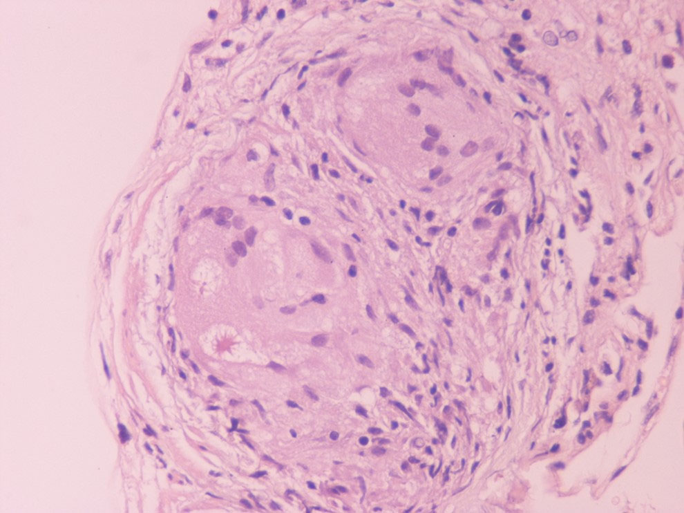
Fig. 1.

A well-formed non-necrotizing granuloma in the heart (Hematoxylin Eosin x400).

5.1 Screening for CS

It is crucial for clinicians to screen for CS in all patients with extra cardiac sarcoidosis. Patients suspected of isolated CS without known sarcoidosis are less frequent; diagnosis in this case requires a high level of suspicion and exclusion of common diagnoses (mainly ischemic heart disease). The screening starts with a detailed history and physical examination and electrocardiogram (ECG) (Dubrey and Falk, 2010; Mantini et al., 2012). Patients with symptoms, an abnormal ECG, or cardiomegaly on chest x-ray without known risk factors should be referred for further testing. Transthoracic echocardiogram and 24h-holter monitoring may add additional information before the patient proceeds with subsequent advanced imaging (Kim et al., 2009). All patients with a pathologic ECG should be further investigated for CS, particularly those who experience clinical symptoms including palpitations, pre-syncope and syncope (From et al., 2011). Mehta et al. showed that the presence of cardiac symptoms (significant palpitations, syncope, or presyncope) and/or an abnormal cardiac test in known sarcoidosis patients had a sensitivity of 100% and a specificity of 87% for CS diagnosis (Mehta et al., 2008). Despite this, the absence of cardiac related symptoms does not exclude the diagnosis of CS, and data on whether and when patients with a negative initial work-up should be rescreened, are lacking (Birnie et al., 2014). Male sex, cardiac-related symptoms, ECG changes, serum NT-proBNP level, multiorgan involvement, and radiological pulmonary progression have been suggested as potential risk factors for cardiac sarcoidosis development (Darlington et al., 2014; Youssef et al., 2011).

5.2 Electrocardiogram (ECG) and Holter monitoring

The electrocardiogram (ECG) result is usually abnormal in patients with clinically evident disease and mostly normal in clinically silent CS. Abnormalities include various degrees of conduction block, such as isolated bundle branch block and fascicular block. Furthermore, QRS complex fragmentation, pathological Q waves (pseudo infarct pattern), ST changes and (rarely) epsilon waves can occur. These ECG findings in patients with extra cardiac sarcoidosis warrant further imaging studies to rule out cardiac sarcoidosis, with which they have shown significant association (Martusewicz-Boros et al., 2016b). Some patients are diagnosed with abnormal ECG features years after the onset of non-cardiac sarcoidosis; thus, ECG is essential for long-term follow-up. Other possible signals might be baseline heart rate and PR interval. Signal averaged ECG and Holter monitoring revealing late potentials might predict cardiac involvement in sarcoidosis (Yodogawa et al., 2018). Most studies show that Holter monitoring can be a predictor of cardiac involvement with sensitivity of 89% and specificity of 21% (Freeman et al., 2013). Holter is also useful to monitor response to treatment with immune-suppressives in conductive abnormalities in serial fashion (Padala et al., 2017). The exact place of Holter monitoring in the screening for CS is not clearly defined; however, it is certainly a comfortable, out-patient assessment follow-up tool in patients suspected of or diagnosed with CS.

5.3 Echocardiography

Similar to ECG, the trans-thoracic echocardiogram (TTE) result is oftenabnormal in manifested disease and normal in clinically silent CS (Kim et al., 2009). Though not pathognomonic, some findings are accepted as major and minor criteria in various diagnostic guidelines (Birnie et al., 2014; Hiraga et al., 1993; Judson et al., 2014; Patel et al., 2009; Terasaki et al., 2019). Echocardiography is not useful in screening for CS, due to its low sensitivity, around 25% in most studies (Kouranos et al., 2017). The most characteristic abnormality is basal interventricular thinning, and less often myocardial wall thickness, isolated wall motion abnormalities, LV and/or RV diastolic and systolic dysfunction and aneurysms are detected (Agarwal et al., 2014; Skold et al., 2002; Burstow et al., 1989; Sun et al., 2011). Newer techniques, including strain rate, might improve echocardiography sensitivity in CS diagnosis (Joyce et al., 2015; Murtagh et al., 2016).

5.4 Biomarkers

Most biomarkers such as angiotensin-converting enzyme, neopterin and troponin are elevated in patients with sarcoidosis; however, they lack sensitivity and specificity and may be affected by concomitant drugs administered (d’Alessandro et al., 2020; Kandolin et al., 2015; Vorselaars et al., 2015). Hypercalciuria, increased serum chitotriosidase and BAL biomarkers (elevated CD4+/CD8+ ratio and KL-6, decreased Natural Killer and CD103+CD4+cells) indicate sarcoidosis diagnosis and disease activity (Cameli et al., 2020; d’Alessandro et al., 2020). Exhaled nitric oxide has failed to prove useful in pulmonary sarcoidosis (Cameli et al., 2016).

5.5 Cardiac magnetic resonance (CMR)

Recent studies have focused on the diagnostic and prognostic value of advanced cardiac imaging, trying to overcome the limitations of the aforementioned exams, in order to achieve an earlier diagnosis, at less advanced stages of the disease (Cheong et al., 2009; Kim et al., 2009; Patel et al., 2009). CMR showing late gadolinium enhancement (LGE) is regarded as the study of choice for diagnosing cardiac involvement in sarcoidosis (Smedema et al., 2005). Apart from LGE, CMR may detect morphologic abnormalities (such as wall thinning and aneurysms) and functional parameters of cardiac chambers (LV and RV). LGE most commonly represents scar tissue, although inflammation may sometimes lead to extracellular expansion leading to LGE. In a study of 321 biopsy-proven sarcoidosis patients, CMR allowed the diagnosis of 44 patients with normal echocardiograms, as well as 15 asymptomatic patients. LGE may independently predict future adverse events, such as atrioventricular block (AVB), ventricular tachycardia (VT), sudden cardiac death (SCD) and heart failure (Nadel et al., 2015). Association of LGE on CMR with adverse cardiac outcomes is shown in other studies as well (Ichinose et al., 2008; Nagai et al., 2014). There is often a multifocal distribution, although a pattern of enhancement is not pathognomonic for CS (Cummings et al., 2009). LGE is mostly seen in basal segments, particularly of the septum and lateral wall, and usually in the midmyocardium and sub-epicardium (non-infarct pattern) as opposed to sub-endocardial scarring in the event of myocardial infarction. Few studies have focused on right ventricular involvement, suggesting that the RV free wall may also be involved in predicting adverse outcomes, particularly ventricular tachyarrhythmias (Crawford et al., 2014; Patel et al., 2011). The presence of LGE in patients with normal or near-normal LVEF greatly increases the likelihood of adverse events (Agoston-Coldea et al., 2016; Coleman et al., 2017; Ekström et al., 2016; Ise et al., 2014; Murtagh et al., 2016; Shafee et al., 2012; Yasuda et al., 2016). Remarkably, CMR identifies small regions of myocardial damage in subjects with preserved LV systolic function, allowing detection of “silent” CS (Pizarro et al., 2016). Interestingly, involvement of the RV in addition to the LV increases the risk of worse outcomes and all-cause mortality (Smedema et al., 2017). CMR with LGE is less sensitive regarding active myocardial inflammation; consequently, it may not properly guide immunosuppressive therapy. Moreover, CMR is not useful for extra-cardiac sarcoidosis compared to CT and PET-CT. Novel CMR techniques utilizing T1 and T2 mapping may allow detection of myocardial inflammation, suggesting a role in monitoring response to treatment (Crouser et al., 2014; Puntmann et al., 2017). The main value of CMR in the diagnostic algorithm of CS is its high negative predictive value, which exceeds 90% (Cheong et al., 2009). Representative CMR images of cardiac sarcoidosis are shown in Fig. 2.
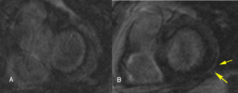
Fig. 2.

Cardiac magnetic resonance images in a 54-year-old male with cardiac sarcoidosis, showing late gadolinium enhancement in the posterior-lateral left ventricle wall sub-epicardially (B, yellow arrows).

5.6 Positron emission tomography (Pet)

Neither CMR nor PET alone are positive in all CS cases. This is why when CMR is found normal – or is not available – further investigation with PET is warranted. Fluorodeoxyglucose (FDG) is a glucose analog that is useful in differentiating between normal and active inflammatory lesions (Pellegrino et al., 2005). In contrast to CMR which detects scar tissue, PET recognizes myocardial inflammation. FDG-PET testing should be performed at experienced centers (Birnie et al., 2014). Its use in assessing cardiac involvement in sarcoidosis requires extensive pre-imaging preparation to suppress physiologic glucose uptake by normal myocardium (Osborne et al., 2017). Although there are no specific imaging findings pathognomonic for CS diagnosis, focal or focal-on diffuse FDG uptake patterns suggest active CS (Ishimaru et al., 2005; Youssef et al., 2012). PET alone compared to Japanese guidelines showed 89% sensitivity and 78% specificity in diagnosing CS (Ishimaru et al., 2005). The presence of FDG uptake and perfusion defects on PET is associated with higher risk of VT or death. RV involvement on PET scan may be a marker of severe disease, a finding observed in CMR studies as well. FDG uptake serves as an excellent tool for early diagnosis and guiding therapy with a follow up approximately every 6 months (Blankstein et al., 2014). Abnormal tracer accumulation, initially considered a minor criterion for diagnosis, is now upgraded as a major criterion for diagnosis in recently published Japanese guidelines (Chareonthaitawee et al., 2017; Kumita et al., 2019; Terasaki and Yoshinaga, 2017; Wicks et al., 2018).

5.7 Combined use of FDG-PET and CMR

Given the high negative predictive value of CMR and the likelihood of 10-20% of false negatives with PET, CMR is given priority as initial diagnostic modality when CS is suspected in patients without contraindications for CMR testing. In a position statement, Slart et al. recommend the addition of FDG-PET to increase the diagnostic accuracy of CMR when disease is highly suspected (Slart et al., 2017). CMR and PET are therefore rather complementary, enabling imaging of the 2 different stages of the disease (i.e., fibrosis and inflammation); combination of the data is encouraged, especially if one of the tests yields inconclusive results (White et al., 2013). It should be noted that when CMR and/or PET reveal regional myocardial anomalies that may be due to sarcoidosis or atherosclerotic coronary artery disease, coronary angiography may be necessary to exclude significant coronary artery defects.

5.8 Recommended diagnostic approach for patients with suspected CS

Three major guidelines exist for the diagnosis of CS. The first is the WASOG organ assessment tool published in 2014 and updated in the recent American Thoracic Society guidelines for sarcoidosis diagnosis (Crouser et al., 2020; Judson et al., 2014). The second is the HRS expert consensus statement in 2014 (Birnie et al., 2014) that suggested two diagnostic pathways: (a) histological diagnosis with the presence of noncaseating granulomas in myocardial tissue or (b) clinical diagnosis, as in histological diagnosis of extra cardiac sarcoidosis and one or more of the following clinical/imaging criteria: (1) cardiomyopathy and/or heart block, (2) unexplained sustained ventricular tachycardia, below 40%, (3) unexplained reduced left ventricular ejection fraction (4) Mobitz type II second-degree AV block or third-degree AV block, (5) late gadolinium enhancement on CMR in a pattern consistent with cardiac sarcoidosis, (6) patchy uptake of cardiac FDG-PET in a pattern consistent with cardiac sarcoidosis, or (7) positive gallium uptake in a pattern consistent with cardiac sarcoidosis.

All of these criteria, as well as immunomodulatory treatment responsive cardiomyopathy and/or AV block are considered features probable of sarcoidosis and in particular cardiac involvement in ATS guidelines (Crouser et al., 2020). When these criteria are present in a patient with known extra cardiac sarcoidosis, cardiac sarcoidosis is probable (> 50% probability). When present in a patient without sarcoidosis diagnosis, these clinical features-criteria constitute compatible with sarcoidosis disease and necessitate histological confirmation (not necessarily from the heart) according to standard diagnostic criteria. Using the HRS criteria, isolated CS can thus only be diagnosed with myocardial tissue, making this challenging diagnosis difficult to confirm (Birnie et al., 2014). No firm criteria on how to manage suspected isolated CS exist.

The third major guideline is the Japanese Ministry of Health and Welfare guidelines, initially described in 1993 and updated in 2017 with the following significant changes: (1) allowing diagnosis of isolated CS without a positive endomyocardial biopsy, (2) adding ventricular arrhythmia to the major criteria along with high-grade atrioventricular block and (3) moving abnormal ventricular wall anatomy, elevated myocardial uptake with FDG-PET and late gadolinium-enhanced CMR imaging to major criteria (Hiraga et al., 1993; Terasaki et al., 2019). Most recently, the Japanese Society of Nuclear Cardiology updated the recommendations regarding the 18FDG PET/CT use in diagnosis of CS (Ishimaru et al., 2005). The Japanese criteria may reflect the increased incidence of CS and its implications in Japan.

The latest ATS sarcoidosis guidelines recommend screening for cardiac sarcoidosis based solely on symptoms or signs and ECG, considering TTE and 24-hour Holter monitoring only in selected cases (conditional recommendation with low quality evidence). Patients with suspected cardiac involvement should undergo initial investigation with cardiac magnetic resonance imaging, and/or PET when CMR is not available, to obtain diagnostic and prognostic information (Crouser et al., 2020). Major recommendations and guidelines are displayed in Tables 1 and 2.

Table 1.Japanese cardiac sarcoidosis and Heart Rhythm Society guidelines (Birnie et al., 2014; Terasaki et al., 2019).
2016 JCS Guideline on Diagnosis and Treatment of Cardiac Sarcoidosis 2014 HRS expert consensus statement on the diagnosis and management of arrhythmias associated with cardiac sarcoidosis
• Histological diagnosis • Histological diagnosis
• Clinical diagnosis group when
A. Epithelioid granulomas are found in organs other than the heart, and two or more major criteria or one major and two or more minor criteria • Clinical diagnosis of cardiac sarcoidosis (probable CS) meaning histological diagnosis of extra cardiac sarcoidosis, exclusion of other diagnoses and one or more of
Major criteria
1. High-grade atrioventricular block (including complete atrioventricular block) or fatal ventricular arrhythmia (e.g., sustained ventricular tachycardia, and ventricular fibrillation) 1. cardiomyopathy and/or heart block unexplained reduced left ventricular ejection fraction (LVEF) of less than 40%
2. Basal thinning of the ventricular septum or abnormal ventricular wall anatomy (ventricular aneurysm, thinning of the middle or upper ventricular septum, regional ventricular wall thickening) 2. unexplained sustained (spontaneous or induced) ventricular tachycardia
3. Left ventricular contractile dysfunction (left ventricular ejection fraction less than 50%) or focal ventricular wall asynergy
4. 67Ga citrate scintigraphy or FDG PET reveals abnormally high tracer accumulation in the heart 3. Mobitz type II second-degree AV block or third-degree AV block
5. Gadolinium-enhanced MRI reveals delayed contrast enhancement of the myocardium
Minor criteria
1. Abnormal ECG findings: Ventricular arrhythmias (nonsustained ventricular tachycardia, multifocal or frequent premature ventricular contractions), bundle branch block, axis deviation, or abnormal Q waves 4. patchy uptake of cardiac FDG-PET in a pattern consistent with cardiac sarcoidosis
2. Perfusion defects on myocardial perfusion scintigraphy (SPECT) 5. late gadolinium enhancement on CMRI in a pattern consistent with cardiac sarcoidosis
3. Endomyocardial biopsy: Monocyte infiltration and moderate or severe myocardial interstitial fibrosis
4. When the patient shows clinical findings strongly suggestive of pulmonary or ophthalmic sarcoidosis; at least 2 of the five characteristic laboratory findings of sarcoidosis and clinical findings strongly suggest cardiac involvement 6. positive gallium uptake in a pattern consistent with cardiac sarcoidosis
Table 2.Probable cardiac sarcoidosis according to World Association of Sarcoidosis and Other Granulomatous diseases (WASOG) criteria (Crouser et al., 2020; Judson et al., 2014).
2014 The WASOG Sarcoidosis Organ Assessment Instrument 2020 Diagnosis and Detection of SarcoidosisAn Official American Thoracic Society Clinical Practice Guideline
Probable criteria Probable criteria
1. Treatment responsive Cardiomyopathy or atrioventricular nodal block 1. Treatment-responsive CM or AVNB
2. Reduced LVEF in the absence of other clinical risk factors 2. Reduced LVEF with no risk factors (echo and MRI)
3. Spontaneous or inducible sustained VT with no other risk factor 3. Spontaneous/inducible VT with no risk factors
4. Mobitz type II or 3rd degree heart block 4. New-onset, third-degree AV block in young or middle-aged adults
5. Patchy uptake on dedicated cardiac PET 5. Increased inflammatory activity in heart (MRI, PET, and gallium)
6. Delayed enhancement on CMR
7. Positive gallium uptake
8. Defect on perfusion scintigraphy or SPECT scan
9. T2 prolongation on CMR
(FDG) PET = (Fluorodeoxyglucose) positron emission tomography; MRI = magnetic resonance imaging; ECG = electrocardiogram; SPECT = Single-photon emission computed tomography; LVEF = left ventricular ejection fraction; VT = ventricular tachycardia; CM = cardiomyopathy; AVNB = atrioventricular node block; AV = atrioventricular
5.9 Isolated cardiac sarcoidosis

When cardiac sarcoidosis is diagnosed or suspected without systemic sarcoidosis diagnosis, the term isolated cardiac sarcoidosis is used (ICS). Since established criteria for diagnosing CS require either extra-cardiac biopsy confirmation or EMB confirmation (a method of limited sensitivity as already shown), the diagnosis of ICS represents a true clinical challenge. Previous reports describe ICS as responsible for 25-65% of total CS (Kandolin et al., 2015; Okada et al., 2018). More recent data using FDG-PET report a more modest prevalence for ICS (9% of total) (Giudicatti et al., 2020). Scarce data indicate that ICS presents with lower LVEF, more frequent VTs, and necessitate ICDs more frequently than CS with systemic sarcoidosis (Kron et al., 2015; Okada et al., 2018).

ICS is rarely diagnosed compared to other causes of arrhythmogenic cardiomyopathy (ACM), often in cardiac explants post-transplant (Chang et al., 2012). Arrhythmogenic right ventricular cardiomyopathy (ARVC) is an inherited disease caused by mutations in desmosome encoding genes. Although HRS 2019 guidelines have established criteria for ARVC diagnosis (inverted T and epsilon waves on ECG, myocardial biopsy with decreased myocytes, VT‘s of LBBB, CMR features and family history, among others), confusion with ICS still occurs (Towbin et al., 2019). Electrophysiology studies have shown that reduced LVEF, a significantly wider QRS, right-sided apical VT, and more inducible forms of monomorphic VT are mainly encountered in ICS (Dechering et al., 2013). Differential diagnosis between ICS and ARVC may only partly be aided by modern diagnostic modalities, as shown in small cohorts (Steckman et al., 2012). Left ventricle scar on CMR points to ICS, while solely RV fibrosis points to ARVC, although both of these clues are not pathognomonic (Macias et al., 2014). The FDG-PET CT scan may test positive in ARVC as well as in ICS, as shown by Protonotarios et al (Protonotarios et al., 2019). Hoogendoorn et al. applied electroanatomical voltage mapping in order to characterize RV scar characteristics and distribution in ICS and ARVC. The authors validated their results in a separate CS population with excellent sensitivity and specificity (Hoogendoorn et al., 2020). Undiagnosed ICS may warrant ICD placement or transplant, as in other causes of ACM. An algorithm including CMR, FDG-PET, electroanatomic mapping (and guided-EMB) in experienced centers may facilitate the diagnosis of ICS in the near future (Muser et al., 2018).

6. Treatment

In CS, clinicians first need to answer the key question whether the disease is only myocardial or affects the conduction system. Secondly, the initiating dose of corticosteroids is significantly lower than the one originally administered a few decades ago. Thirdly, it remains questionable whether asymptomatic patients should be treated, since reducing the risk of future adverse events should be weighed along with the treatment’s side effects (Patel et al., 2009). There is a lack of evidence whether treatment should be started at the presence of active lesions or the presence of clinical symptomatology (Birnie et al., 2014; Soejima and Yada, 2009). Many experts recommend treatment of asymptomatic patients, to prevent disease progression to fibrosis (Doughan and Williams, 2006; Ohira et al., 2008; Orii et al., 2015; Shafee et al., 2012).

6.1 Immunosuppression

Corticosteroids have been used for decades as first line treatment of cardiac sarcoidosis (Aryal and Nathan, 2019; Grutters and van den Bosch, 2006). Steroid-sparing immunomodulators have been used or tested as second line treatment for refractory disease. A large international multicenter registry found that 26.4% of CS patients were on steroids alone, 22.6% were on steroids and a steroid-sparing agent, and 9.7% were on steroid-sparing agent alone, with methotrexate being the first steroid-sparing choice (Kron et al., 2017). Retrospective studies have shown that corticosteroid treatment led to a reduction in VT, reversal of atrioventricular block, improvement in left ventricular ejection fraction and survival benefit (Banba et al., 2007; Furushima et al., 2004; Hulten et al., 2016; Kusano, 2013; Stees et al., 2011). Corticosteroid therapy in cardiac sarcoidosis is mainly responsible for an improvement in ejection fraction in patients with mild to moderate LV dysfunction, or in patients with severe dysfunction and LVEF < 35% as shown elsewhere (Sadek et al., 2013).

Long-term use of high doses of corticosteroids adversely affects patients’ quality of life and has been associated with life-threatening side effects (Baughman et al., 2006; Cox et al., 2004; Nagai et al., 2014; Uthman et al., 2007). A prednisone dose of 40mg daily appears to be as effective and less toxic than higher doses administered in the past. A commonly used initial dose for those receiving prednisone only is 40mg daily, while for those patients receiving an additional immunosuppressive agent, the initial dose is often
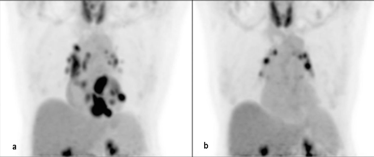
 20 mg daily. Based on response to treatment after 1 to 3 months, prednisone should be tapered to 5 to 15 mg/day for an additional 9 to 12 months, while follow up should comprise at least 3 years in case of disease relapse (Fussner et al., 2018) Silent CS patients are difficult to handle, since a clear benefit on survival lacks. Hence, as mentioned above, the balancing between suppression of the disease and side effects can be challenging, while optimal dosage and duration of therapy remain unclear (Hamzeh et al., 2012; Chapelon-Abric et al., 2004; Chiu et al., 2005; Sperry et al., 2018). Regarding asymptomatic cardiac sarcoidosis, some centers will assess and monitor, while others will initiate therapy with a dose of 30-40 mg prednisolone (Beegle et al., 2013). The effect of corticosteroids is variable, depending on whether the mechanism is related to myocardial scar formation or active inflammation (Schutt et al., 2010). Methotrexate is often used as a second-line agent, with a study showing that 80% of physicians consider it as a first-choice second-line treatment (Nagai et al., 2014). The usual dose of methotrexate is 10-25 mg once weekly with a 5 mg folic acid supplement (Nagai et al., 2014). Other second line therapies include mycophenolate mofetil, azathioprine, cyclophosphamide and lately infliximab (Demeter, 1988; Müller-Quernheim et al., 1999). Methotrexate and/or adalimumab have shown efficacy in maintaining radiographic disease quiescence after a tapering-off of prednisone, although patients who discontinue immunosuppression have increased risk of radiologic recurrence and recurrent VT (Rosenthal et al., 2019). Furthermore, immunosuppression with prednisone and methotrexate showed lower risk for recurrence, lower steroid dose and less side effects compared to treatment solely with prednisone (Ballul et al., 2019). The use of immunosuppression to prevent disease reactivation is prolonged, and close follow up with PET scans is useful when treatment is stopped (Fig. 3) (Bremer et al., 2018; Mankad et al., 2019). Studies on cardiac sarcoidosis treatment are shown in Table 3.

Fig. 3.

FDG-PET of a 63-year-old female on methotrexate for cardiac sarcoidosis before (a) and during (b) therapy with methotrexate for 2 years, exhibiting a significant decrease in FDG uptake in the heart suggesting adequate response to therapy. The scan continues to show FDG uptake in mediastinal lymph nodes (Bremer et al., 2018)

Table 3.Studies on cardiac sarcoidosis treatment.
Authors Number of patients Year of study High dose Immunosuppresion Tapering Low dose Immunosuppresion
Yazaki et al., 2001 95 2001 Prednisone 30-60 mg/d Variable Prednisone 5 to 15 mg/d
Chapelon-Abric et al., 2004 41 2004 IV prednisolone 15mg/kg × 3d Variable Not reported
Chiu et al., 2005 43 2005 Prednisone 60 mg 2 months Prednisone 10 mg/ d
Nagai et al., 2014 17, 67 2014, 2015 Prednisone 30 to 40 mg/d Variable Prednisolone 5 to 15 mg/d
Ballul et al., 2019 36 2016 Prednisone 60 mg/d Not reported Not reported
Fussner et al., 2018 91 2018 Prednisone 40-60 mg/d Variable Variable
6.2 Ventricular arrhythmias

Corticosteroids and antiarrhythmic drugs are initiated together for VTs when active inflammation is evident (Jefic et al., 2009), while catheter ablation and cardioverter defibrillator implantation are suggested if VT cannot be controlled (Naruse et al., 2014; Segawa et al., 2016). Amiodarone and sotalol are classic choices, although VTs are frequently resistant. The potential side effects of amiodarone include pneumonitis and pulmonary fibrosis, so a long-term treatment is not indicated for young patients (Birnie et al., 2014). Ablation therapy can be an option in VTs refractory to medical therapy. Despite a successful ablation, the recurrence rate remains high, although the total arrythmia burden may be reduced in up to 88% of cases with few major procedure related complications (Muser et al., 2016; Okada et al., 2018; Papageorgiou et al., 2018). CMR and PET findings could help in selecting more suitable patients for ablation therapy (Sohn et al., 2018). Yalagudri et al. stratified sarcoidosis patients with unexplained VT by FDG-PET results. They found that patients with increased myocardial FDG uptake were treated with immunosuppression, antiarrhythmics, and received ICD placement. If they had recurrence and had increased myocardial FDG uptake on their repeat FDG-PET CT scan, their immunosuppression was intensified. Patients without myocardial FDG uptake on initial PET CT scan were treated with antiarrhythmics and ICD without immunosuppression; if they lacked good clinical response, they later underwent catheter ablation. 13/14 patients in the inflammation cohort remained free of VT at their last follow-up visit (mean 38.2 months). All 4 patients in the scar cohort, despite the use of ICD and antiarrhythmics, experienced VT recurrence and underwent catheter ablation (Yalagudri et al., 2017). This study illustrates the fact that myocardial disease is more responsive to immunosuppressive treatment while arrythmias, though also partially responsive, will need risk assessment and electrophysiology study for ICD placement. In a large series, all patients with atrioventricular block at presentation underwent device placement (Fussner et al., 2018). According to HRS statement, implantable cardioverter defibrillators (ICDs) and cardiac resynchronization therapy defibrillators (CRT-Ds) are the preferred options compared to conventional pacemakers for cardiac sarcoidosis patients presenting with heart block (Birnie et al., 2014). Heart transplantation is seldom indicated, but may be of use in young refractory cases with non-responsive ventricular tachycardia or severe heart failure (Fussner et al., 2018).

6.3 Risk stratification for sudden cardiac death and indications for ICD implantation

Sudden cardiac death (SCD) is considered responsible for the majority of deaths in cardiac sarcoidosis (Roberts et al., 1977). HRS 2014 guidelines highlight that in CS with conduction abnormalities, device implantation and an implantable cardioverter-defibrillator are useful, along with immunosuppression when 2nd or 3rd degree AV block are present (Birnie et al., 2014). Atrial arrythmias require anti-coagulation, electrophysiological study (EPS) in cases other than atrial fibrillation and avoidance of class I anti-arrhythmic drugs (propafenone, flecainide, procainamide). On the other hand, ventricular arrythmias may require ablation and anti-arrhythmics in addition to systemic treatment. EPS is recommended in CS in cases of LVEF > 35% despite immunosuppression.

ICD is recommended in case of 1) spontaneous sustained ventricular arrhythmias and 2) in cases of LVEF < 35% despite optimal immunosuppression treatment. ICD may be useful in cases of inducible ventricular arrythmias, unexplained syncope, when a pacemaker placement is indicated, and in cases of LVEF between 36-49%. ICD is not recommended when EF is normal, without syncope history, with normal CMR and EPS study, or with no indication for pacemaker use (Birnie et al., 2014).

Identifying patients at risk for SCD is difficult (Ekstrom et al., 2016; Mehta et al., 2011; Vignaux et al., 2002). CS patients who have had sustained VT/ VF or LVEF < 35% have a high risk of recurrent events, SCD, and a class I indication/recommendation for ICD (Betensky et al., 2012; Kron et al., 2013; Schuller et al., 2012). In general ICD is used in CS with low LVEF, as shown in a multicenter study of 235 CS patients, where patients who received ICD therapy had a lower mean LVEF compared with those who did not receive such therapy (38.1% versus 48.5%) (Kron et al., 2013).

CS patients with AV block and preserved LV function are shown to carry a 9% risk of SCD and 24% risk of SCD/VT at 5 years (Nordenswan et al., 2018). This is in line with the above suggestion to implant an ICD when a pacemaker is recommended, even with preserved LVEF (Birnie et al., 2014). The 2018 ACC/AHA/HRS Guideline agreed with the recommendation for ICD implantation in cardiac sarcoidosis patients with an indication for pacing (Kusumoto et al., 2019). This recommendation includes patients with second- or third-degree AV block who are on antiarrhythmic or beta blocker therapy and immunosuppression.

EPS is recommended in relatively preserved LVEF. This is because it has not shown more predictive value than LVEF, whereas the inducibility of sustained ventricular arrhythmias is inversely correlated with LVEF (Aizer et al., 2005). We have already discussed the prognostic significance of CMR and PET on VAs, VTs, and SCD (Yasuda et al., 2016; Youssef et al., 2012).

A recent study showed that adverse cardiovascular events in cardiac sarcoidosis (VA, SCD, and heart transplantation) occur even if LVEF is moderately affected (Rosenthal et al., 2020). This is in accordance with other studies showing that Electro-physiology study had good predictive value for future VA as well in patients with LVEF > 35% that don’t meet primary indication for ICD therapy; this subgroup is mainly indicated for EPS to guide management (Freeman et al., 2013; Hamzeh et al., 2015; Okada et al., 2019; Suzuki et al., 1994). A suggested management algorithm of cardiac sarcoidosis is proposed in Fig. 4.

Fig. 4.

Suggested management algorithm for patients with probable cardiac sarcoidosis. ATS, American thoracic society criteria, biopsy of extra cardiac site; ECG, electrocardiogram; CMR, cardiac magnetic resonance, PET, positron emission tomography; VA, ventricular arrythmias; LVEF, left ventricle ejection fraction; EPS, electrophysiologic study; ICD, implantable cardioverter defibrillator.

7. Conclusions

Cardiac sarcoidosis should be adequately recognized and treated in a timely fashion, since it represents the second most frequent cause of death from sarcoidosis. Cardiac sarcoidosis may be asymptomatic, present with conduction abnormalities/advanced blocks, tachyarrhythmias or heart failure, be the cause of sudden cardiac death, or be diagnosed post-mortem. The diagnosis of cardiac sarcoidosis has become significantly more facilitated in recent years, as new imaging modalities (CMR and FDG PET) obviate the need for endomyocardial biopsy and provide functional/prognostic information. Ischemic heart disease sometimes has to be excluded. Diagnosis of cardiac sarcoidosis isolated at the heart is puzzling, however. Treatment with corticosteroids as the gold standard is imperative.The recognition of patients in risk for arrhythmias is crucial; these latter may need further electrophysiological studies and device placement, with improved outcome. Monitoring of CS, diagnosing probable cases (especially of isolated cardiac sarcoidosis), and the management of clinically silent cases are issues that must be addressed in the future.

Authors’ contributions

EM, AA, EA and IP equally contributed in drafting the manuscript.

Acknowledgments

We would like to express my gratitude to all those who helped me during the writing of this manuscript.

Conflict of interest

The authors declare no conflicts of interest statement.

References
[1]
Abe, C., Iwai, K., Mikami, R. and Hosoda, Y. (1984) Frequent isolation of propionibacterium acnes from sarcoidosis lymph nodes. Zentralblatt FÜR Bakteriologie, Mikrobiologie Und Hygiene. 1. Abt. Originale. a, Medizinische Mikrobiologie, Infektionskrankheiten Und Parasitologie 256, 541-547.
[2]
Abe, S., Yamaguchi, E., Makimura, S., Okazaki, N., Kunikane, H. and Kawakami, Y. (1987) Association of HLA-DR with sarcoidosis. Chest 92, 488-490.
[3]
Agarwal, A., Sulemanjee, N. Z., Cheema, O., Downey, F. X. and Tajik, A. J. (2014) Cardiac sarcoid: A chameleon masquerading as hypertrophic cardiomyopathy and dilated cardiomyopathy in the same patient. Echocardiography 31, E138-E141.
[4]
Agoston-Coldea, L., Kouaho, S., and Sacre, K. (2016) High mass (> 18 g) of late gadolinium enhancement on CMR imaging is associated with major cardiac events on long-term outcome in patients with biopsy proven extra-cardiac sarcoidosis. International Journal of Cardiology 222, 950-956.
[5]
Aizer, A., Stern, E. H., Gomes, J. A., Teirstein, A. S., Eckart, R. E. and Mehta, D. (2005) Usefulness of programmed ventricular stimulation in predicting future arrhythmic events in patients with cardiac sarcoidosis. American Journal of Cardiology 96, 276-282.
[6]
Arkema, E. V. and Cozier, Y. C. (2018) Epidemiology of sarcoidosis: current findings and future directions. Therapeutic Advances in Chronic Disease 9, 227-240.
[7]
Aryal, S. and Nathan, S. D. (2019) Contemporary optimized practice in the management of pulmonary sarcoidosis. Therapeutic Advances in Respiratory Disease 13, 1753466619868935.
[8]
Asakawa, N., Uchida, K., Sakakibara, M., Omote, K., Noguchi, K., Tokuda, Y., Kamiya, K., Hatanaka, K. C., Matsuno, Y., Yamada, S., Asakawa, K., Fukasawa, Y., Nagai, T., Anzai, T., Ikeda, Y., Ishibashi-Ueda, H., Hirota, M., Orii, M., Akasaka, T., Uto, K., Shingu, Y., Matsui, Y., Morimoto, S., Tsutsui, H. and Eishi, Y. (2017) Immunohistochemical identification of propionibacterium acnes in granuloma and inflammatory cells of myocardial tissues obtained from cardiac sarcoidosis patients. Plos One 12, e0179980.
[9]
Ballul, T., Borie, R., Crestani, B., Daugas, E., Descamps, V., Dieude, P., Dossier, A., Extramiana, F., van Gysel, D., Papo, T, and Sacre, K. (2019) Treatment of cardiac sarcoidosis: a comparative study of steroids and steroids plus immunosuppressive drugs. International Journal of Cardiology 276, 208-211.
[10]
Banba, K., Kusano, K. F., Nakamura, K., Morita, H., Ogawa, A., Ohtsuka, F., Ogo, K. O., Nishii, N., Watanabe, A., Nagase, S., Sakuragi, S. and Ohe, T. (2007) Relationship between arrhythmogenesis and disease activity in cardiac sarcoidosis. Heart Rhythm journal 4, 1292-1299.
[11]
Bargagli, E. and Prasse, A. (2018) Sarcoidosis: a review for the internist. Internal and Emergency Medicine 16.
[12]
Baughman, R. P., Drent, M., Kavuru, M., Judson, M. A., Costabel, U., du Bois, R., Albera, C., Brutsche, M., Davis, G., Donohue, J. F., Mueller-Quernheim, J., Schlenker-Herceg, R., Flavin, S., Lo, K. H., Oemar, B. and Barnathan, E. S. (2006) Sarcoidosis I. infliximab therapy in patients with chronic sarcoidosis and pulmonary involvement. American Journal of Respiratory and Critical Care Medicine 174, 795-802.
[13]
Baughman, R. P., Lower, E. E. and du Bois, R. M. (2003) Sarcoidosis. The Lancet 361, 1111-1118.
[14]
Baughman, R., Teirstein, A., Judson, M., Rossman, M., Yeager, H., Bresnitz, E., DePalo, L., Hunninghake, G., Iannuzzi, M., Johns, C., McLennan, G., Moller, D., Newman, L., Rabin, D., Rose, C., Rybicki, B., Weinberger, S., Terrin, M., Knatterud, G., Cherniak, R. and Group, A. E. (2001) Clinical characteristics of patients in a case control study of sarcoidosis. American Journal of Respiratory and Critical Care Medicine 164, 1885-1889.
[15]
Beegle, S. H., Barba, K., Gobunsuy, R. and Judson, M. A. (2013) Current and emerging pharmacological treatments for sarcoidosis: a review. Drug Design Development and Therapy journal 7, 525-538.
[16]
Berlin, M., Fogdell-Hahn, A., Olerup, O., Eklund, A. and Grunewald, J. (1997) HLA-DR predicts the prognosis in scandinavian patients with pulmonary sarcoidosis. American Journal of Respiratory and Critical Care Medicine 156, 1601-1605.
[17]
Bernstein, M. (1929) Boeck’s sarcoid. Archives of Internal Medicine 44, 721.
[18]
Betensky, B. P., Tschabrunn, C. M., Zado, E. S., Goldberg, L. R., Marchlinski, F. E. and Garcia, F. C. (2012) Long-term follow-up of patients with cardiac sarcoidosis and implantable cardioverter-defibrillators. Heart Rhythm journal 9, 884-891.
[19]
Birnbaum, A. D., Oh, F. S., Ckakrabarti, A., Tessler, H. H., and Goldstein, D. A. (2011) Clinical features and diagnostic evaluation of biopsy-proven ocular sarcoidosis. Archives of Ophthalmology 129, 409-413.
[20]
Birnie, D. H., Nery, P. B., Ha, A. C. and Beanlands, R. S. B. (2016) Cardiac sarcoidosis. Journal of the American College of Cardiology 68, 411-421.
[21]
Birnie, D. H., Sauer, W. H., Bogun, F., Cooper, J. M., Culver, D. A., Duvernoy, C. S., Judson, M. A., Kron, J., Mehta, D., Cosedis Nielsen, J., Patel, A. R., Ohe, T., Raatikainen, P. and Soejima, K. (2014) HRS expert consensus statement on the diagnosis and management of arrhythmias associated with cardiac sarcoidosis. Heart Rhythm 11, 1304-1323.
[22]
Blankstein, R., Osborne, M., Naya, M., Waller, A., Kim, C. K., Murthy, V. L., Kazemian, P., Kwong, R. Y., Tokuda, M., Skali, H., Padera, R., Hainer, J., Stevenson, W. G., Dorbala, S., and Di Carli, M. F. (2014) Cardiac positron emission tomography enhances prognostic assessments of patients with suspected cardiac sarcoidosis. Journal of the American College of Cardiology 63, 329-336.
[23]
Bremer, W., Sweiss, N. J.and Lu, Y. (2018) Serial FDG-PET/CT imaging in the management of cardiac sarcoidosis. Clinical Nuclear Medicine 43, e50-e52.
[24]
Brennan, N. J., Crean, P., Long, J. P. and Fitzgerald, M. X. (1984) High prevalence of familial sarcoidosis in an Irish population. Thorax 39, 14-18.
[25]
Brewerton, D. A., Cockburn, C., James, D. C., James, D. G. and Neville, E. (1977) HLA antigens in sarcoidosis. Clinical and Experimental Immunology 27, 227-229.
[26]
Brito-Zerón, P., Sellarés, J., Bosch, X., Hernández, F., Kostov, B., Sisó-Almirall, A., Lopez Casany, C., Santos, J. M., Paradela, M., Sánchez, M., Ramírez, J., Xaubet, A., Agustí, C. and Ramos-Casals, M. (2016) Epidemiologic patterns of disease expression in sarcoidosis: age, gender and ethnicity-related differences. Clinical and Experimental Rheumatology 34, 380-388.
[27]
Burstow, D. J., Tajik, A. J., Bailey, K. R., DeRemee, R. A. and Taliercio, C. P. (1989) Two-dimensional echocardiographic findings in systemic sarcoidosis. The American Journal of Cardiology 63, 478-482.
[28]
Byg, K. E., Milman, N., and Hansen, S. (2003) Sarcoidosis in Denmark 1980-1994: a registry-based incidence study comprising 5536 patients. Sarcoidosis Vasculitis and Diffuse Lung Diseases journal 20, 46-52.
[29]
Cameli, P., Barbagli, E. and Rottoli, P. (2016) Exhaled nitric oxide is not increased in pulmonary sarcoidosis. Sarcoidosis, Vasculitis, and Diffuse Lung Diseases: Official Journal of WASOG 33, 39-40.
[30]
Cameli, P., Gonnelli, S., Bargagli, E., d’Alessandro, M., Bergantini, L., Favetta, V., Tomai Pitinca, M., Lisi, E., Refini, R., Pieroni, M., Sestini, P. and Caffarelli, C. (2020) The role of urinary calcium and chitotriosidase in a cohort of chronic sarcoidosis patients. Respiration 99, 207-212.
[31]
Chang, T. I., Chi, N. H., Chou, N. K., Tsao, C. I., Yu, H. Y., Chen, Y. S., and Wang, S. S. (2012) Isolated cardiac sarcoidosis in heart transplantation. Transplantation Proceedings 44, 903-906.
[32]
Chapelon-Abric, C., de Zuttere, D., Duhaut, P., Veyssier, P., Wechsler, B., Huong, D. L., de Gennes, C., Papo, T., Bletry, O., Godeau, P. and Piette, J. C. (2004) Cardiac sarcoidosis-a retrospective study of 41 cases. Medicine 83, 315-334.
[33]
Chareonthaitawee, P., Beanlands, R. S., Chen, W., Dorbala, S., Miller, E. J., Murthy, V. L., Birnie, D. H., Chen, E. S., Cooper, L. T., Tung, R. H., White, E. S., Borges-Neto, S., Di Carli, M. F., Gropler, R. J., Ruddy, T. D., Schindler, T. H., and Blankstein, R. (2017) Joint SNMMI-ASNC expert consensus document on the role of 18F-FDG PET/ CT in cardiac sarcoid detection and therapy monitoring. Journal of Nuclear Cardiology 24, 1741-1758.
[34]
Chen, E. S. and Moller, D. R. (2008) Etiology of sarcoidosis. Clinics in Chest Medicine 29, 365-377.
[35]
Cheong, B. Y., Muthupillai, R., Nemeth, M., Lambert, B., Dees, D., Huber, S., Castriotta, R., and Flamm S. D. (2009) The utility of delayed enhancement magnetic resonance imaging for identifying nonischemic myocardial fibrosis in asymptomatic patients with biopsy-proven systemic sarcoidosis. Sarcoidosis Vasculitis and Diffuse Lung Diseases Journal 26, 39-46.
[36]
Chiu, C. Z., Nakatani, S., Zhang, G., Tachibana, T., Ohmori, F., Yamagishi, M., Kitakaze, M., Tomoike, H. and Miyatake, K. (2005) Prevention of left ventricular remodeling by long-term corticosteroid therapy in patients with cardiac sarcoidosis. American Journal of Cardiology 95, 143-146.
[37]
Coleman, G. C., Shaw, P. W., Balfour, P. C. Jr., Gonzalez, J. A., Kramer, C. M., Patel, A. R., and Salerno, M. (2017) Prognostic value of myocardial scarring on CMR in patients with cardiac sarcoidosis. JACC Cardiovascular Imaging 10, 411-420.
[38]
Costabel, U. (2001) Sarcoidosis: clinical update. European Respiratory Journal 32, 56s-68s.
[39]
Hunninghake, G. W., Costabel, U., Ando, M., Baughman, R., Cordier, J. F., du Bois, R., Eklund, A., Kitaichi, M., Lynch, J., Rizzato, G., Rose, C., Selroos, O., Semenzato, G., and Sharma, O. P. (1999) ATS/ERS/WASOG statement on sarcoidosis. American Thoracic Society/European Respiratory Society/World Association of Sarcoidosis and other Granulomatous Disorders. Sarcoidosis Vasculitis and Diffuse Lung Diseases: Official Journal of WASOG 16, 149-173.
[40]
Cox, C. E., Donohue, J. F., Brown, C. D., Kataria, Y. P. and Judson, M. A. (2004) Health-related quality of life of persons with sarcoidosis. Chest Journal 125, 997-1004.
[41]
Cozier, Y. C. (2016) Assessing the worldwide epidemiology of sarcoidosis: challenges and future directions. European Respiratory Journal 48, 1545-1548.
[42]
Cozier, Y. C., Berman, J. S., Palmer, J. R., Boggs, D. A., Serlin, D. M. and Rosenberg, L. (2011) Sarcoidosis in Black Women in the United States. Chest 139, 144-150.
[43]
Cozier, Y. C., Coogan, P. F., Govender, P., Berman, J. S., Palmer, J. R. and Rosenberg, L. (2015) Obesity and weight gain in relation to incidence of sarcoidosis in US black women. Chest 147, 1086-1093.
[44]
Cozier, Y. C., Ruiz-Narvaez, E. A., McKinnon, C. J., Berman, J. S., Rosenberg, L. and Palmer, J. R. (2012) Fine-mapping in African-American women confirms the importance of the 10p12 locus to sarcoidosis. Genes & Immunity 13, 573-578.
[45]
Cozier, Y., Ruiz-Narvaez, E., McKinnon, C., Berman, J., Rosenberg, L. and Palmer, J. (2013) Replication of genetic loci for sarcoidosis in US black women: data from the Black Women’s Health Study. Human Genetics 132, 803-810.
[46]
Crawford, T., Mueller, G., Sarsam, S., Prasitdumrong, H., Chaiyen, N., Xioukui, G., Schuller, J., Kron, J., Nour, K. A., Cheng, A., Ji, S. Y., Feinstein, S., Gupta, S., Ilg, K., Sinno, M., Abu-Hashish, S., Al-Mallah, M., Sauer, W. H., Ellenbogen, K., Morady, F., and Bogun, F. (2014) Magnetic resonance imaging for identifying patients with cardiac sarcoidosis and preserved or mildly reduced left ventricular function at risk of ventricular arrhythmias. Circulation Arrhythmia and Electrophysiology Journal 7, 1109-1115.
[47]
Crouser, E. D., Maier, L. A., Wilson, K. C., Bonham, C. A., Morgenthau, A. S., Patterson, K. C., Abston, E., Bernstein, R. C., Blankstein, R., Chen, E. S., Culver, D. A., Drake, W., Drent, M., Gerke, A. K., Ghobrial, M., Govender, P., Hamzeh, N., James, W. E., Judson, M. A., Kellermeyer, L., Knight, S., Koth, L. L., Poletti, V., Raman, S. V., Tukey, M. H., Westney, G. E., and Baughman, R. P. (2020) Diagnosis and detection of sarcoidosis: an official American Thoracic Society clinical practice guideline. American Journal of Respiratory and Critical Care Medicine 201, e26‐e51.
[48]
Crouser, E. D., Ono, C., Tran, T., He, T., and Raman, S. V. (2014) Improved detection of cardiac sarcoidosis using magnetic resonance with myocardial T2 mapping. American Journal of Respiratory and Critical Care Medicine 189, 109-112.
[49]
Crowley, L. E., Herbert, R., Moline, J. M., Wallenstein, S., Shukla, G., Schechter, C., Skloot, G. S., Udasin, I., Luft, B. J., Harrison, D., Shapiro, M., Wong, K., Sacks, H. S., Landrigan, P. J. and Teirstein, A. S. (2011) “Sarcoid like” granulomatous pulmonary disease in World Trade Center disaster responders. American Journal of Industrial Medicine 54, 175-184.
[50]
Judson, M. A., Boan, A. D., Lackland, D. T. (2012) The clinical course of sarcoidosis: presentation, diagnosis, and treatment in a large white and black cohort in the United States. Sarcoidosis Vasculitis and Diffuse Lung Diseases Journal 29, 119-127.
[51]
Cummings, K. W., Bhalla, S., Javidan-Nejad, C., Bierhals, A. J., Gutierrez, F. R., and Woodard, P. K. (2009) A pattern-based approach to assessment of delayed enhancement in nonischemic cardiomyopathy at MR imaging. Radiographics 29, 89-103.
[52]
d’Alessandro, M., Carleo, A., Cameli, P., Bergantini, L., Perrone, A., Vietri, L., Lanzarone, N., Vagaggini, C., Sestini, P. and Bargagli, E. (2020) BAL biomarkers’ panel for differential diagnosis of interstitial lung diseases. Clinical and Experimental Medicine 20, 207-216.
[53]
d’Alessandro, M., Bergantini, L., Perrone, A., Cameli, P., Cameli, M., Prasse, A., Plataroti, D., Sestini, P. and Bargagli, E. (2020) Serial investigation of angiotensin-converting enzyme in sarcoidosis patients treated with angiotensin-converting enzyme inhibitor. European Journal of Internal Medicine 78, 58-62.
[54]
Darlington, P., Gabrielsen, A., Sörensson, P., Cederlund, K., Eklund, A. and Grunewald, J. (2014) Cardiac involvement in Caucasian patients with pulmonary sarcoidosis. Respiratory Research 15, 15.
[55]
Darlington, P., Haugom-Olsen, H., von Sivers, K., Wahlström, J., Runold, M., Svjatoha, V., Porwit, A., Eklund, A. and Grunewald, J. (2012) T-cell phenotypes in bronchoalveolar lavage fluid, blood and lymph nodes in pulmonary sarcoidosis - indication for an airborne antigen as the triggering factor in sarcoidosis. Journal of Internal Medicine 272, 465-471.
[56]
Dechering, D. G., Kochhäuser, S., Wasmer, K., Zellerhoff, S., Pott, C., Köbe, J., Spieker, T., Piers, S. R., Bittner, A., Mönnig, G., Breithardt, G., Wichter, T., Zeppenfeld, K., and Eckardt, L. (2013) Electrophysiological characteristics of ventricular tachyarrhythmias in cardiac sarcoidosis versus arrhythmogenic right ventricular cardiomyopathy. Heart Rhythm journal 10, 158-164.
[57]
Demeter, S. L. (1988) Myocardial sarcoidosis unresponsive to steroids. Treatment with cyclophosphamide. Chest journal 94, 202-203.
[58]
Demirkok, S. S., Basaranoglu, M., Çoker, E. and Karayel, T. (2007) Seasonality of the onset of symptoms, Tuberculin test anergy and Kveim positive reaction in a large cohort of patients with sarcoidosis. Respirology 12, 591-593.
[59]
Deubelbeiss, U., Gemperli, A., Schindler, C., Baty, F. and Brutsche, M. H. (2010) Prevalence of sarcoidosis in Switzerland is associated with environmental factors. European Respiratory Journal 35, 1088-1097.
[60]
Hiraga, H., Yuwai, K., and Hiroe, M. (2007) Diagnostic standard and guidelines for sarcoidosis. Jpn J Sarcoidosis Granulomatous Disord 27, 89-102.
[61]
Doughan, A. R. and Williams, B. R. (2006) Cardiac sarcoidosis. Heart journal 92, 282‐288.
[62]
Dubrey, S. W. and Falk, R. H. (2010) Diagnosis and management of cardiac sarcoidosis. Progress in Cardiovascular Diseases 52, 336-346.
[63]
Ekstrom, K., Lehtonen, J., Hanninen, H., Kandolin, R., Kivisto, S. and Kupari, M. (2016) Magnetic resonance imaging as a predictor of survival free of life-threatening arrhythmias and transplantation in cardiac sarcoidosis. Journal of the American Heart Association 5, e003040.
[64]
Ekström, K., Lehtonen, J., Hänninen, H., Kandolin, R., Kivistö, S., and Kupari, M. (2016) Magnetic resonance imaging as a predictor of survival free of life-threatening arrhythmias and transplantation in cardiac sarcoidosis. Journal of the American Heart Association 5, e003040.
[65]
Erdal, B. S., Clymer, B. D., Yildiz, V. O., Julian, M. W. and Crouser, E. D. (2012) Unexpectedly high prevalence of sarcoidosis in a representative U.S. Metropolitan population. Respiratory Medicine 106, 893-899.
[66]
Foreman, M. G., Mannino, D. M., Kamugisha, L. and Westney, G. E. (2006) Hospitalization for patients with sarcoidosis: 1979-2000. Sarcoidosis, Vasculitis, and Diffuse Lung Diseases: Official Journal of WASOG 23, 124-129.
[67]
Freeman, A. M., Curran-Everett, D., Weinberger, H. D., Fenster, B. E., Buckner, J. K., Gottschall, E. B., Sauer, W. H., Maier, L. A. and Hamzeh, N. Y. (2013) Predictors of cardiac sarcoidosis using commonly available cardiac studies. American Journal of Cardiology 112, 280-285.
[68]
From, A. M., Maleszewski, J. J. and Rihal, C. S. (2011) Current Status of Endomyocardial Biopsy. Mayo Clinic Proceedings 86, 1095-1102.
[69]
Furushima, H., Chinushi, M., Sugiura, H., Kasai, H., Washizuka, T. and Aizawa, Y. (2004) Ventricular tachyarrhythmia associated with cardiac sarcoidosis: its mechanisms and outcome. Clinical Cardiology 27, 217-222.
[70]
Fussner, L. A., Karlstedt, E., Hodge, D. O., Fine, N. M., Kalra, S., Carmona, E. M., Utz, J. P., Isaac, D. L. and Cooper, L. T. (2018) Management and outcomes of cardiac sarcoidosis: a 20-year experience in two tertiary care centers. European Journal of Heart Failure 20, 1713-1720.
[71]
Gerke, A. K. (2014) Morbidity and mortality in sarcoidosis. Current Opinion in Pulmonary Medicine 20, 472-478.
[72]
Giudicatti, L., Marangou, J., Nolan, D., Dembo, L., Baumwol, J., and Dwivedi, G. (2020) The utility of whole body 18F-FDG PET-CT in diagnosing isolated cardiac sarcoidosis: the western Australian cardiac sarcoid study. Heart Lung Circulation journal 29, e1-e6.
[73]
Greulich, S., Deluigi, C. C., Gloekler, S., Wahl, A., Zürn, C., Kramer, U., Nothnagel, D., Bültel, H., Schumm, J., Grün, S., Ong, P., Wagner, A., Schneider, S., Nassenstein, K., Gawaz, M., Sechtem, U., Bruder, O. and Mahrholdt, H. (2013) CMR imaging predicts death and other adverse events in suspected cardiac sarcoidosis. JACC: Cardiovascular Imaging 6, 501-511.
[74]
Gribbin, J., Hubbard, R. B., Le Jeune, I., Smith, C. J. P., West, J. and Tata, L. J. (2006) Incidence and mortality of idiopathic pulmonary fibrosis and sarcoidosis in the UK. Thorax 61, 980-985.
[75]
Grubić, Z., Peros-Golubicić, T., Stingl, K., and Zunec, R. (2011) The investigation of HLA microsatellites influence in predisposition to sarcoidosis among Croatians. Sarcoidosis Vasculitis and Diffuse Lung Diseases journal 28, 18‐26.
[76]
Grunewald, J., Eklund, A. and Olerup, O. (2004) Human leukocyte antigen class I Alleles and the disease course in sarcoidosis patients. American Journal of Respiratory and Critical Care Medicine 169, 696-702.
[77]
Grutters, J. C. and van den Bosch, J. M. (2006) Corticosteroid treatment in sarcoidosis. European Respiratory Journal 28, 627-636.
[78]
Gupta, D., Agarwal, R. and Aggarwal, A. N. (2013) Seasonality of sarcoidosis: the ’heat’ is on…. Sarcoidosis, Vasculitis, and Diffuse Lung Diseases: Official Journal of WASOG 30, 241-243.
[79]
Hamzeh, N. Y., Wamboldt, F. S. and Weinberger, H. D. (2012) Management of cardiac sarcoidosis in the United States: a Delphi study. Chest journal 141, 154-162.
[80]
Hamzeh, N., Steckman, D. A., Sauer, W. H. and Judson, M. A. (2015) Pathophysiology and clinical management of cardiac sarcoidosis. Nature Reviews Cardiology 12, 278‐288.
[81]
Hattori, T., Konno, S., Shijubo, N., Yamaguchi, T., Sugiyama, Y., Honma, S., Inase, N., Ito, Y. M. and Nishimura, M. (2018) Nationwide survey on the organ-specific prevalence and its interaction with sarcoidosis in Japan. Scientific Reports 8, 9440.
[82]
Hedfors, E., and Lindstrom, F. (1983) HLA-B8/DR3 in sarcoidosis: correlation to acute onset disease with arthritis. Tissue Antigens 22, 200-203.
[83]
Henke, C. E., Henke, G., Elveback, L. R., Beard, C. M., Ballard, D. J. and Kurland, L. T. (1986) The epidemiology of sarcoidosis in Rochester, Minnesota: a population-based study of incidence and survival. American Journal of Epidemiology 123, 840-845.
[84]
Hillerdal, G., Nöu, E., Osterman, K. and Schmekel, B. (1984) Sarcoidosis: epidemiology and prognosis. A 15-year European study. The American Review of Respiratory Disease 130, 29-32.
[85]
Hiraga, H., Yuwa, K., and Hiroe, M. (1993) Guideline for diagnosis of cardiac sarcoidosis: study report on diffuse pulmonary diseases. In Japanese: The Japanese ministry of health, labour and welfare, Tokyo, Japan, 23-24.
[86]
Homma, J. Y., Abe, C., Chosa, H., Ueda, K., Saegusa, J., Nakayama, M., Homma, H., Washizaki, M. and Okano, H. (1978) Bacteriological investigation on biopsy specimens from patients with sarcoidosis. The Japanese Journal of Experimental Medicine 48, 251-255.
[87]
Honda, Y., Nagai, T., Ikeda, Y., Sakakibara, M., Asakawa, N., Nagano, N., Nakai, M., Nishimura, K., Sugano, Y., Ohta‐Ogo, K., Asaumi, Y., Aiba, T., Kanzaki, H., Kusano, K., Noguchi, T., Yasuda, S., Tsutsui, H., Ishibashi‐Ueda, H. and Anzai, T. (2016) Myocardial immunocompetent cells and macrophage phenotypes as histopathological surrogates for diagnosis of cardiac sarcoidosis in japanese. Journal of the American Heart Association 5, e004019.
[88]
Hoogendoorn, J. C., Sramko, M., Venlet, J., Siontis, Κ. C., Kumar, S., Singh, R., Nakajima. I., Piers, S. R., de Riva Silva, M., Glashan, C. A., Crawford, T., Tedrow, U. B., Stevenson, W. G., Bogun, F. and Zeppenfeld, K. (2020) Electroanatomical voltage mapping to distinguish right-sided cardiac sarcoidosis from arrhythmogenic right ventricular cardiomyopathy. JACC Clinical Electrophysiology journal 6, 696-707.
[89]
Huang, H., Lu, Z., Jiang, C., Liu, J., Wang, Y. and Xu, Z. (2013) Imbalance between Th17 and Regulatory T-Cells in sarcoidosis. International Journal of Molecular Sciences 14, 21463-21473.
[90]
Hulten, E., Agarwal, V., Cahill, M., Cole, G., Vita, T., Parrish, S., Bittencourt, M. S., Murthy, V. L., Kwong, R., Di Carli, M. F. and Blankstein, R. (2016) Presence of late gadolinium enhancement by cardiac magnetic resonance among patients with suspected cardiac sarcoidosis is associated with adverse cardiovascular prognosis: a systematic review and meta-analysis. Circulation Cardiovascular Imaging 9, e005001.
[91]
Iannuzzi, M. (2007) Genetics of sarcoidosis. Seminars in Respiratory and Critical Care Medicine 28, 015-021.
[92]
Iannuzzi, M. C., and Fontana, J. R. (2011) Sarcoidosis: clinical presentation, immunopathogenesis, and therapeutics. Journal of the American Medical Association 305, 391-399.
[93]
Iannuzzi, M. C., Maliarik, M. J., Poisson, L. M. and Rybicki, B. A. (2003) Sarcoidosis susceptibility and resistance HLA-DQB1 Alleles in African Americans. American Journal of Respiratory and Critical Care Medicine 167, 1225-1231.
[94]
Iannuzzi, M. C., Rybicki, B. A. and Teirstein, A. S. (2007) Sarcoidosis. New England Journal of Medicine 357, 2153-2165.
[95]
Ichinose, A., Otani, H., Oikawa, M., Akase, K., Saito, H., Shimokawa, H., and Takahashi, S. (2008) MRI of cardiac sarcoidosis: basal and subepicardial localization of myocardial lesions and their effect on left ventricular function. American Journal of Roentgenology 191, 862-869.
[96]
Ise, T., Hasegawa, T., Morita, Y., Yamada, N., Funada, A., Takahama, H., Amaki, M., Kanzaki, H., Okamura, H., Kamakura, S., Shimizu, W., Anzai, T., and Kitakaze, M. (2014) Extensive late gadolinium enhancement on cardiovascular magnetic resonance predicts adverse outcomes and lack of improvement in LV function after steroid therapy in cardiac sarcoidosis. Heart journal 100, 1165-1172.
[97]
Ishige, I., Usui, Y., Takemura, T. and Eishi, Y. (1999) Quantitative PCR of mycobacterial and propionibacterial DNA in lymph nodes of Japanese patients with sarcoidosis. The Lancet 354, 120-123.
[98]
Ishihara, M., Ohno, S., Ishida, T., Ando, H., Naruse, T., Nose, Y. and Inoko, H. (1994) Molecular genetic studies of HLA class II alleles in sarcoidosis. Tissue Antigens 43, 238-241.
[99]
Ishimaru, S., Tsujino, I., Sakaue, S., Oyama, N., Takei, T., Tsukamoto, E., Tamaki, N., and Nishimura, M. (2005) Combination of 18F-fluoro-2-deoxyglucose positron emission tomography and magnetic resonance imaging in assessing cardiac sarcoidosis. Sarcoidosis Vasculitis and Diffuse Lung Diseases journal 22, 234-235.
[100]
Ishimaru, S., Tsujino, I., Takei, T., Tsukamoto, E., Sakaue, S., Kamigaki, M., Ito, N., Ohira, H., Ikeda, D., Tamaki, N., and Nishimura, M. (2005) Focal uptake on 18F-fluoro-2-deoxyglucose positron emission tomography images indicates cardiac involvement of sarcoidosis. European Heart Journal 26, 1538-1543.
[101]
Iwai, K., Sekiguti, M., Hosoda, Y., DeRemee, R. A., Tazelaar, H. D., Sharma, O. P., Maheshwari, A. and Noguchi, T. I. (1994) Racial difference in cardiac sarcoidosis incidence observed at autopsy. Sarcoidosis 11, 26-31.
[102]
Iwai, K., Tachibana, T., Takemura, T., Matsui, Y., Kitalchi, M. and Kawabata, Y. (1993) Pathological studies on sarcoidosis autopsy. I. Epidemiological features of 320 cases in Japan. Pathology International 43, 372-376.
[103]
Izumi, T. (1992) Symposium: population differences in clinical features and prognosis of sarcoidosis throughout the world. Sarcoidosis journal 9, S105-S118.
[104]
Jain, R., Yadav, D., Puranik, N., Guleria, R. and Jin, J. (2020) Sarcoidosis: Causes, diagnosis, clinical features, and treatments. Journal of Clinical Medicine 9, 1081.
[105]
James, D. G., and Hosoda, Y. (1994) Epidemiology. In: Sarcoidosis and Other Granulomatous Disorders. Edited by: James. New York, Marcel Dekker, 729-743.
[106]
James, D. G., Neville, E., Siltzbach, L. E., Turiaf, J., Battesti, J. P., Sharma, O. P., Hosoda, Y., Mikami, R., Odaka, M., Villar, T. G., Djurič, B., Douglas, A. C., Middleton, W., Karlish, A., Blasi, A., Olivieri, D. and Press, P. (1976) A worldwide review of sarcoidosis. Annals of the New York Academy of Sciences 278, 321-334.
[107]
Jefic, D., Joel, B., Good, E., Morady, F., Rosman, H., Knight, B. and Bogun, F. (2009) Role of radiofrequency catheter ablation of ventricular tachycardia in cardiac sarcoidosis: report from a multi-centre registry. Heart Rhythm journal 6, 189-195.
[108]
Joyce, E., Department of Advanced Heart Disease; Brigham and Womens Hospital; Boston MA USA, Ninaber, M. K., Katsanos, S., Debonnaire, P., Department of Cardiology; Sint-Jan Hospital Bruges; Bruges Belgium, Kamperidis, V., Bax, J. J., Taube, C., Delgado, V. and Ajmone Marsan, N. (2015) Subclinical left ventricular dysfunction by echocardiographic speckle-tracking strain analysis relates to outcome in sarcoidosis. European Journal of Heart Failure 17, 51-62.
[109]
Judson, M. A., Costabel, U., Drent, M., Wells, A., Maier, L., Koth, L., Shigemitsu, H., Culver, D. A., Gelfand, J., Valeyre, D., Sweiss, N., Crouser, E., Morgenthau, A. S., Lower, E. E., Azuma, A., Ishihara, M., Morimoto, S., Yamaguchi, T., Shijubo, N., Grutters, J. C., Rosenbach, M., Li, H. P., Rottoli, P., Inoue, Y., Prasse, A., Baughman, R. P., and Organ Assessment Instrument Investigators TW. (2014) The WASOG sarcoidosis organ assessment instrument: an update of a previous clinical tool. Sarcoidosis Vasculitis and Diffuse Lung Diseases journal 31, 19-27.
[110]
Judson, M. A., Hirst, K., Iyengar, S. K., Rybicki, B. A., El Ghormli, L., Baughman, R. P., Donohue, J. F., Elston, R. C., Kavuru, M. S., Moller, D. R., Newman, L. S., Rabin, D. L., Rossman, M. D., Teirstein, A. S. and Iannuzzi, M. C. (2006) Comparison of sarcoidosis phenotypes among affected African-American siblings. Chest 130, 855-862.
[111]
Kandolin, R., Lehtonen, J., Airaksinen, J., Vihinen, T., Miettinen, H., Kaikkonen, K., Haataja, P., Kerola, T. and Kupari, M. (2015) Usefulness of cardiac troponins as markers of early treatment response in cardiac sarcoidosis. The American Journal of Cardiology 116, 960-964.
[112]
Kandolin, R., Lehtonen, J., Airaksinen, J., Vihinen, T., Miettinen, H., Ylitalo, K., Kaikkonen, K., Tuohinen, S., Haataja, P., Kerola, T., Kokkonen, J., Pelkonen, M., Pietilä-Effati, P., Utrianen, S., and Kupari, M. (2015) Cardiac sarcoidosis: epidemiology, characteristics, and outcome over 25 years in a nationwide study. Circulation 131, 624-632.
[113]
Kandolin, R., Lehtonen, J., Graner, M., Schildt, J., Salmenkivi, K., Kivistö, S. M. and Kupari, M. (2011) Diagnosing isolated cardiac sarcoidosis. Journal of Internal Medicine 270, 461-468.
[114]
Kim, J. S., Judson, M. A., Donnino, R., Gold, M., Cooper, L. T., Prystowsky, E. N. and Prystowsky, S. (2009) Cardiac sarcoidosis. American Heart Journal 157, 9-21.
[115]
Korsten, P., Tampe, B., Konig, M. F. and Nikiphorou, E. (2018) Sarcoidosis and autoimmune diseases. Current Opinion in Pulmonary Medicine 24, 504-512.
[116]
Kouranos, V., Tzelepis, G. E., Rapti, A., Mavrogeni, S., Aggeli, K., Douskou, M., Prasad, S., Koulouris, N., Sfikakis, P., Wells, A. and Gialafos, E. (2017) Complementary role of CMR to conventional screening in the diagnosis and prognosis of cardiac sarcoidosis. JACC: Cardiovascular Imaging 10, 1437-1447.
[117]
Kouranos, V., Ward, S., Kokosi, M. A., Castillo, D., Chua, F., Judge, E. P., Thomas, S., Van Tonder, F., Devaraj, A., Nicholson, A. G., Maher, T. M., Renzoni, E. A., and Wells, A. U. (2020) Mixed ventilatory defects in pulmonary sarcoidosis: prevalence and clinical features. Chest Journal (in press)
[118]
Kowalska, M., Niewiadomska, E. and Zejda, J. E. (2014) Epidemiology of sarcoidosis recorded in 2006-2010 in the Silesian voivodeship on the basis of routine medical reporting. Annals of Agricultural and Environmental Medicine: AAEM 21, 55-58.
[119]
Kreider, M. E., Christie, J. D., Thompson, B., Newman, L., Rose, C., Barnard, J., Bresnitz, E., Judson, M. A., Lackland, D. T. and Rossman, M. D. (2005) Relationship of environmental exposures to the clinical phenotype of sarcoidosis. Chest 128, 207-215.
[120]
Kron, J., Chicos, A., & Bogun, F. (2017) Treatment of cardiac sarcoidosis with steroids and steroid-sparing immunosuppressants: findings from the Cardiac Sarcoidosis Consortium. Heart Rhythm Society 38th Annual Scientific Sessions. Boston.
[121]
Kron, J., Chicos, A., & Bogun, F. (2017) Treatment of cardiac sarcoidosis with steroids and steroid-sparing immunosuppressants: findings from the Cardiac Sarcoidosis Consortium. Heart Rhythm Society 38th Annual Scientific Sessions. Boston.
[122]
Kron, J., Sauer, W., Mueller, G., Schuller, J., Bogun, F., Sarsam, S., Rosenfeld, L., Mitiku, T. Y., Cooper, J. M., Mehta, D., Greenspon, A. J., Ortman, M., Delurgio, D. B., Valadri, R., Narasimhan, C., Swapna, N., Singh, J. P., Danik, S., Markowitz, S. M., Almquist, A. K., Krahn, A. D., Wolfe, L. G., Feinstein, S., Ellenbogen, K. A., and Crawford, T. (2015) Outcomes of patients with definite and suspected isolated cardiac sarcoidosis treated with an implantable cardiac defibrillator. Journal of Interventional Cardiac Electrophysiology 43, 55-64.
[123]
Kron, J., Sauer, W., Schuller, J., Bogun, F., Crawford, T., Sarsam, S., Rosenfeld, L., Mitiku, T. Y., Cooper, J. M., Mehta, D., Greenspon, A. J., Ortman, M., Delurgio, D. B., Valadri, R., Narasimhan, C., Swapna, N., Singh, J. P., Danik, S., Markowitz, S. M., Almquist, A. K., Krahn, A. D., Wolfe, L. G., Feinstein, S., Ellenbogen, K. (2013) Efficacy and safety of implantable cardiac defibrillators for treatment of ventricular arrhythmias in patients with cardiac sarcoidosis. Europace 15, 347-354.
[124]
Kucera, G. P., Rybicki, B. A., Kirkey, K. L., Coon, S. W., Major, M. L., Maliarik, M. J. and Iannuzzi, M. C. (2003) Occupational Risk Factors for Sarcoidosis in African-American Siblings. Chest 123, 1527-1535.
[125]
Kumita, S., Yoshinaga, K., Miyagawa, M., Momose, M., Kiso, K., Kasai, T., Naya, M., and Committee for diagnosis of cardiac sarcoidosis using 18F-FDG PET, Japanese Society of Nuclear Cardiology. (2019) Recommendations for 18F-fluorodeoxyglucose positron emission tomography imaging for diagnosis of cardiac sarcoidosis-2018 update: Japanese Society of Nuclear Cardiology recommendations. Journal of Nuclear Cardiology 26, 1414‐1433.
[126]
Kusano, K. F. (2013) Effect of corticosteroid on arrhythmic events in patients with cardiac sarcoidosis. Journal of Cardiology 62, 326-327.
[127]
Kusumoto, F. M., Schoenfeld, M. H., Barrett, C., Edgerton, J. R., Ellenbogen, K. A., Gold, M. R., Goldschlager, N. F., Hamilton, R. M., Joglar, J. A., Kim, R. J., Lee, R., Marine, J. E., McLeod, C. J., Oken, K. R., Patton, K. K., Pellegrini, C. N., Selzman, K. A., Thompson, A. and Varosy, P. D. (2019) 2018 ACC/AHA/HRS guideline on the evaluation and management of patients with bradycardia and cardiac conduction delay. Circulation 140, e382-e482.
[128]
Lill, H., Kliiman, K., Altraja, A. and Department of Pulmonary Medicine; University of Tartu; Tartu Estonia (2016) Factors signifying gender differences in clinical presentation of sarcoidosis among Estonian population. The Clinical Respiratory Journal 10, 282-290.
[129]
Lio, D., Candore, G., Romano, G. C., D’Anna, C., Gervasi, F., Di Lorenzo, G., Modica, M. A., Potestio, M. and Caruso, C. (1997) Modification of cytokine patterns in subjects bearing the HLA-B8DR3 phenotype: implications for autoimmunity. Cytokines, Cellular and Molecular Therapy 3, 217-224.
[130]
Loddenkemper, R., Kloppenborg, A., Schoenfeld, N., Grosser, H., and Costabel, U. (1998) Clinical findings in 715 patients with newly detected pulmonary sarcoidosis-results of a cooperative study in former West Germany and Switzerland. WATL Study Group. Wissenschaftliche Arbeitsgemeinschaft für die Therapie von Lungenkrankheitan. Sarcoidosis Vasculitis and Diffuse Lung Diseases journal 15, 178-182.
[131]
Macias, C., Nakamura, K., Tung, R., Boyle, N. G., Kalyanam, S. and Bradfield, J. S. (2014) Importance of delayed enhanced cardiac MRI In idiopathic RVOT-VT: differentiating mimics including early stage ARVC and cardiac sarcoidosis. Journal of Atrial Fibrillation 7, 1097.
[132]
Maliarik, M., Chen, K., Major, M., Sheffer, R., Popovich, J., Rybicki, B. and Iannuzzi, M. (1998) Analysis of HLA-DPB1 polymorphisms in African-Americans with sarcoidosis. American Journal of Respiratory and Critical Care Medicine 158, 111-114.
[133]
Mankad, P., Mitchell, B., Birnie, D. and Kron, J. (2019) Cardiac sarcoidosis. Current Cardiology Reports 21, 152.
[134]
Mantini, N., Williams Jr., B., Stewart, J., Rubinsztain, L. and Kacharava, A. (2012) Cardiac sarcoid: A clinician’s review on how to approach the patient with cardiac sarcoid. Clinical Cardiology 35, 410-415.
[135]
Martinetti, M., Tinelli, C., Kolek, V., Cuccia, M., Salvaneschi, L., Pasturenzi, L., Semenzato, G., Cipriani, A., Bartova, A. and Luisetti, M. (1995) American Journal of Respiratory and Critical Care Medicine 152, 557-564.
[136]
Martusewicz-Boros, M. M., Boros, P. W., Wiatr, E., Kempisty, A., Piotrowska-Kownacka, D. and Roszkowski-Śliż, K. (2016a) Cardiac Sarcoidosis: Is it More Common in Men? Lung 194, 61-66.
[137]
Martusewicz-Boros, M. M., Boros, P. W., Wiatr, E., Zych, J., Kempisty, A., Kram, M., Piotrowska-Kownacka, D., Wesołowski, S., Baughman, R. P. and Roszkowski-Sliż, K. (2019) Cardiac sarcoidosis. Medicine 98, e18037.
[138]
Martusewicz-Boros, M. M., Boros, P. W., Wiatr, E., Zych, J., Piotrowska-Kownacka, D. and Roszkowski-Śliż, K. (2016b) Prevalence of cardiac sarcoidosis in white population. Medicine 95, e4518.
[139]
Mc Ardle, B. A., Birnie, D. H., Klein, R., de Kemp, R. A., Leung, E., Renaud, J., DaSilva, J., Wells, G. A., Beanlands, R. S., and Nery, P. B. (2013) Is there an association between clinical presentation and the location and extent of myocardial involvement of cardiac sarcoidosis as assessed by (1)(8)F-Fluorodeoxyglucose positron emission tomography? Circulation Cardiovascular Imaging 6, 617-626.
[140]
McGrath, D. S., Daniil, Z., Foley, P., du Bois, J. L., Lympany, P. A., Cullinan, P., and du Bois, R. M. (2000) Epidemiology of familial sarcoidosis in the UK. Thorax 55, 751-754.
[141]
Mehta, D., Lubitz, S. A., Frankel, Z., Wisnivesky, J. P., Einstein, A. J., Goldman, M., Machac, J. and Teirstein, A. (2008) Cardiac involvement in patients with sarcoidosis. Chest 133, 1426-1435.
[142]
Mehta, D., Mori, N., Goldbarg, S. H., Lubitz, S., Wisnivesky, J. P. and Teirstein, A. (2011) Primary prevention of sudden cardiac death in silent cardiac sarcoidosis: role of programmed ventricular stimulation. Circulation Arrhythmia Electrophysiology 4, 43-48.
[143]
Mirsaeidi, M., Machado, R. F., Schraufnagel, D., Sweiss, N. J. and Baughman, R. P. (2015) Racial difference in sarcoidosis mortality in the United States. Chest 147, 438-449.
[144]
Moller, W., Bath, W., Pohlit, W., Rust, M., Siekmeier, R., Stahlhofen, W., and Heyder, J. (1996) Smoking impairs alveolar macrophage activation after inert dust exposure. Toxicology Letters 88, 131-137.
[145]
Morimoto, T., Azuma, A., Abe, S., Usuki, J., Kudoh, S., Sugisaki, K., Oritsu, M. and Nukiwa, T. (2008) Epidemiology of sarcoidosis in Japan. European Respiratory Journal 31, 372-379.
[146]
Müller-Quernheim, J., Kienast, K., Held, M., Pfeifer, S. and Costabel, U. (1999) Treatment of chronic sarcoidosis with an azathioprine/ prednisolone regimen. European Respiratory Journal 14, 1117-1122.
[147]
Murtagh, G., Laffin, L. J., Beshai, J. F., Maffessanti, F., Bonham, C. A., Patel, A. V., Yu, Z., Addetia, K., Mor-Avi, V., Moss, J. D., Hogarth, D. K., Sweiss, N. J., Lang, R. M.,and Patel, A. R. (2016) Prognosis in myocardial damage in sarcoidosis patients with preserved left ventricular ejection fraction: risk stratification using cardiac magnetic resonance. Circulation Cardiovascular Imaging 9, e003738.
[148]
Murtagh, G., Laffin, L. J., Patel, K. V., Patel, A. V., Bonham, C. A., Yu, Z., Addetia, K., El-Hangouche, N., Maffesanti, F., Mor-Avi, V., Hogarth, D. K., Sweiss, N. J., Beshai, J. F., Lang, R. M. and Patel, A. R. (2016) Improved detection of myocardial damage in sarcoidosis using longitudinal strain in patients with preserved left ventricular ejection fraction. Echocardiography 33, 1344-1352.
[149]
Muser, D., Santangeli, P., Liang, J. J., Castro, S. A., Magnani, S., Hayashi, T., Garcia, F. C., Frankel, D. S., Dixit, S., Zado, E. S., Lin, D., Desjardins, B., Callans, D. J., Alavi, A. and Marchlinski, F. E. (2018) Characterization of the electroanatomic substrate in cardiac sarcoidosis: correlation with imaging findings of scar and inflammation. JACC Clinical Electrophysiology journal 4, 291-303.
[150]
Muser, D., Santangeli, P., Pathak, R. K., Castro, S. A., Liang, J. J., Magnani, S., Hayashi, T., Garcia, F. C., Hutchinson, M. D., Supple, G. E., Frankel, D. S., Riley, M. P., Lin, D., Schaller, R. D., Desjardins, B., Dixit, S., Callans, D. J., Zado, E. S. and Marchlinski, F. E. (2016) Long-term outcomes of catheter ablation of ventricular tachycardia in patients with cardiac sarcoidosis. Circulation Arrhythmia Electrophysiology 9, 113.
[151]
Nadel, J., Lancefield, T., Voskoboinik, A., and Taylor, A. J. (2015) Late gadolinium enhancement identified with cardiac magnetic resonance imaging in sarcoidosis patients is associated with long-term ventricular arrhythmia and sudden cardiac death.European heart journal cardiovascular Imaging 16, 634-641.
[152]
Nagai, S., Yokomatsu, T., Tanizawa, K., Ikezoe, K., Handa, T., Ito, Y., Ogino, S. and Izumi, T. (2014) Treatment with methotrexate and low-dose corticosteroids in sarcoidosis patients with cardiac lesions. Internal Medicine 53, 427-433.
[153]
Nagai, T., Kohsaka, S., Okuda, S., Anzai, T., Asano, K., Fukuda, K. (2014) Incidence and prognostic significance of myocardial late gadolinium enhancement in patients with sarcoidosis without cardiac manifestation. Chest journal 146, 1064-1072.
[154]
Nagai, T., Nagano, N., Sugano, Y., Asaumi, Y., Aiba, T., Kanzaki, H., Kusano, K., Noguchi, T., Yasuda, S., Ogawa, H., Department of Cardiovascular Medicine, National Cerebral and Cardiovascular Center and Anzai, T. (2015) Effect of corticosteroid therapy on long-term clinical outcome and left ventricular function in patients with cardiac sarcoidosis. Circulation Journal 79, 1593-1600.
[155]
Naruse, T. K., Matsuzawa, Y., Ota, M., Katsuyama, Y., Matsumori, A., Hara, M., Nagai, S., Morimoto, S., Sasayama, S. and Inoko, H. (2000) HLA-DQB1*0601 is primarily associated with the susceptibility to cardiac sarcoidosis. Tissue Antigens 56, 52-57.
[156]
Naruse, Y., Sekiguchi, Y., Nogami, A., Okada, H., Yamauchi, Y., Machino, T., Kuroki, K., Ito, Y., Yamasaki, H., Igarashi, M., Tada, H., Nitta, J., Xu, D., Sato, A. and Aonuma, K. (2014) Systematic treatment approach to ventricular tachycardia in cardiac sarcoidosis. Circulation Arrhythmia Electrophysiology 7, 407-413.
[157]
Negi, M., Takemura, T., Guzman, J., Uchida, K., Furukawa, A., Suzuki, Y., Iida, T., Ishige, I., Minami, J., Yamada, T., Kawachi, H., Costabel, U. and Eishi, Y. (2012) Localization of propionibacterium acnes in granulomas supports a possible etiologic link between sarcoidosis and the bacterium. Modern Pathology 25, 1284-1297.
[158]
Nery, P. B., Beanlands, R. S., Nair, G. M., Green, M., Yang, J., Mcardle, B. A., Davis, D., Ohira, H., Gollob, M. H., Leung, E., Healey, J. S. and Birnie, D. H. (2014a) Atrioventricular block as the initial manifestation of cardiac sarcoidosis in middle-aged adults. Journal of Cardiovascular Electrophysiology 25, 875-881.
[159]
Nery, P. B., Keren, A., Healey, J., Leug, E., Beanlands, R. S. and Birnie, D. H. (2013) Isolated cardiac sarcoidosis: establishing the diagnosis with electroanatomic mapping-guided endomyocardial biopsy. Canadian Journal of Cardiology 29, 1015.e1-1015.e3.
[160]
Nery, P. B., Mc Ardle, B. A., Redpath, C. J., Leung, E., Lemery, R., Dekemp, R., Yang, J., Keren, A., Beanlands, R. S. and Birnie, D. H. (2014b) Prevalence of cardiac sarcoidosis in patients presenting with monomorphic ventricular tachycardia. Pacing and Clinical Electrophysiology 37, 364-374.
[161]
Neville, E., Walker, A. N. and James, D. G. (1983) Prognostic factors predicting the outcome of sarcoidosis: an analysis of 818 patients. The Quarterly Journal of Medicine 52, 525-533.
[162]
Newman, L. S., Rose, C. S., Bresnitz, E. A., Rossman, M. D., Barnard, J., Frederick, M., Terrin, M. L., Weinberger, S. E., Moller, D. R., McLennan, G., Hunninghake, G., DePalo, L., Baughman, R. P., Iannuzzi, M. C., Judson, M. A., Knatterud, G. L., Thompson, B. W., Teirstein, A. S., Yeager, H., Johns, C. J., Rabin, D. L., Rybicki, B. A. and Cherniack, R. (2004) A Case Control Etiologic Study of Sarcoidosis. American Journal of Respiratory and Critical Care Medicine 170, 1324-1330.
[163]
Nordenswan, H. K., Lehtonen, J., Ekström, K., Kandolin, R., Simonen, P., Mäyränpää, M., Vihinen, T., Miettinen, H., Kaikkonen, K., Haataja, P., Kerola, T., Rissanen, T. T., Kokkonen, J., Alatalo, A., Pietilä-Effati, P., Utriainen, S. and Kupari. M. (2018) Outcome of cardiac sarcoidosis presenting with high-grade atrioventricular block. Circulation Arrhythmia Electrophysiology 11, e006145.
[164]
Oe, Y., Department of Pathology National Cerebral and Cardiovascular Center Osaka Japan, Ishibashi‐Ueda, H., Matsuyama, T., Kuo, Y., Nagai, T., Department of Cardiovascular Medicine National Cerebral and Cardiovascular Center Osaka Japan, Ikeda, Y., Ohta‐Ogo, K., Noguchi, T., Anzai, T. and Department of Cardiovascular Medicine National Cerebral and Cardiovascular Center Osaka Japan. (2019) Lymph vessel proliferation on cardiac biopsy may help in the diagnosis of cardiac sarcoidosis. Journal of the American Heart Association 8.
[165]
Ohira, H., Tsujino, I., Ishimaru, S., Oyama, N., Takei, T., Tsukamoto, E., Miura, M., Sakaue, S., Tamaki, N. and Nishimura, M. (2008) Myocardial imaging with 18F-fluoro-2-deoxyglucose positron emission tomography and magnetic resonance imaging in sarcoidosis. European Journal of Nuclear Medicine and Molecular Imaging 35, 933-941.
[166]
Okada, D. R., Bravo, P. E., Vita, T., Agarwal, V., Osborne, M. T., Taqueti, V. R., Skali, H., Chareonthaitawee, P., Dorbala, S., Stewart, G., Di Carli, M., and Blankstein, R. (2016) Isolated cardiac sarcoidosis: a focused review of an under-recognized entity [published correction appears in J Nucl Cardiol. 2016 Sep 28;]. Journal of Nuclear Cardiology 25, 1136-1146.
[167]
Okada, D. R., Smith, J., Derakhshan, A., Gowani, Z., Zimmerman, S. L., Misra, S., Berger, R. D., Calkins, H., Tandri, H. and Chrispin, J. (2019) Electrophysiology study for risk stratification in patients with cardiac sarcoidosis and abnormal cardiac imaging. International Journal of Cardiology Heart and Vasculature 23, 100342.
[168]
Okada, D. R., Smith, J., Derakhshan, A., Gowani, Z.,Misra, S.and Berger, R. D. (2018) Ventricular arrhythmias in cardiac sarcoidosis. Circulation 138, 1253-1264.
[169]
Okumura, W., Iwasaki, T., Toyama, T., Iso, T., Arai, M., Oriuchi, N., Endo, K., Yokoyama, T., Suzuki, T. and Kurabayashi, M. (2004) Usefulness of fasting 18F-FDG PET in identification of cardiac sarcoidosis. Journal of Nuclear Medicine : Official Publication, Society of Nuclear Medicine 45, 1989-1998.
[170]
Orii, M., Hirata, K., Tanimoto, T., Ota, S., Shiono, Y., Yamano, T., Matsuo, Y., Ino, Y., Yamaguchi, T., Kubo, T., Tanaka, A. and Akasaka, T. (2015) Comparison of cardiac MRI and 18F-FDG positron emission tomography manifestations and regional response to corticosteroid therapy in newly diagnosed cardiac sarcoidosis with complete heart block. Heart Rhythm journal 12, 2477-2485.
[171]
Osborne, M. T., Hulten, E. A., Murthy, V. L., Skali, H., Taqueti, V. R., Dorbala, S., DiCarli, M. F., and Blankstein, R. (2017) Patient preparation for cardiac fluorine-18 fluorodeoxyglucose positron emission tomography imaging of inflammation. Journal of Nuclear Cardiology 24, 86‐99.
[172]
Padala, S. K., Peaslee, S., Sidhu, M. S., Steckman, D. A. and Judson, M. A. (2017) Impact of early initiation of corticosteroid therapy on cardiac function and rhythm in patients with cardiac sarcoidosis. International Journal of Cardiology 227, 565-570.
[173]
Papageorgiou, N., Providência, R., Bronis, K., Dechering, D. G., Srinivasan, N. and Eckardt, L. (2018) Catheter ablation for ventricular tachycardia in patients with cardiac sarcoidosis: a systematic review. Europace journal 20, 682-691.
[174]
Parkes, S. A., Baker, S. B., Bourdillon, R. E., Murray, C. R., Rakshit, M., Sarkies, J. W., Travers, J. P. and Williams, E. W. (1985) Incidence of sarcoidosis in the Isle of man. Thorax 40, 284-287.
[175]
Pasadhika, S. and Rosenbaum, J. T. (2015) Ocular sarcoidosis. Clinics in Chest Medicine 36, 669-683.
[176]
Patel, A. R., Klein, M. R., Chandra, S., Spencer, K.T., Decara, J. M., Lang, R. M., Burke, M. C., Garrity, E. R., Hogarth, K. D., Archer, S. L., Sweiss, N. J., and Beshai, J. F. (2011) Myocardial damage in patients with sarcoidosis and preserved left ventricular systolic function: an observational study. European Journal of Heart Failure 13, 1231-1237.
[177]
Patel, M. R., Cawley, P. J., Heitner, J. F., Klem, I., Parker, M. A., Jaroudi, W. A., Meine, T. J., White, J. B., Elliott, M. D., Kim, H. W., Judd, R. M. and Kim, R. J. (2009) Detection of myocardial damage in patients with sarcoidosis. Circulation 120, 1969-1977.
[178]
Patel, N., Kalra, R., Doshi, R., Arora, H., Bajaj, N. S., Division of Cardiovascular Medicine, Brigham and Women’s Hospital and Harvard Medical School, Boston, MA, Department of Radiology, Brigham and Women’s Hospital and Harvard Medical School, Boston, MA, Arora, G., Arora, P. and Section of Cardiology, Birmingham Veterans Affairs Medical Center, Birmingham, AL (2018) Hospitalization rates, prevalence of cardiovascular manifestations, and outcomes associated with sarcoidosis in the United States. Journal of the American Heart Association 7.
[179]
Pellegrino, D., Bonab, A. A., Dragotakes, S. C., Pitman, J. T., Mariani, G., Carter, E. A. (2005) Inflammation and infection: imaging properties of 18F-FDG-labeled white blood cells versus 18F-FDG. Journal of Nuclear Medicine 46, 1522-1530.
[180]
Pereira, C. A. C., Dornfeld, M. C., Baughman, R. and Judson, M. A. (2014) Clinical phenotypes in sarcoidosis. Current Opinion in Pulmonary Medicine 20, 496-502.
[181]
Perry, A. and Vuitch, F. (1995) Causes of death in patients with sarcoidosis. A morphologic study of 38 autopsies with clinicopathologic correlations. Archives of Pathology &Amp; Laboratory Medicine 119, 167-172.
[182]
Pierre-Louis, B., Prasad, A. and Frishman, W. H. (2009) Cardiac Manifestations of Sarcoidosis and Therapeutic Options. Cardiology in Review 17, 153-158.
[183]
Pietinalho, A., Ohmichi, M., Hiraga, Y., Löfroos, A. B. and Selroos, O. (1996) The mode of presentation of sarcoidosis in Finland and Hokkaido, Japan. A comparative analysis of 571 Finnish and 686 Japanese patients. Sarcoidosis, Vasculitis, and Diffuse Lung Diseases: Official Journal of WASOG 13, 159-166.
[184]
Pizarro, C., Goebel, A., Dabir, D., Hammerstingl, C., Pabst, S., Grohé, C., Fimmers, R., Stoffel-Wagner, B., Nickenig, G., Schild, H., Skowasch, D., Thomas, D. (2016) Cardiovascular magnetic resonance-guided diagnosis of cardiac affection in a Caucasian sarcoidosis population. Sarcoidosis Vasculitis and Diffuse Lung Diseases journal 32, 325-335.
[185]
Prasse, A., Katic, C., Germann, M., Buchwald, A., Zissel, G. and Müller-Quernheim, J. (2008) Phenotyping Sarcoidosis from a Pulmonary Perspective. American Journal of Respiratory and Critical Care Medicine 177, 330-336.
[186]
Procaccini, C., Jirillo, E. and Matarese, G. (2012) Leptin as an immunomodulator. Molecular Aspects of Medicine 33, 35-45.
[187]
Protonotarios, A., Wicks, E., Ashworth, M., Stephenson, E., Guttmann, E., Savvatis, K., Sekhri, N., Mohiddin, S. A., Syrris, P., Menezes, L. and Elliott, P. (2019) Prevalence of 18F-fluorodeoxyglucose positron emission tomography abnormalities in patients with arrhythmogenic right ventricular cardiomyopathy. International Journal of Cardiology 284, 99-104.
[188]
Puntmann, V. O., Isted, A., Hinojar, R., Foote, L., Carr-White, G., and Nagel, E. (2017) T1 and T2 mapping in recognition of early cardiac involvement in systemic sarcoidosis. Radiology 285, 63-72.
[189]
Rabin, D. L. (2004) Sarcoidosis: social predictors of severity at presentation. European Respiratory Journal 24, 601-608.
[190]
Rabin, D. L., Richardson, M. S. A., Stein, S. R. and Yeager Jr, H. (2001) Sarcoidosis severity and socioeconomic status. European Respiratory Journal 18, 499-506.
[191]
Rao, D. A. and Dellaripa, P. F. (2013) Extrapulmonary manifestations of sarcoidosis. Rheumatic Disease Clinics of North America 39, 277-297.
[192]
Reynolds, H. Y. (2002) Sarcoidosis: impact of other illnesses on the presentation and management of multi-organ disease. Lung 180, 281-299.
[193]
Rivera, N. V., Ronninger, M., Shchetynsky, K., Franke, A., Nöthen, M. M., Müller-Quernheim, J., Schreiber, S., Adrianto, I., Karakaya, B., van Moorsel, C. H. M., Navratilova, Z., Kolek, V., Rybicki, B. A., Iannuzzi, M. C., Petrek, M., Grutters, J. C., Montgomery, C., Fischer, A., Eklund, A., Padyukov, L. and Grunewald, J. (2016) High-Density Genetic mapping identifies new susceptibility variants in sarcoidosis phenotypes and shows genomic-driven phenotypic differences. American Journal of Respiratory and Critical Care Medicine 193, 1008-1022.
[194]
Roberts, W. C., McAllister, H. A., and Ferrans, V. J. (1977) Sarcoidosis of the heart: a clinicopathologic study of 35 necropsy patients (group I) and review of 78 previously described necropsy patients (group II). American Journal of Medicine 63, 86-108.
[195]
Rosenthal, D. G., Cheng, R. K., Petek, B. J., Masri, S. C., Mikacenic, C., Raghu, G. and Patton, K. K. (2020) Risk of adverse cardiovascular events in cardiac sarcoidosis independent of left ventricular function. American Journal of Cardiology 127, 142-148.
[196]
Rosenthal, D. G., Parwani, P., Murray, T. O., Petek, B. J., Benn, B. S., De Marco, T., Gerstenfeld, E. P., Janmohamed, M., Klein, L., Lee, B. K., Moss, J. D., Scheinman, M. M., Hsia, H. H., Selby, V., Koth, L. L., Pampaloni, M. H., Zikherman, J. and Vedantham, V. (2019) Long-term corticosteroid-sparing immunosuppresion for cardiac sarcoidosis. Journal of the American Heart Association 8, e010952.
[197]
Rossman, M. D., Thompson, B., Frederick, M., Maliarik, M., Iannuzzi, M. C., Rybicki, B. A., Pandey, J. P., Newman, L. S., Magira, E., Beznik-Cizman, B. and Monos, D. (2003) HLA-DRB1*1101: A Significant Risk Factor for Sarcoidosis in Blacks and Whites. The American Journal of Human Genetics 73, 720-735.
[198]
Rybicki, B. A., and the SAGA Study Consortium, Sinha, R., Iyengar, S., Gray-McGuire, C., Elston, R. C. and Iannuzzi, M. C. (2007) Genetic linkage analysis of sarcoidosis phenotypes: the sarcoidosis genetic analysis (SAGA) study. Genes & Immunity 8, 379-386.
[199]
Rybicki, B. A., Major, M., Popovich, J., Maliank, M. J. and lannuzzi, M. C. (1997) Racial differences in sarcoidosis incidence: A 5-Year study in a health maintenance organization. American Journal of Epidemiology 145, 234-241.
[200]
Rybicki, B. A., Maliarik, M. J., Poisson, L. M., Sheffer, R., Chen, K. M., Major, M., Chase, G. A. and Iannuzzi, M. C. (2003) The major histocompatibility complex gene region and sarcoidosis susceptibility in African Americans. American Journal of Respiratory and Critical Care Medicine 167, 444-449.
[201]
Rybicki, B. and Iannuzzi, M. (2007) Epidemiology of sarcoidosis: Recent advances and future prospects. Seminars in Respiratory and Critical Care Medicine 28, 022-035.
[202]
Sadek, M. M., Yung, D., Birnie, D. H., Beanlands, R. S. and Nery, P. B. (2013) Corticosteroid therapy for cardiac sarcoidosis: a systematic review. Canadian Journal of Cardiology 29, 1034-1041.
[203]
Sakthivel, P. and Bruder, D. (2017) Mechanism of granuloma formation in sarcoidosis. Current Opinion in Hematology 24, 59-65.
[204]
Sato, H., Woodhead, F. A., Ahmad, T., Grutters, J. C., Spagnolo, P., van den Bosch, J. M. M., Maier, L. A., Newman, L. S., Nagai, S., Izumi, T., Wells, A. U., du Bois, R. M. and Welsh, K. I. (2010) Sarcoidosis HLA class II genotyping distinguishes differences of clinical phenotype across ethnic groups. Human Molecular Genetics 19, 4100-4111.
[205]
Sawahata, M., Sugiyama, Y., Nakamura, Y., Nakayama, M., Mato, N., Yamasawa, H. and Bando, M. (2015) Age-related and historical changes in the clinical characteristics of sarcoidosis in Japan. Respiratory Medicine 109, 272-278.
[206]
Schuller, J. L., Olson, M. D., Zipse, M. M., Schneider, P. M., Aleong, R. G., Wienberger, H. D., Varosy, P. D. and Sauer, W. H. (2011) Electrocardiographic characteristics in patients with pulmonary sarcoidosis indicating cardiac involvement. Journal of Cardiovascular Electrophysiology 22, 1243-1248.
[207]
Schuller, J. L., Zipse, M., Crawford, T., Bogun, F., Beshai, J., Patel, A. R., Sweiss, N. J., Nguyen, D. T., Aleong, R. G., Varosy, P. D., Weinberger, H. D. and Sauer, W. H. (2012) Implantable cardioverter defibrillator therapy in patients with cardiac sarcoidosis. Journal of Cardiovascular Electrophysiology 23, 925-929.
[208]
Schupp, J. C., Freitag-Wolf, S., Bargagli, E., Mihailović-Vučinić, V., Rottoli, P., Grubanovic, A., Müller, A., Jochens, A., Tittmann, L., Schnerch, J., et al. (2018) Phenotypes of organ involvement in sarcoidosis. European Respiratory Journal 51, 1700991.
[209]
Schurmann, M., Lympany, P. A., Reichel, P., Muller-Myhsok, B., Wurm, K., Schlaak, M., Muller-Quernheim, J., du Bois, R. M., and Schwinger, E. (2000) Familial sarcoidosis is linked to the major histocompatibility complex region. American Journal of Respiratory and Critical Care Medicine 162, 861-864.
[210]
Schurmann, M., Reichel, P., Muller-Myhsok, B., Schlaak, M., Muller-Quernheim, J., and Schwinger, E. (2001) Results from a genome-wide search for predisposing genes in sarcoidosis. American Journal of Respiratory and Critical Care Medicine 164, 840-846.
[211]
Schutt, A. C., Bullington, W. M. and Judson, M. A. (2010) Pharmacotherapy for pulmonary sarcoidosis: a Delphi consensus study. Respiratory Medicine 104, 717-723.
[212]
Segawa, M., Fukuda, K., Nakano, M., Kondo, M., Satake, H., Hirano, M. and Shimokawa, H. (2016) Time course and factors correlating with ventricular tachyarrhythmias after introduction of steroid therapy in cardiac sarcoidosis. Circulation Arrhythmia Electrophysiology 9, e003353.
[213]
Selroos, O. (1969) The frequency, clinical picture and prognosis of pulmonary sarcoidosis in Finland. Acta Medica Scandinavica Supplement 503, 3-73.
[214]
Shafee, M. A., Fukuda, K., Wakayama, Y., Nakano, M., Kondo, M., Hasebe, Y., Kawana, A., and Shimokawa, H. (2012) Delayed enhancement on cardiac magnetic resonance imaging is a poor prognostic factor in patients with cardiac sarcoidosis. Journal of Cardiology 60, 448-453.
[215]
Siltzbach, L. E., James, D. G., Neville, E., Turiaf, J., Battesti, J. P., Sharma, O. P., Hosoda, Y., Mikami, R. and Odaka, M. (1974) Course and prognosis of sarcoidosis around the world. The American Journal of Medicine 57, 847-852.
[216]
Silverman, K. J., Hutchins, G. M. and Bulkley, B. H. (1978) Cardiac sarcoid: a clinicopathologic study of 84 unselected patients with systemic sarcoidosis. Circulation 58, 1204-1211.
[217]
Silverstein, M. D., Lashner, B. A. and Hanauer, S. B. (1994) Cigarette smoking and ulcerative colitis: A case-control study. Mayo Clinic Proceedings 69, 425-429.
[218]
Skold, C. M., Larsen, F. F., Rasmussen, E., Pehrsson, S. K. and Eklund, A. G. (2002) Determination of cardiac involvement in sarcoidosis by magnetic resonance imaging and Doppler echocardiography. Journal of Internal Medicine 252, 465-471.
[219]
Slart, R. H., Glaudemans, A. W., Lancellotti, P., and Writing group; Document reading group; EACVI Reviewers: This document was reviewed by members of the EACVI Scientific Documents Committee for 2014-2016 and 2016-2018. (2017) A joint procedural position statement on imaging in cardiac sarcoidosis: from the Cardiovascular and Inflammation and Infection Committees of the European Association of Nuclear Medicine, the European Association of Cardiovascular Imaging, and the American Society of Nuclear Cardiology. European Heart Journal of Cardiovascular Imaging 18, 1073-1089.
[220]
Smedema, J. P., van Geuns, R. J., Ainslie, G., Ector, J., Heidbuchel, H., Crijns, H. J. GM. (2017) Right ventricular involvement in cardiac sarcoidosis demonstrated with cardiac magnetic resonance. ESC Heart Failure 4, 535‐544.
[221]
Smedema, J., Snoep, G., van Kroonenburgh, M. P. G., van Geuns, R., Dassen, W. R. M., Gorgels, A. P. M. and Crijns, H. J. G. M. (2005) Evaluation of the accuracy of gadolinium-enhanced cardiovascular magnetic resonance in the diagnosis of cardiac sarcoidosis. Journal of the American College of Cardiology 45, 1683-1690.
[222]
Smith, M. J., Turton, C. W., Mitchell, D. N., Turner-Warwick, M., Morris, L. M. and Lawler, S. D. (1981) Association of HLA B8 with spontaneous resolution in sarcoidosis. Thorax 36, 296-298.
[223]
Soejima, K. and Yada, H. (2009) The Work-Up and management of patients with apparent or subclinical cardiac sarcoidosis: with emphasis on the associated heart rhythm abnormalities. Journal of Cardiovascular Electrophysiology 20, 578-583.
[224]
Sohn, D. W., Park, J. B., Lee, S. P., Kim, H. K. and Kim, Y. J. (2018) Viewpoints in the diagnosis and treatment of cardiac sarcoidosis: proposed modification of current guidelines. Clinical Cardiology 41, 1386-1394.
[225]
Spagnolo, P. and Grunewald, J. (2013) Recent advances in the genetics of sarcoidosis. Journal of Medical Genetics 50, 290-297.
[226]
Sperry, B. W., Tamarappoo, B. K., Oldan, J. D., Javed, O., Culver, D. A., Brunken, R., Cerqueira, M. D. and Hachamovitch, R. (2018) Prognostic impact of extent, severity, and heterogeneity of abnormalities on (18) F-FDG PET scans for suspected cardiac sarcoidosis. JACC Cardiovascular Imaging 11, 336-345.
[227]
Steckman, D. A., Schneider, P. M., Schuller, J. L., Aleong, R. G., Nguyen, D. T., Sinagra, G., Vitrella, G., Brun, F., Cova, M. A., Pagnan, L., Mestroni, L., Varosy, P. D. and Sauer, W. (2012) Utility of cardiac magnetic resonance imaging to differentiate cardiac sarcoidosis from arrhythmogenic right ventricular cardiomyopathy. American Journal of Cardiology 110, 575-579.
[228]
Stees, C. S., Khoo, M. S. C., Lowery, C. M. and Sauer, W. H. (2011) Ventricular tachycardia storm successfully treated with immunosuppression and catheter ablation in a patient with cardiac sarcoidosis. Journal of Cardiovascular Electrophysiology 22, 210-213.
[229]
Sun, B. J., Lee, P. H., Choi, H. O., Ahn, J., Seo, J., Kim, D., Song, J., Choi, K. J., Kang, D. and Song, J. (2011) Prevalence of echocardiographic features suggesting cardiac sarcoidosis in patients with pacemaker or implantable cardiac defibrillator. Korean Circulation Journal 41, 313.
[230]
Suzuki, T., Kanda, T., Kubota, S., Imai, S. and Murata, K. (1994) Holter monitoring as a noninvasive indicator of cardiac involvement in sarcoidosis. Chest journal 106, 1021-1024.
[231]
Swigris, J. J., Olson, A. L., Huie, T. J., Fernandez-Perez, E. R., Solomon, J., Sprunger, D. and Brown, K. K. (2011) Sarcoidosis-related Mortality in the United States from 1988 to 2007. American Journal of Respiratory and Critical Care Medicine 183, 1524-1530.
[232]
Ten Berge, B., Paats, M. S., Bergen, I. M., Van Den Blink, B., Hoogsteden, H. C., Lambrecht, B. N., Hendriks, R. W. and KleinJan, A. (2012) Increased IL-17A expression in granulomas and in circulating memory T cells in sarcoidosis. Rheumatology 51, 37-46.
[233]
Terasaki, F., and Yoshinaga, K. (2017) New guidelines for diagnosis of cardiac sarcoidosis in Japan. Annals of Nuclear Cardiology 3, 42-45.
[234]
Terasaki, F., Azuma, A., Anzai, T., Ishizaka, N., Ishida, Y., Isobe, M., Inomata, T., Ishibashi-Ueda, H., Eishi, Y., Kitakaze, M., et al. (2019) JCS 2016 Guideline on diagnosis and treatment of cardiac sarcoidosis ― Digest version ―. Circulation Journal 83, 2329-2388.
[235]
Thomeer, M., Demedts, M., Vandeurzen, K. and VRGT Working group on Interstitial Lung Diseases. (2001) Registration of interstitial lung diseases by 20 centres of respiratory medicine in flanders. Acta Clinica Belgica 56, 163-172.
[236]
Towbin, J. A., McKenna, W. J., Abrams, D. J., Ackerman, M. J., Calkins, H., Darrieux, F. C. C., Daubert, J. P., de Chillou, C., DePasquale, E. C., Desai, M. Y., Estes, N. A. M. 3rd., Hua, W., Indik, J. H., Ingles, J., James, C. A., John, R. M., Judge, D. P., Keegan, R., Krahn, A. D., Link, M. S., Marcus, F. I., McLeod, C. J., Mestroni, L., Priori, S. G., Saffitz, J. E., Sanatani, S., Shimizu, W., van Tintelen, J. P., Wilde, A. A. M., and Zareba, W. et al. (2019) 2019 HRS expert consensus statement on evaluation, risk stratification, and management of arrhythmogenic cardiomyopathy. Heart Rhythm journal 16, e301-e372.
[237]
Tung, R., Bauer, B., Schelbert, H., Lynch 3rd, J. P., Auerbach, M., Gupta, P., Schiepers, C., Chan, S., Ferris, J., Barrio, M., Ajijola, O., Bradfield, J., and Shivkumar, K. (2015) Incidence of abnormal positron emission tomography in patients with unexplained cardiomyopathy and ventricular arrhythmias: the potential role of occult inflammation in arrhythmogenesis. Heart Rhythm journal 12, 2488-2498.
[238]
Uemura, A., Morimoto, S., Hiramitsu, S., Kato, Y., Ito, T. and Hishida, H. (1999) Histologic diagnostic rate of cardiac sarcoidosis: Evaluation of endomyocardial biopsies. American Heart Journal 138, 299-302.
[239]
Ungprasert, P., Carmona, E. M., Utz, J. P., Ryu, J. H., Crowson, C. S. and Matteson, E. L. (2016a) Epidemiology of sarcoidosis 1946-2013. Mayo Clinic Proceedings 91, 183-188.
[240]
Ungprasert, P., Crowson, C. S. and Matteson, E. L. (2016b) Seasonal variation in incidence of sarcoidosis: a population-based study, 1976-2013. Thorax 71, 1164-1166.
[241]
Ungprasert, P., Crowson, C. S. and Matteson, E. L. (2017a) Epidemiology and clinical characteristics of sarcoidosis: an update from a population-based cohort study from Olmsted County, Minnesota. Reumatismo 69, 16.
[242]
Ungprasert, P., Crowson, C. S. and Matteson, E. L. (2017b) Influence of gender on epidemiology and clinical manifestations of sarcoidosis: a population-based retrospective cohort study 1976-2013. Lung 195, 87-91.
[243]
Uthman, I., Touma, Z, and Khoury, M. (2007) Cardiac sarcoidosis responding to monotherapy with infliximab. Clinical Rheumatology 26, 2001-2003.
[244]
Valeyre, D., Soler, P., Clerici, C., Pre, J., Battesti, J. P., Georges, R. and Hance, A. J. (1988) Smoking and pulmonary sarcoidosis: effect of cigarette smoking on prevalence, clinical manifestations, alveolitis, and evolution of the disease. Thorax 43, 516-524.
[245]
Verleden, G. M., du Bois, R. M., Bouros, D., Drent, M., Millar, A., Müller-Quernheim, J, Semenzato, G., Johnson, S., Sourvino. G., Olivier, D., Pietinalho, A., and Xaubet, A. (2001) Genetic predisposition and pathogenetic mechanisms of interstitial lung diseases of unknown origin. European Respiratory Journal, Supplement 32, 17s-29s.
[246]
Vignaux, O., Dhote, R., Duboc, D., Blanche, P., Devaux, J. Y., Weber, S. and Legmann, P. (2002) Detection of myocardial involvement in patients with sarcoidosis applying T2-weighted, contrast-enhanced, and cine magnetic resonance imaging initial results of a prospective study. Journal of Computer Assisted Tomography 26, 762-767.
[247]
Vihlborg, P., Bryngelsson, I., Andersson, L. and Graff, P. (2017) Risk of sarcoidosis and seropositive rheumatoid arthritis from occupational silica exposure in Swedish iron foundries: a retrospective cohort study. Bmj Open 7, e016839.
[248]
Viskum, K. and Vestbo, J. (1993) Vital prognosis in intrathoracic sarcoidosis with special reference to pulmonary function and radiological stage. The European Respiratory Journal 6, 349-353.
[249]
Voortman, M., Hendriks, C. M. R., Elfferich, M. D. P., Bonella, F., Moller, J., De Vries, J., Costabel, U. and Drent, M. (2019) The burden of sarcoidosis symptoms from a patient perspective. Lung 197, 155-161.
[250]
Vorselaars, A. D. M., van Moorsel, C. H. M., Zanen, P., Ruven, H. J. T., Claessen, A. M. E., van Velzen-Blad, H. and Grutters, J. C. (2015) ACE and sIL-2R correlate with lung function improvement in sarcoidosis during methotrexate therapy. Respiratory Medicine 109, 279-285.
[251]
White, J. A., Rajchl, M., Butler, J., Thompson, T. R., Prato, F. S., and Wisenberg, G. (2013) Active cardiac sarcoidosis: first clinical experience of simultaneous positron emission tomography— magnetic resonance imaging for the diagnosis of cardiac disease. Circulation 127, 639-641.
[252]
Wicks, E. C., Menezes, L. J., Barnes, A., Mohiddin, S. A., Sekhri, N., Porter, J. C., Booth, H. L., Garrett, E., Patel, R. S., Pavlou, M., Groves, A. M., and Elliott, P. M. (2018) Diagnostic accuracy and prognostic value of simultaneous hybrid 18 F-fluorodeoxyglucose positron emission tomography/magnetic resonance imaging in cardiac sarcoidosis. European Heart Journal of Cardiovascular Imaging 19, 757-767.
[253]
Wozniak, S. E., Gee, L. L., Wachtel, M. S. and Frezza, E. E. (2009) Adipose tissue: The new endocrine organ? A review article. Digestive Diseases and Sciences 54, 1847-1856.
[254]
Xiao, L., Kookana, A., McClure, R. and Heraganahally, S. (2018) Sarcoid-resembling granulomatous lung disease secondary to occupational magnetite iron dust exposure. Respirology Case Reports 6, e00331.
[255]
Yalagudri, S., Zin, T. N., Devidutta, S., Saggu, D., Thachil, A, and Chennapragada, S. (2017) Tailored approach for management of ventricular tachycardia in cardiac sarcoidosis. Journal of Cardiovascular Electrophysiology 28, 893-902.
[256]
Yamamoto, M., Sharma, O. P., and Hosoda, Y. (1992) The 1991 descriptive definition of sarcoidosis. In Proceedings of the XII world congress on sarcoidosis. Sarcoidosis 9, 33-34.
[257]
Yanardag, H., Pamuk, N. and Karayel, T. (2003) Cutaneous involvement in sarcoidosis: analysis of the features in 170 patients. Respiratory Medicine 97, 978-982.
[258]
Yanardag, H., Tetikkurt, C., Bilir, M., Demirci, S. and Iscimen, A. (2013) Diagnosis of cutaneous sarcoidosis; clinical and the prognostic significance of skin lesions. Multidisciplinary Respiratory Medicine 8, 26-31.
[259]
Yasuda, M., Iwanaga, Y., Kato, T., Izumi, T., Inuzuka, Y., Nakamura, T., Miyaji, Y., Kawamura, T., Ikeguchi, S., Inoko, M., Kurita, T., and Miyazaki, S. (2016) Risk stratification for major adverse cardiac events and ventricular tachyarrhythmia’s by cardiac MRI in patients with sarcoidosis. Open Heart journal 3, e000437.
[260]
Yazaki, Y., Isobe, M., Hiroe, M., Morimoto, S., Hiramitsu, S., Nakano, T., Izumi, T. and Sekiguchi, M. (2001) Prognostic determinants of long-term survival in Japanese patients with cardiac sarcoidosis treated with prednisone. The American Journal of Cardiology 88, 1006-1010.
[261]
Yodogawa, K., Seino, Y., Ohara, T., Iwasaki, Y., Hayashi, M., Miyauchi, Y., Azuma, A. and Shimizu, W. (2018) Prognostic significance of ventricular late potentials in patients with pulmonary sarcoidosis. Heart Rhythm 15, 798-802.
[262]
Youssef, G., Beanlands, R. S. B., Birnie, D. H. and Nery, P. B. (2011) Cardiac sarcoidosis: applications of imaging in diagnosis and directing treatment. Heart 97, 2078-2087.
[263]
Youssef, G., Leung, E., Mylonas, I., Nery, P., Williams, K., Wisenberg, G., Gulenchyn, K. Y., Dekemp, R. A., Dasilva, J., Birnie, D., Wells, G. A., and Beanlands, R. S. (2012) The use of 18F-FDG PET in the diagnosis of cardiac sarcoidosis: a systematic review and metaanalysis including the Ontario experience. Journal of Nuclear Medicine 53, 241-248.
Share
Back to top