IMR Press / RCM / Volume 22 / Issue 1 / DOI: 10.31083/j.rcm.2021.01.299
Open Access Original Research
Trimethylamine N-oxide is associated with coronary atherosclerotic burden in non-ST-segment myocardial infarction patients: SZ-NSTEMI prospective cohort study
Show Less
1 Department of Cardiology, Fuwai Hospital Chinese Academy of Medical Sciences Shenzhen, 518055 Shenzhen, P. R. China
2 Shenzhen Key Laboratory of Biomimetic Materials and Cellular Immunomodulation, Institute of Biomedicine and Biotechnology, Shenzhen Institute of Advanced Technology, Chinese Academy of Sciences, 518055 Shenzhen, P. R. China
3 Department of Cardiology, The First Affiliated Hospital of Dalian Medical University, 116011 Dalian, P. R. China
4 Guangdong Cardiovascular Institute, Guangdong General Hospital, Guangdong Academy of Medical Sciences, 510080 Guangzhou, P. R. China
5 Tianjin Key Laboratory of Ionic-Molecular Function of Cardiovascular Disease, Department of Cardiology, Tianjin Institute of Cardiology, Second Hospital of Tianjin Medical University, 300211 Tianjin, P. R. China
6 Department of Cardiology, Shanghai General Hospital, Shanghai Jiao Tong University School of Medicine, 200080 Shanghai, P. R. China
7 Department of Cardiology, Rehman Medical Institute, 25000 Peshawar, Pakistan
*Correspondence: wanglilifuwai@outlook.com (Li-Li Wang); jl.chang@siat.ac.cn (Jun-Lei Chang)
Academic Editor: Peter A. McCullough
Rev. Cardiovasc. Med. 2021, 22(1), 231–238; https://doi.org/10.31083/j.rcm.2021.01.299
Submitted: 7 January 2021 | Revised: 11 March 2021 | Accepted: 12 March 2021 | Published: 30 March 2021
Copyright: © 2021 The Authors. Published by IMR Press.
This is an open access article under the CC BY 4.0 license (https://creativecommons.org/licenses/by/4.0/).
Abstract

Trimethylamine N-oxide (TMAO) is reported to accelerate atherosclerosis and the development of adverse cardiac outcomes. Relationship between coronary atherosclerotic burden and TMAO has been examined in stable coronary artery disease and ST-segment elevation myocardial infarction, but not in non-ST-segment elevation myocardial infarction (NSTEMI). We examined the association between TMAO and coronary atherosclerotic burden in NSTEMI. In this prospective cohort study, two groups including NSTEMI (n = 73) and age-sex matched Healthy (n = 35) individuals were enrolled between 2019 and 2020. Coronary atherosclerotic burden was stratified based on the number of diseased coronary vessels and clinical risk scores including SYNTAX and GENSINI. Fasting plasma TMAO was measured by isotope dilution high-performance liquid chromatography. The median plasma TMAO levels were significantly higher in the NSTEMI group than in the Healthy group, respectively (0.59 μM; interquartile range [IQR]: 0.43-0.78 versus 0.42 μM; IQR: 0.33-0.64; P = 0.006). Within the NSTEMI group, higher TMAO levels were observed in the multivessel disease (MVD) versus single vessel disease (P = 0.002), and intermediate-high risk (score 23) versus low risk (score < 23) of SYNTAX (P = 0.003) and GENSINI (P = 0.005). TMAO level remained an independent predictor of MVD (odds ratio [OR]: 5.94, P = 0.005), intermediate-high risk SYNTAX (OR: 3.61, P = 0.013) and GENSINI scores (OR: 4.60, P = 0.008) following adjustment for traditional risk factors. Receiver operating characteristic curve (AUC) analysis for TMAO predicted MVD (AUC: 0.73, 95% confidence interval [Cl]: 0.60-0.86, P = 0.002), intermediate-high SYNTAX score (AUC: 0.70, 95% Cl: 0.58-0.82, P = 0.003) and GENSINI score (AUC: 0.70, 95% Cl: 0.57-0.83, P = 0.005). In all, TMAO levels are independently associated with high coronary atherosclerotic burden in NSTEMI.

Keywords
Trimethylamine N-oxide
Non-ST-segment elevation myocardial infarction
Coronary atherosclerotic burden
SYNTAX score
GENSINI score
Multivessel disease
1. Introduction

Cardiovascular diseases (CVDs) remain a leading cause of death worldwide and their prevalence is increasing in the general population [1, 2]. The non-ST-segment elevation myocardial infarction (NSTEMI) is a frequent type of CVDs undergoing coronary intervention and reported with two-fold higher long-term adverse outcomes than patients with ST-segment elevation myocardial infarction (STEMI) [3, 4]. Nevertheless, sufficient consideration attributed to the reduction of the traditional risk factors including hyperlipidemia, smoking, hypertension, diabetes mellitus, and treatment with novel pharmacotherapies only decreased 30% of adverse outcomes related with CVDs [5, 6, 7]. Hence, identification of novel pathogenic risk factor related to CVDs has important public health significance for disease prevention and early stratification [8].

Recently, trimethylamine N-oxide (TMAO) metabolite gained widespread attention and purposed to play a potential role in adverse outcomes and pathogenesis for CVDs [9, 10, 11, 12, 13]. A significant association of the TMAO with the atherosclerotic burden in coronary vessels has been reported in STEMI and stable coronary artery disease (CAD) [14, 15]. However, the relationship between high coronary atherosclerotic burden and TMAO is yet to be explored in NSTEMI patients. In present study, we examined the relationship of the plasma TMAO levels with metrics that reflect coronary atherosclerotic burden, including the number of diseased coronary vessels and clinical risk scores including the SYNTAX and GENSINI scores in newly diagnosed NSTEMI patients.

2. Materials and methods
2.1 Study design

This prospective cohort is a registered clinical study (ChiCTR1900022366) and received Ethics approval (YN201901) at the Fuwai Hospital Chinese Academy of Medical Sciences Shenzhen. All procedures were in accordance with the Declaration of Helsinki and each participant provided informed consent.

2.2 Study population

The main study protocol has been published in detail previously [16]. In brief, two groups of individuals were prospectively enrolled including NSTEMI group (n = 73) and age-sex matched Healthy group (n = 35) between 2019 and 2020. The NSTEMI group included patients with newly diagnosed NSTEMI, between 18 to 75 years old, underwent coronary angiography (CAG) within 24 hours, with significant lesions ( 70% stenosis) in a single or multiple coronary lesions vessels. Patients were excluded those having previous history of CAD including NSTEMI, STEMI, stable or unstable CAD or percutaneous coronary intervention or heart surgery, with < 70% stenosis lesions in a single coronary or any extent of a lesion in the left main coronary artery, current presentation with STEMI, stable or unstable CAD, having a history severe renal and hepatic diseases, atrial fibrillation, valvular heart disease, peripheral vascular disease, severe heart failure, chronic pulmonary disease, acute or chronic inflammatory disease, active cancer, past three months history with cerebrovascular event, pregnancy, or refused participation. The age-sex matched Healthy group individuals were those who visited our hospital for routine annual health examination and selected after completed screening with no CVD or/and non-CVD. The Healthy group main purpose was to compare their TMAO reference interval concentrations with NSTEMI group.

NSTEMI diagnosis was confirmed accordingly to European Society of Cardiology guidelines including the presence of specific symptoms, changes in the electrocardiogram (ECG) and myocardial biomarkers [1, 3, 4]. The symptoms were specific angina related symptoms (chest discomfort or dyspnea) lasting more than 30 minutes without accompanying persistent ST-segment elevation; changes in ECG presented with ST-segment depression 0.05 mV, T-wave inversion 0.3 mV or flat T wave, or transient ST-segment elevation 0.05 mV or normal ECG; and increased levels of high sensitive troponin T and I as defined by values exceeding the upper limits of normal [1, 3, 4].

2.3 Study procedures

The following baseline information including demographic, 12-lead ECG, physical examination, detailed present and past medical history, and blood samples were obtained in all individuals.

In the NSTEMI group, standard Fuwai hospital protocol was used to perform the diagnostic CAG within 24 hours following admission via the redial approach. A series of diagnostic angiogram projections were obtained including 4 views for the left anterior descending (LAD) and left circumflex coronary (Cx), and 2 views for the right coronary artery. All patients received dual antiplatelet therapy [300 mg of aspirin (follow by 75 to 100 mg daily) and 600 mg of clopidogrel (follow by 75 mg daily for 12 months)], and additionally unfractionated heparin infusion (70-100 IU/kg) during the intervention.

The coronary atherosclerotic burden was calculated using SYNTAX and GENSINI scores by two expert cardiologists separately who were independent from this study. The SYNTAX score was calculated by (www.syntaxscore.com). The GENSINI score was obtained accordingly 1 point (1% to 25% stenosis), 2 points (26% to 50% stenosis), 4 points (51% to 75% stenosis), 8 points (76% to 90% stenosis), 16 points (91% to 99% stenosis), and 32 points for a complete occlusion [17]. These points were further multiplied according to the importance of the coronary artery as 2.5 for proximal LAD artery and proximal Cx, 1.5 for mid-LAD stenosis, and 1 for distal LAD, mid/distal Cx, and right coronary artery stenosis [17]. Additionally, SYNTAX and GENSINI scores were further categorized into intermediate-high risk (score 23) and low risk (score < 23). If any difference were present in calculated scores, then a third interventional cardiologist opinion was obtained. Moreover, the number of diseased coronary vessels were categorized into single vessel disease (SVD) or multiple vessel disease (MVD) by one or more major coronary arteries having 70% lesion following CAG respectively.

2.4 Blood sampling and biomarkers measurement

The blood samples were obtained from the peripheral veins with the subjects in the fasting state on the morning after the day of admission, or prior any procedure. Additionally, an extra EDTA sample was also collected at the same time and immediately centrifuged at 2500 g at room temperature for 10 minutes and obtained plasma sample was incubated at -80 C for TMAO measurement. The isotope dilution high-performance liquid chromatography with online tandem mass spectrometry using a QTRAP 4500 triple quadrupole mass spectrometer (AB SCIEX, Framingham, MA) with a d9-(trimethyl)-labeled internal standard was used for the TMAO measurement as described previously [14, 15, 18]. An estimated glomerular filtration rate (eGFR) assessment was obtained from modified Diet in Renal Disease study. Laboratory personnel were blinded to individual characteristics who analyzed plasma TMAO and other routine laboratory tests.

2.5 Statistical analysis

The continuous and categorical data were displayed as mean ± standard deviation or median with interquartile range or percentages. Kolmogorov-Smirnov was performed for normality. Comparing two groups were analyzed by Mann-Whitney U test or t-tests or Fisher exact tests or Pearson χ2 tests as appropriate. Spearmen’s correlation was applied to test the correlation between the number of diseased coronary vessels, SYNTAX, and GENSINI scores. Logistic regression was applied to analyse the association of TMAO with the presence of MVD, and intermediate-high risk SYNTAX and GENISI scores following adjustment of traditional risk factors including [age, sex, hypertension, diabetes mellitus, smoking, low-density lipoprotein cholesterol (LDL), triglyceride (TG), eGFR, high sensitivity C reactive protein (hs-CRP), and body mass index (BMI)]. Moreover, an area under receiver-operating curves (AUC) was examined for TMAO predictability for high coronary atherosclerotic burden as defined above. SPSS 25 (IBM) was used for all statistical analyses with P-value 0.05 as a significant.

3. Results

In this prospective cohort, the mean age (60.1 ± 12 versus 56 ± 7.7, P = 0.083) and male (83.6% versus 77.1%, P = 0.290) were noted in NSTEMI and Healthy groups respectively.

The baseline characteristics of the NSTEMI group stratified by MVD, SYNTAX and GENSINI scores are shown in Table 1. The fifty-two patients (71.2%) had MVD and twenty-one patients (28.7%) had SVD. The median SYNTAX score 23 (IQR: 11.5-29) and median GENSINI score 38 (IQR: 20-64) were noted. Comparable baseline characteristics were seen between MVD versus SVD, and intermediate-high risk (score 23) versus low risk (score < 23) SYNTAX and GENSINI scores (P > 0.05).

The median plasma TMAO level (0.59 μM; interquartile range [IQR]: 0.43-0.78) was significantly higher in the NSTEMI group than the individuals in Healthy group (0.42 μM; IQR: 0.33-0.64; P = 0.006; Fig. 1). Within the NSTEMI group, significantly higher plasma TMAO levels in the MVD versus SVD (P = 0.002), and intermediate-high risk (score 23) versus low risk (score < 23) of SYNTAX (P = 0.003) and GENSINI (P = 0.005) were observed (Fig. 2 and Table 1). There was a strong positive correlation between plasma TMAO levels and SYNTAX score (Spearman’s correlation: r = 0.28, P = 0.016), GENSINI score (Spearman’ correlation: r = 0.26, P = 0.025), and the number of coronary vessels affected (Spearman’s correlation: r = 0.25, P = 0.034).

Fig. 1.

TMAO levels in NSTEMI and Healthy groups. TMAO, trimethylamine N-oxide.
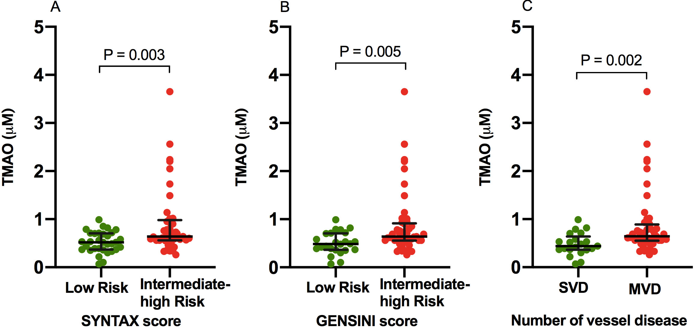
Fig. 2.

Comparison of TMAO levels in Low versus Intermediate-High Atherosclerotic Burden. TMAO levels in low risk versus intermediate-high risk SYNTAX (A) and GENSINI (B) scores, and number of vessel disease (C). MVD, multivessel disease; SVD, single vessel disease; TMAO, trimethylamine N-oxide.

Table 1.Baseline characteristics of NSTEMI Group stratified by SYNTAX and GENSINI scores, and MVD
Variables Total (n = 73) SYNTAX Score GENSINI Score MVD
< 23 (n = 32) 23 (n = 41) P value < 23 (n = 24) 23 (n = 49) P value NO (n = 21) YES (n = 52) P value
Age (Years) 60.1 ± 12.1 58.2 ± 11.1 61.6 ± 12.8 0.237 59 ± 11.5 60.6 ± 12.5 0.598 58.7 ± 11.4 60.7 ± 12.4 0.535
Men 61 (69.3%) 27 (84.4%) 34 (83.9%) 0.564 20 (83.3%) 41 (83.7%) 0.607 17 (81%) 44 (84.6%) 0.473
BMI (kg/m2) 25 ± 3.3 25.6 ± 3.2 24.5 ± 3.3 0.159 25.5 ± 3.5 24.8 ± 3.2 0.370 25.6 ± 3.6 24.8 ± 3.2 0.364
SBP 136.3 ± 19.4 134.6 ± 21.1 137.6 ± 18.1 0.517 135.3 ± 22.8 136.8 ± 17.7 0.759 133.8 ± 21.2 137.3 ± 18.8 0.508
DBP 83.4 ± 12.8 83.9 ± 15.1 83 ± 10.8 0.785 83.5 ± 16.5 83.3 ± 10.7 0.947 83.4 ± 16.4 83.4 ± 11.1 0.997
Heart rate (bpm) 71 (65-71) 70 (65.2-76) 72 (65-79) 0.656 69 (64.2-75.7) 73 (65-77) 0.301 69 (63.5-74.5) 73.5 (65-79.5) 0.137
NSTEMI Symptoms (days) 3 (1-7) 4 (1-7) 2 (1-9.5) 0.391 4 (2-6) 2 (1-9.5) 0.479 4 (1.5-6.5) 2 (1-8.7) 0.592
Hypertension 37 (50.7%) 14 (43.8%) 23 (56.1%) 0.209 9 (37.5%) 28 (57.1%) 0.092 7 (33.3%) 30 (57.7%) 0.051
Diabetes mellitus 20 (27.4%) 7 (22%) 13 (31.7%) 0.253 5 (20.8%) 15 (30.6%) 0.278 4 (19%) 15 (30.8%) 0.237
Previous IS/TIA 2 (2.7%) 0 2 (4.9%) 0.312 0 3 (4.1%) 0.447 0 2 (3.8%) 0.505
Hyperlipidemia 34 (46.6%) 13 (40.6%) 21 (51.2%) 0.254 9 (37.5%) 25 (51%) 0.201 8 (38.1%) 26 (50%) 0.254
Smoker 24 (32.9%) 14 (43.8%) 10 (24.4%) 0.067 10 (41.7%) 14 (28.6%) 0.196 10 (47.7%) 14 (26.9%) 0.078
LVEF (%) 60 (56.5-60) 50 (59-60) 60 (55-60) 0.895 60 (60-60) 60 (55-60) 0.581 60 (59-60) 60 (55-60) 0.761
eGFR (mL/min/1.73 m2) 85.3 ± 20.2 86.4 ± 19.1 84.3 ± 21.2 0.661 85.2 ± 19.3 85.3 ± 20.8 0.992 81.6 ± 17.2 86.7 ± 21.2 0.332
TMAO (mM) 0.59 (0.43-0.78) 0.52 (0.37-0.70) 0.64 (0.56-0.98) 0.003 0.48 (0.36-0.70) 0.64 (0.55-0.91) 0.005 0.44 (0.36-0.64) 0.64 (0.55-0.88) 0.002
hs-CRP (mg/L) 2.8 (0.9-9.8) 2.5 (0.9-9) 3.3 (0.8-10.7) 0.839 2.9 (1-8.4) 2.7 (0.8-10) 0.821 2.4 (0.9-6.2) 3.3 (0.9-10.1) 0.728
D-dimer (mg/L) 0.3 (0.2-0.5) 0.3 (0.2-0.5) 0.3 (0.2-0.49) 0.281 0.3 (0.2-0.49) 0.3 (0.2-0.5) 0.198 0.2 (0.2-0.5) 0.3 (0.2-0.5) 0.675
hs-TnI (ng/mL) 0.8 (0.1-4.5) 1.4 (0.2-4.3) 0.3 (0.07-4.6) 0.266 1.3 (0.3-2.9) 0.3 (0.07-5.8) 0.336 0.8 (0.3-2.9) 0.3 (0.07-6.5) 0.457
hs-TnT (ng/mL) 0.1 (0.04-0.5) 0.2 (0.05-0.5) 0.1 (0.038-0.6) 0.347 0.2 (0.06-0.5) 0.1 (0.03-0.6) 0.354 0.1 (0.06-0.6) 0.1 (0.03-0.58) 0.330
NT-proBNP (pg/mL) 333 (128-623) 197 (105-495) 425 (181-673) 0.053 232 (81-621) 360 (174-624) 0.139 256 (68-589) 336 (144-629) 0.167
TC (mmol/L) 4.4 (3.7-5.2) 4.4 (3.7-5.2) 4.4 (3.7-5.3) 0.947 4.5 (3.9-5.3) 4.1 (3.6-5.2) 0.354 4.2 (3.8-5) 4.4 (3.6-5.3) 0.622
TG (mmol/L) 1.7 (1.1-2.3) 1.7 (1.1-2.5) 1.6 (1-2.2) 0.339 1.6 (1.1-2.28) 1.7 (1.1-2.48) 0.729 1.6 (1.1-2.7) 1.7 (1.1-2.3) 0.992
LDL (mmol/L) 2.8 (2.4-3.7) 2.9 (2.5-3.8) 2.4 (2.4-3.6) 0.420 3 (2.6-4) 2.8 (2.4-3.6) 0.243 2.8 (2.4-3.6) 2.9 (2.4-3.7) 0.807
HDL (mmol/L) 1 (0.9-1.2) 1 (0.8-1.1) 1 (0.9-1.2) 0.158 1 (0.9-1.2) 1 (0.8-1.2) 0.851 1 (0.9-1.2) 1 (0.8-1.1) 0.961
Continues data are expressed as means ± SD or medians (interquartile ranges), and categorical variables are expressed as counts (%). Hyperlipidemia was defined as a fasting total cholesterol level 5.2 (mmol/L), or/and triglyceride level 1.7 (mmol/L), or/and low density lipoprotein > 2.6 (mmol/L). BMI, body mass index; DBP, diastolic blood pressure; eGFR, estimated glomerular filtration rate; HDL, high density lipoprotein; hs-CRP, high sensitivity C Reactive protein; hs-TnI, high sensitivity Troponin I; hs-TnT, high sensitivity Troponin T; IS/TIA, ischemic stroke/ transient ischemic attack; LDL, low density lipoprotein; LVEF, left ventricular ejection fraction; MVD, multivessel disease; NT-proBNP, N-terminal pro-B-type natriuretic peptide; SBP, systolic blood pressure; TC, total cholesterol; TG, triglyceride; TMAO, trimethylamine N-oxide.

Multivariate logistic regression adjusting analysis found that plasma TMAO level was independently associated with MVD (odds ratio [OR]: 5.94, P = 0.005), and intermediate-high risk (score
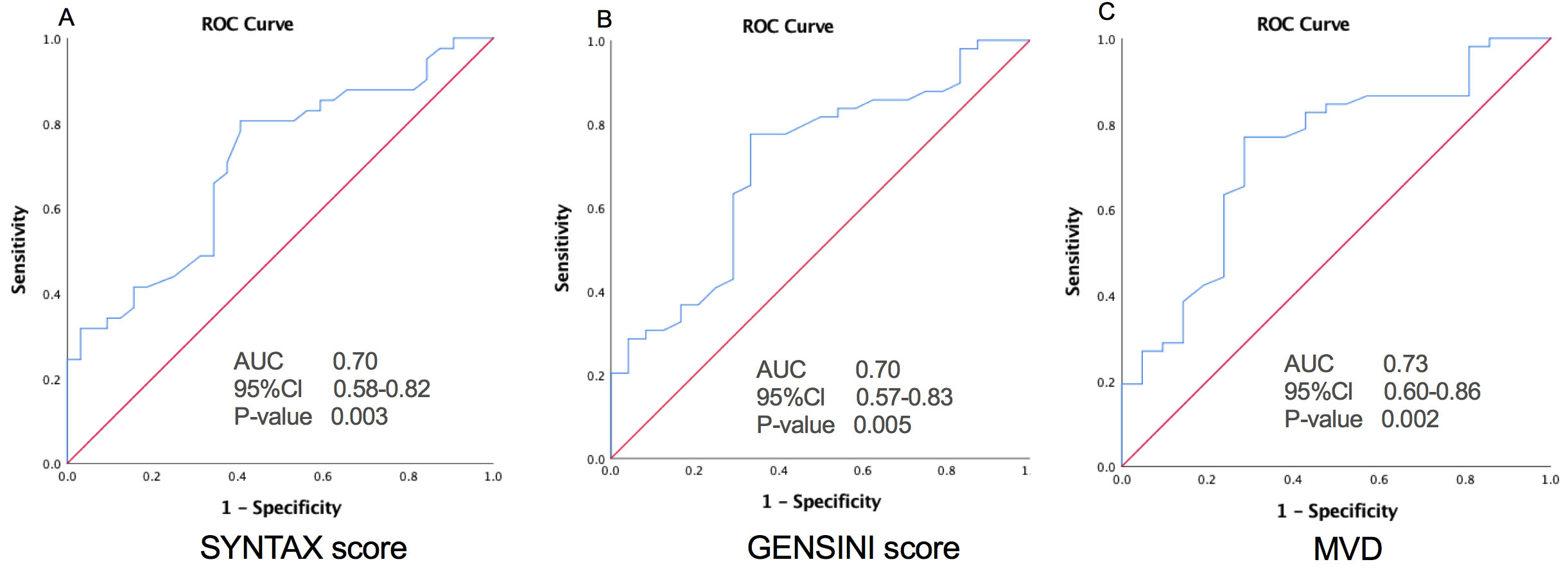
 23) SYNTAX (OR: 3.61, P = 0.013) and GENSINI scores (OR: 4.60, P = 0.008) following adjustment of traditional risk factors (including age, sex, hypertension, diabetes mellitus, smoking, LDL, TG, eGFR, hs-CRP, and BMI) as displayed in Table 2. Additionally, the AUC for TMAO predicted high atherosclerotic burden as MVD (AUC: 0.73, P = 0.002), and intermediate-high risk (score 23) SYNTAX (AUC: 0.70, P = 0.003) and GENSINI scores (AUC: 0.70, P = 0.005) as illustrated in Fig. 3.

Fig. 3.

Relationship of TMAO with High Atherosclerotic Burden. The ROC curves of TMAO for predicting a high atherosclerotic burden as intermediate-high risk SYNTAX (A) and GENSINI (B) scores, and MVD (C). AUC, area under the receiver-operating characteristic curve; Cl, confidence interval; MVD, multivessel disease.

Table 2.Association between TMAO with SYNTAX and GENSINI scores, and MVD
SYNTAX score GENSINI score MVD
OR (95% Cl) P value OR (95% Cl) P value OR (95% Cl) P value
TMAO (mM) Unadjusted 2.85 (0.66-5.04) 0.011 3.03 (0.59-5.47) 0.015 3.63 (0.883-6.38) 0.010
Adjusted* 3.61 (0.74-6.47) 0.013 4.60 (1.19-8.00) 0.008 5.94 (1.79-10.10) 0.005
*Adjusted for traditional risk factors including age, sex, diabetes, hypertension, smoking, TG, LDL, BMI, hs-CRP, eGFR.
Cl, confidence interval; OR, odds ratio; and other abbreviations are listed in Table 1.
4. Discussion

The main finding of this study was a significant association between plasma TMAO levels and high coronary atherosclerotic burden as stratified by number of disease vessels and clinical risk scores including SYNTAX and GENSINI scores in NSTEMI patients. Moreover, TMAO independently predicted the MVD, and intermediate-high risk (score 23) SYNTAX and GENSINI scores, even following traditional risk factors adjustment. In all, TMAO predicts high coronary atherosclerotic burden as quantified above.

Atherosclerosis is a significant clinical problem leading to ischemia in different parts of the vasculature [19, 20, 21]. In this occurs in the coronary vessels, then CAD with calcification [22], partial or total vessel occlusion [23, 24, 25], leading to gradual reduction or absent coronary blood flow [21, 26]. This can manifest as silent asymptomatic disease, angina pectoris [27, 28], NSTEMI or STEMI [29, 30, 31], with adverse consequences such as arrhythmias, heart failure, and death [32]. Therefore, there is an increasing interest to develop predictive risk models for accurate risk stratification, which may include clinical signs or symptoms, imaging results and biomarkers [33]. The TMAO is a gut metabolite, generated from dietary nutrients such as choline, L-carnitine, betaine, and phosphatidylcholine [9, 34, 35]. There is growing appreciation that TMAO has mechanistic links to proatherogenic effect via increased macrophage foam cell formation, activation of the inflammatory and platelet hyperactivity pathways, impaired cholesterol and bile acid transport [9, 10, 11, 12, 34, 35, 36, 37]. These all impaired factors are associated with high-risk CVDs including stroke, atherosclerosis, atrial fibrillation, heart failure, and chronic kidney disease [9, 10, 11, 12, 20, 34, 35, 36, 37, 38]. Based on experimental animal studies, TMAO accelerates atherosclerotic progression by inhibiting reverse cholesterol transport, enhancing platelet activity and thrombosis, and activating macrophages, while targeting production of TMAO inhibit pathogenesis of atherosclerotic [9, 10, 34, 35, 36, 37]. Recent clinical data demonstrated a close relationship of TMAO with markedly high short and long-term adverse outcomes related to CVDs even following adjustment for confounding risk variables [11, 12, 13, 38]. More recently, Yoriko et al. further appraised that long-term increases in TMAO were linked to a higher risk for CVDs and repeated evaluation of TMAO over 10 years increased the early identification of individuals with a greater CVD-related risk [39].

This study is the first, to the best of the authors’ knowledge, to evaluate the relationship between TMAO and coronary atherosclerotic burden in NSTEMI patients. Our study further contributes to earlier findings those suggested a significant link between plasma TMAO and atherosclerotic burden in STEMI and stable CAD patients [14, 15]. Sheng et al. showed a significant association between plasma TMAO levels and high atherosclerotic burden as ascertained by the SYNTAX score and MVD in STEMI [14]. Senthong et al. noted that strong predictability of TMAO for high coronary atherosclerotic burden as evaluated via SYNTAX score stable CAD [15]. Lately, Guo et al. also observed a positive linked of TMAO level with of a high atherosclerotic burden particularly severe coronary artery lesion ( 90% stenosis) compared to mild coronary artery lesion in CAD patients [40].

The strength of our study that TMAO was evaluated in multiple parameters for quantification and stratification of coronary atherosclerotic burden including clinical risk scores such as SYNTAX and GENSINI, and MVD as compared to prior studies evaluated TMAO in only SYNTAX score and/or MVD [14, 15]. A high SYNTAX and GENSINI scores, and MVD are associated with poor prognosis and considered as clinical markers of coronary atherosclerotic burden [3, 17, 41]. Notably, the median SYNTAX score 23 (IQR: 11.5-29) in NSTEMI patients recruited in the present cohort were seemingly higher than STEMI patients described by Sheng et al. 18 (IQR: 11-23.5) and stable CAD patients by Senthong et al. 11 (IQR: 4-18.5), which might be partially attributed to higher proportion of patients (56.1%) in intermediate-high risk SYNTAX score category in the present cohort compared to Sheng et al. (27.8%) and Senthong et al. (18.1%) [14, 15]. Moreover, our study included NSTEMI patients with significant coronary lesion ( 70% stenosis) as compared to prior studies 50% [14, 15] and that might further strengthened our observation for association of TMAO with significant coronary lesion ( 70% stenosis). In fact, Guo et al. reported strong correlation of TMAO for server coronary artery stenosis compared to mild coronary artery stenosis [40]. Furthermore, our study observed a lower concentration of TMAO in patients with NSTEMI than previously reported studies in other types of CAD [14, 15, 40]. Indeed, Wang et al. reported a wide range of TMAO concentrations (lower limit 0.05 μM to upper limit 200 μM) with 95% accuracy determination by isotope dilution high-performance liquid chromatography among 4000 subjects including both healthy individuals and cardiovascular patients [18]. These differences may be caused by many factors associated with different populations particularly demographic characteristics including age, sex, and ethnicity, different types of CAD and criteria for severity, Eastern and Western dietary habits, and composition of gut microbiota [42, 43, 44, 45, 46]. Furthermore, Liu et al. reported significant alteration in gut microbiota profiles in different CAD subtypes of patients and atherosclerotic severity, however they did not observe these changes in TMAO but purposed accordingly to their findings [42]. Therefore, more investigations that cover all these points are required for further explanation.

Recently, there is accumulating evidence to suggest TMAO is a clinically relevant target or marker for risk stratification and short or long-term prognostic outcome related in CVDs [11, 12, 14, 15]. Moreover, inhibition of TMAO has been identified as a novel therapeutic approach for the prevention of atherosclerosis progression compared with existing traditional treatment [10, 11, 12, 37, 39, 43, 44, 45, 46]. In light of these promising findings, implication of TMAO may establish early diagnosis, risk stratification, optimal treatment, and preventive strategies for CVDs [10, 11, 12, 37, 39, 43, 44, 45, 46].

4.1 Limitations

Firstly, we were unable to assess the nutritional and diet status before enrollment for each individual that may bias results as TMAO may be influenced by nutritional status as previously reported [43, 44, 45, 46]. Secondly, we could not exclude selection bias as the present cohort is a single-center study with a comparatively small number of NSTEMI patients, particularly in all sub-categorized groups. Thirdly, the COVID-19 pandemic unable us to continue and complete our original SZ-NSTEMI trial including enrollment of NSTEMI patients about two hundred participants for the evaluation of TMAO in regards to short and long-term prognostic outcomes after primary percutaneous coronary intervention as previously described [16]. Therefore, we recommended a large prospective randomized control trial to evaluate the association between TMAO and coronary atherosclerotic burden as well as a prognostic biomarker for adverse cardiac outcomes in NSTEMI patients undergoing coronary intervention.

5. Conclusions

In conclusion, our study demonstrated that plasma TMAO levels were significantly associated with a high coronary atherosclerotic burden as quantified by the number of diseased vessels, and clinical risk scores including SYNTAX and GENSINI scores in patients with NSTEMI. We encouraged and warranted future large, randomized trial to evaluate our observed results in NSTEMI patients for the clinical endpoints.

Abbreviations

CAD, coronary artery disease; CAG, coronary angiography; CVDs, cardiovascular diseases; ECG, electrogram; MVD, multiple vessels disease; NSTMEI, non-ST-segment myocardial infarction; STEMI, ST-segment myocardial infarction; SVD, single vessel disease; SYNTAX, SYNergy between percutaneous coronary intervention with TAXus and cardiac surgery; TMAO, trimethylamine N-oxide.

Author contributions

Lili W, Junlei C, and Bin Waleed K were involved to research design, protocol, submission to ethical committee, and trial registration. Bin Waleed K, Pengnong C, Tu H, Ding L, Yangkong L, Houqing Z, Qiying C, and Aimei S contributed to literature review, patients selection accordingly to inclusion and exclusion criteria, informed consent, and data collection and interpretation of data. Yunlong X, Shulin W, Tse G, Xintao L, and Afrasyab A underwent statistical analysis and manuscript writing. All authors are involved in final draft of the manuscript, read and approved the final manuscript.

Ethics approval and consent to participate

The study was conducted in accordance with the Declaration of Helsinki and the Ethics Committee of Fuwai Hospital Chinese Academy of Medical Sciences Shenzhen (Ethics approval number: YN201901) and registered clinical study (ChiCTR1900022366) as SZ-NSTEMI. The study can be identified http://www.chictr.org.cn/showprojen.aspx?proj=37821. Written informed consent was obtained from all participants.

Acknowledgment

We thanked all participated individuals in this study.

Funding

This study was funded by the Science Technology and Innovation Commission of Shenzhen Municipality (ZDSYS20190902093409851), Guangdong Innovation Platform of Translational Research for Cerebrovascular Diseases, and National Natural Science Foundation of China (81971634).

Conflict of interest

The authors declare no conflicts of interest.

References
[1]
Damman P, van ’t Hof AW, Ten Berg JM, Jukema JW, Appelman Y, Liem AH, et al. 2015 ESC guidelines for the management of acute coronary syndromes in patients presenting without persistent ST-segment elevation: comments from the Dutch ACS working group. Netherlands Heart Journal. 2017; 25: 181-185.
[2]
Roever L, Tse G, Biondi-Zoccai G. Trends in cardiovascular disease in Australia and in the world. European Journal of Preventive Cardiology. 2018; 25: 1278-1279.
[3]
Neumann FJ, Sousa-Uva M, Ahlsson A, Alfonso F, Banning AP, Benedetto U, et al. 2018 ESC/EACTS Guidelines on myocardial revascularization. The Task Force on myocardial revascularization of the European Society of Cardiology (ESC) and European Association for Cardio-Thoracic Surgery (EACTS). Giornale Italiano di Cardiologia. 2019; 20:1S-61S.
[4]
Thygesen K, Alpert JS, Jaffe AS, Chaitman BR, Bax JJ, Morrow DA, et al. Fourth universal definition of myocardial infarction. Circulation. 2018; 138: e618-e651.
[5]
Mozaffarian D, Benjamin EJ, Go AS, Arnett DK, Blaha MJ, Cushman M, et al. Heart disease and stroke statistics-2016 update: a report from the American Heart Association. Circulation. 2016; 133: e38-360.
[6]
Savonitto S, Ardissino D, Granger CB, Morando G, Prando MD, Mafrici A, et al. Prognostic value of the admission electrocardiogram in acute coronary syndromes. JAMA. 1999; 281: 707-713.
[7]
Winter M, Wiesbauer F, Blessberger H, Pavo N, Sulzgruber P, Huber K, et al. Lipid profile and long-term outcome in premature myocardial infarction. European Journal of Clinical Investigation. 2018; 48: e13008.
[8]
Li CK, Xu Z, Ho J, Lakhani I, Liu YZ, Bazoukis G, et al. Association of NPAC score with survival after acute myocardial infarction. Atherosclerosis. 2020; 301: 30-36.
[9]
Wang Z, Klipfell E, Bennett BJ, Koeth R, Levison BS, Dugar B, et al. Gut flora metabolism of phosphatidylcholine promotes cardiovascular disease. Nature. 2011; 472: 57-63.
[10]
Wang Z, Roberts AB, Buffa JA, Levison BS, Zhu W, Org E, et al. Non-lethal inhibition of gut microbial trimethylamine production for the treatment of atherosclerosis. Cell. 2015; 163: 1585-1595.
[11]
Schiattarella GG, Sannino A, Toscano E, Giugliano G, Gargiulo G, Franzone A, et al. Gut microbe-generated metabolite trimethylamine-N-oxide as cardiovascular risk biomarker: a systematic review and dose-response meta-analysis. European Heart Journal. 2017; 38: 2948-2956.
[12]
Li XS, Obeid S, Klingenberg R, Gencer B, Mach F, Räber L, et al. Gut microbiota-dependent trimethylamine N-oxide in acute coronary syndromes: a prognostic marker for incident cardiovascular events beyond traditional risk factors. European Heart Journal. 2017; 38: 814-824.
[13]
Tan Y, Zhou J, Liu C, Zhou P, Sheng Z, Li J, et al. Association between plasma trimethylamine n-oxide and neoatherosclerosis in patients with very late stent thrombosis. Canadian Journal of Cardiology. 2020; 36: 1252-1260.
[14]
Sheng Z, Tan Y, Liu C, Zhou P, Li J, Zhou J, et al. Relation of circulating trimethylamine N-Oxide with coronary atherosclerotic burden in patients with ST-segment elevation myocardial infarction. The American Journal of Cardiology. 2019; 123: 894-898.
[15]
Senthong V, Li XS, Hudec T, Coughlin J, Wu Y, Levison B, et al. Plasma trimethylamine N-Oxide, a gut microbe-generated phosphatidylcholine metabolite, is associated with atherosclerotic burden. Journal of the American College of Cardiology. 2016; 67: 2620-2628.
[16]
Bin Waleed K, Lu Y, Liu Q, Zeng F, Tu H, Wei Y, et al. Association of trimethylamine N-oxide with coronary atherosclerotic burden in patients with non-ST-segment elevation myocardial infarction. Medicine. 2020; 99: e20794.
[17]
Rampidis GP, Benetos G, Benz DC, Giannopoulos AA, Buechel RR. A guide for Gensini Score calculation. Atherosclerosis. 2019; 287: 181-183.
[18]
Wang Z, Levison BS, Hazen JE, Donahue LM, Li X, Hazen SL. Measurement of trimethylamine-N-oxide by stable isotope dilution liquid chromatography tandem mass spectrometry. Analytical Biochemistry. 2014; 455: 35-40.
[19]
Lee YT, Laxton V, Lin HY, Chan YWF, Fitzgerald-Smith S, To TLO, et al. Animal models of atherosclerosis. Biomedical Reports. 2017; 6: 259-266.
[20]
Lee YT, Lin HY, Chan YWF, Li KHC, To OTL, Yan BP, et al. Mouse models of atherosclerosis: a historical perspective and recent advances. Lipids in Health and Disease. 2017; 16: 12.
[21]
Hu Z, Chen Z, Wang Y, Jiang J, Tse G, Xu W, et al. Effects of granulocyte colony‑stimulating factor on rabbit carotid and porcine heart models of chronic obliterative arterial disease. Molecular Medicine Reports. 2019; 19: 4569-4578.
[22]
Sawant AC, Panchal H, Radadiya D, Pomakov A, Tse G, Liu T, et al. Comparison of rotational with orbital atherectomy during percutaneous coronary intervention for coronary artery calcification: a systematic review and meta-analysis. Cardiovascular Revascularization Medicine. 2020; 21: 501-507.
[23]
Chi WK, Liu T, Nombela-Franco L, Tse G. The impact of chronic total occlusion in non-infarct-related coronary arteries. EuroIntervention. 2019; 15: e297-e298.
[24]
Li KHC, Wong KHG, Gong M, Liu T, Li G, Xia Y, et al. Percutaneous coronary intervention versus medical therapy for chronic total occlusion of coronary arteries: a systematic review and meta-analysis. Current Atherosclerosis Reports. 2019; 21: 42.
[25]
Chen Q, Wang M, Shao S, Liu H, Xia X, Tse G, et al. Mechanisms of intravascular linear ablation induced restenosis in rabbit abdominal aorta. BioMed Research International. 2018; 2018: 1-11.
[26]
Yeung C, Baranchuk A, Tse G, Liu T. The importance of measuring coronary blood flow for clinical decision making. Current Cardiology Reviews. 2019; 15: 320-321.
[27]
Shao S, Shi Z, Tse G, Wang X, Ni Y, Liu H, et al. Effects of trimetazidine pretreatment on endothelial dysfunction and myocardial injury in unstable angina patients undergoing percutaneous coronary intervention. Cardiology Research and Practice. 2019; 2019: 4230948.
[28]
Wang W, Zhang X, Chen K, Yin L, Gong M, Liu Y, et al. Effects of nicorandil infusion on ECG parameters in patients with unstable angina pectoris and percutaneous coronary intervention. Annals of Noninvasive Electrocardiology. 2020; 25: e12736.
[29]
Tse G, Gong M, Nunez J, Sanchis J, Li G, Ali-Hasan-Al-Saegh S, et al. Frailty and mortality outcomes after percutaneous coronary intervention: a systematic review and meta-analysis. Journal of the American Medical Directors Association. 2017; 18: 1097.e1-1097.e10.
[30]
Lampropoulos K, Megalou A, Bazoukis G, Tse G, Manolis A. Pre-loading therapy with statins in patients with angina and acute coronary syndromes undergoing PCI. Journal of Interventional Cardiology. 2017; 30: 507-513.
[31]
Roever L, Tse G, Biondi-Zoccai G. Improvement of LDL cholesterol target achievement rates through cardiac rehabilitation after myocardial infarction. European Journal of Preventive Cardiology. 2019; 26: 791-792.
[32]
Li X, Guan B, Wang Y, Tse G, Zou F, Khalid BW, et al. Association between high-density lipoprotein cholesterol and all-cause mortality in the general population of northern China. Scientific Reports. 2019; 9: 14426.
[33]
Roever L, Tse G, Versaci F, Biondi-Zoccai G. Admission glucagon-like peptide-1 levels in acute myocardial infarction: is this a new biomarker of cardiovascular risk? European Heart Journal. 2020; 41: 890-891.
[34]
Koeth RA, Wang Z, Levison BS, Buffa JA, Org E, Sheehy BT, et al. Intestinal microbiota metabolism of L-carnitine, a nutrient in red meat, promotes atherosclerosis. Nature Medicine. 2013; 19: 576-585.
[35]
Tang WHW, Wang Z, Levison BS, Koeth RA, Britt EB, Fu X, et al. Intestinal microbial metabolism of phosphatidylcholine and cardiovascular risk. The New England Journal of Medicine. 2013; 368: 1575-1584.
[36]
Komaroff AL. The microbiome and risk for atherosclerosis. JAMA. 2018; 319: 2381-2382.
[37]
Roberts AB, Gu X, Buffa JA, Hurd AG, Wang Z, Zhu W, et al. Development of a gut microbe-targeted nonlethal therapeutic to inhibit thrombosis potential. Nature Medicine. 2018; 24: 1407-1417.
[38]
Yu D, Shu XO, Rivera ES, Zhang X, Cai Q, Calcutt MW, et al. Urinary levels of trimethylamine-n-oxide and incident coronary heart disease: a prospective investigation among urban Chinese adults. Journal of the American Heart Association. 2019; 8: e010606.
[39]
Heianza Y, Ma W, DiDonato JA, Sun Q, Rimm EB, Hu FB, et al. Long-term changes in gut microbial metabolite trimethylamine n-oxide and coronary heart disease risk. Journal of the American College of Cardiology. 2020; 75: 763-772.
[40]
Guo F, Zhou J, Li Z, Yu Z, Ouyang D. The association between trimethylamine N-Oxide and its predecessors choline, L-carnitine, and betaine with coronary artery disease and artery stenosis. Cardiology Research and Practice. 2020; 2020: 5854919.
[41]
Neeland IJ, Patel RS, Eshtehardi P, Dhawan S, McDaniel MC, Rab ST, et al. Coronary angiographic scoring systems: an evaluation of their equivalence and validity. American Heart Journal. 2012; 164: 547-552.e1.
[42]
Liu H, Chen X, Hu X, Niu H, Tian R, Wang H, et al. Alterations in the gut microbiome and metabolism with coronary artery disease severity. Microbiome. 2019; 7: 68.
[43]
Witkowski M, Weeks TL, Hazen SL. Gut microbiota and cardiovascular disease. Circulation Research. 2020; 127: 553-570.
[44]
Zhu Y, Li Q, Jiang H. Gut microbiota in atherosclerosis: focus on trimethylamine N‐oxide. Acta Pathologica, Microbiologica et Immunologica Scandinavica. 2020; 128: 353-366.
[45]
Kazemian N, Mahmoudi M, Halperin F, Wu JC, Pakpour S. Gut microbiota and cardiovascular disease: opportunities and challenges. Microbiome. 2020; 8: 36.
[46]
Xu J, Yang Y. Implications of gut microbiome on coronary artery disease. Cardiovascular Diagnosis and Therapy. 2020; 10: 869-880.
Share
Back to top