IMR Press / CEOG / Volume 49 / Issue 2 / DOI: 10.31083/j.ceog4902037
Open Access Case Report
Prenatal identification of novel HSPG2 variants associated with dyssegmental dysplasia Silverman-Handmaker type
Show Less
1 Department of Obstetrics, Shenzhen Futian Maternal and Child Health Hospital, 518045 Shenzhen, Guangdong, China
2 Department of Obstetrics, Shenzhen Maternal and Child Health Hospital, 518000 Shenzhen, Guangdong, China
*Correspondence: wang-yunxia@163.com (Yunxia Wang); 13823106635@163.com (Hui Wang)
Academic Editor: Giovanni Monni
Clin. Exp. Obstet. Gynecol. 2022, 49(2), 37; https://doi.org/10.31083/j.ceog4902037
Submitted: 8 December 2020 | Revised: 22 February 2021 | Accepted: 18 March 2021 | Published: 9 February 2022
Copyright: © 2022 The Author(s). Published by IMR Press.
This is an open access article under the CC BY 4.0 license.
Abstract

Background: We aimed to analyze mutations of the pathogenic gene in dyssegmental dysplasia Silverman-Handmaker (DDSH) type associated with the Heparin sulfate proteoglycan 2 (HSPG2) gene. Case: Prenatal testing for genetic mutations associated with fetal DDSH were performed on a pregnant woman with previous history of carrying a fetus with short limb malformation at the 17th week of gestation. DNA was extracted from amniotic fluid and next-generation sequencing-based deep panel sequencing was performed on the Illumina NextSeq platform to identify possible causative mutations of DDSH. Results: Two novel heterozygous mutations in HSPG2 gene, c.6001dupC (p. R2001pfs*19) and c.11207G>A (p. R373Q), were identified and associated with the DDSH diagnosis. Conclusion: This is the first report to prenatally identify novel mutations in HSPG2 that confirms a DDSH diagnosis.

Keywords
Heparin sulfate proteoglycan 2
Dyssegmental dysplasia Silverman-Handmaker
Gene mutation
Prenatal diagnosis
1. Introduction

Fetal short limb deformity is one of the most common congenital birth defects. While its etiology is diversified, it is primarily related to exposure to drugs, radiation, decoration pollution, chemicals such as hair dyes, benzene, mercury, lead and other heavy metals during pregnancy. Several trials point to recessive genetic diseases of chromosome [1, 2]. Fetal short limb deformities include fatal and non-fatal cases [3]. Related Ultrasound screening is mainly carried out in the second and third trimesters of pregnancy. However, for non-fatal skeletal dysplasia with atypical ultrasonographic features, it is difficult to make a definite diagnosis and classification, resulting in a high probability of misdiagnosis and missed diagnosis. Such failure in prenatal screening can result in disability due to skeletal deformity after birth and even complications of the nervous system that threaten great mental and economic burden to family and society [4, 5, 6, 7]. Accordingly, timely and accurate prenatal diagnosis is of utmost importance [8, 9, 10].

DDSH is a lethal autosomal recessive form of dwarfism with characteristic anisospondylic micromelia. Clinical features of DDSH include dwarfism, short and bowed limbs, flat facial features, anisospondyly, and encephalocele. The neonatal lethal condition DDSH is caused by functional null mutations to HSPG2, which completely prevent perlecan secretion into the extrcellular matrix [11].

The HSPG2 gene is an important, highly conserved gene whose expression affects many developmental processes, including the formation of the heart and brain systems, together with cartilage, bone marrow, and skeletal muscle [12]. A large gene encompassing 97 exons, it maps to chromosome 1p36.1 [11]. HSPG2 encodes the heparin sulfate proteoglycan 2 protein, perlecan, a co-receptor for basic fibroblast growth factor. Perlecan is a major component of basement membranes and is present in many tissues, including cartilage [13]. A type of skeletal dysplasia, DDSH is typically caused by functional mutations in the gene HSPG2, specifically due to a functional deletion mutation of the perlecan gene. In Silverman-Handmaker syndrome, the mutant perlecan molecules were unstable and cannot secreted into the extracellular matrix, and was degraded to smaller fragments with in the cells. Therefore, DDSH is caused by a functional null mutation of HSPG2 [14, 15].

Fewer than 40 cases have been reported in the literature, and only four of these cases were detected antenatally [16].

2. Materials and methods
2.1 Patients

This study was carried out with the approval of the ethics committee (number: SZFTFY-20191108). A Chinese pregnant woman, G3P1A1, 32 years old, was admitted to the prenatal diagnosis center for genetic counseling (16+3 weeks of gestation). The fetus of prior pregnancy was diagnosed with short limb malformation at the 17th week of gestation. Hence, the woman requested a prenatal diagnosis on the 16th week of current pregnancy to determine whether the present fetus was affected. Both parents had no physical deformities.

2.2 Medical history of the pregnant woman

Family history: The first fetus was diagnosed with short limb malformation at the 17th week of gestation. According to the B-scan ultrasound examination report of the fetus, the biparietal diameter was 4 cm, abdominal circumference was 12.5 cm, femur length was 1.7 cm (
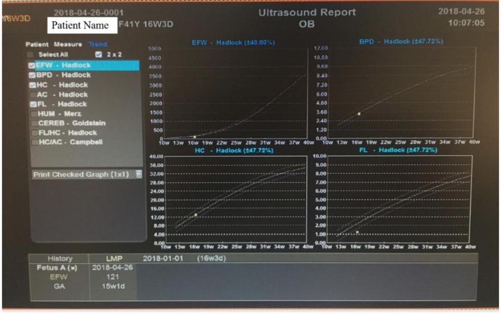
<2.5 percentile), humerus length was 1.7 cm (<2.5 percentile), and the thoracic spine was poor in calcification. Therefore, the pregnancy was terminated by induced labor. The second fetus was a female infant, aged 4, with a height of 100 cm. Examination of the third (and present) fetus at 16 weeks reported biparietal diameter of 3.4 cm, abdominal circumference of 10.5 cm, femur length of 1.2 cm (<2.5 percentile), and humerus length of 1.4 cm (<2.5 percentile). The maximum amniotic fluid depth of the patient was measured by B-mode ultrasonography (Figs. 1,2).

Fig. 1.

The B-ultrasound report.

Fig. 2.

Ultrasound Atlas figures. (a) fetal humerus (14 w). (b) femur length (14 w). (c) fetal humerus (15 w). (d) biparietal diameter (14 w).

2.3 DNA isolation and quantification of the amniotic fluid cell

Amniotic fluid was extracted from the midline (about 1 cm right) of the lower abdomen of the pregnant woman (we determined the puncture point and collected amniotic fluid accurately under the guidance of abdominal ultrasound), then was detected by high through put sequencing. Subsequently, DNA was extracted by Gentra Puregene Blood Kit (Qiagen, Hilden, Germany). NanoDrop 2000 protein nucleic acid analyzer was used to detect DNA concentration and purity.

2.4 DNA isolation and quantification of the parents’ cells

The venous blood of the couple was collected for high throughput sequencing. DNA was extracted by Gentra Puregene Blood Kit (Qiagen Company). NanoDrop 2000 protein nucleic acid analyzer was used to detect DNA concentration and purity. Positive mutation sites found by high throughput sequencing were validated using Sanger sequencing.

2.5 Gene sequencing and analysis

1 μg genomic DNA was sheared using Q800R ultrasonic breaker. The resulting DNA fragments, determined by electrophoresis, were less than 500 bp, with the majority approximately 350 bp. After the construction of the library, the terminal repair was carried out under the action of enzyme. A was added at the 3 ’end, and a specific sequence connector was added at both ends. The barcode sequence was connected. The library with a junction was amplified by PCR. Qubit was used to determine the yield of the constructed libraries, and 300 ng of each sample was mixed. After 24 hours of hybridization, Streptavid in Dynabeads were used to capture the target region and purify the captured products. Post-PCR was used to enrich the target region genes. The size of the library was determined by agarose gel electrophoresis, and the concentration of the library was determined by Qubit3.0 and PCR. The captured libraries were sequenced on a Next Seq 500 sequencer (Illumina, San Diego, CA, USA), with sample dilution and sequencing carried out according to Illumina specifications. The sequencing results were analyzed by bioinformatics methods.

The identified positive mutation sites were validated in DNA extracted from parental blood and amniotic fluid by Sanger sequencing. The primer of HSPG2 gene was designed by the on-line primer design software. Subsequently, the primer was amplified by PCR and the target bands were identified by electrophoresis. The amplified products were sequenced by Sanger. The results of Sanger sequencing were analyzed by Mutation Surveyor V5.0.1 software (SoftGenetics, State College, PA, USA).

3. Results
3.1 Gene mutations and their pathogenicity prediction analysis

350 related genes were detected in this analysis. Therein, 5311 coding regions contained 835,566 base groups. The average coverage depth was 230+/–236x. Therein, 98.2% of the coverage area was greater than 10x and 96.8% of the coverage area was greater than 20x.

Two pathogenic mutations of HSPG2 gene were identified by high throughput sequencing and validated by Sanger sequencing. Two heterozygous mutations of HSPG2 gene were detected as c.6001dupC (p. R2001pfs*19) and c.11207G>A (p. R373Q). The mutation of c.11207G>A (p. R3736Q) was carried out via pathogenicity prediction analysis by HumVar software. The result shows a high pathogenicity possibility for c.11207G>A (p. R3736Q) mutation with a score of 0.994. These two rare variants are closely associated with clinical manifestations.

3.2 Sanger sequencing

Next generation sequencing results were confirmed by Sanger sequencing. Heterozygous mutation c.6001dupC (p. R2001pfs*19) and c.11207G
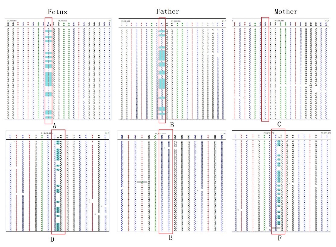
>A (p. R373Q) could be detected in both parents. This result indicates that the two gene variants in the fetus were inherited from its parents (both heterozygous) (Fig. 3A–F).

Fig. 3.

Gene mutation map of fetus and parents. Mutation site: high throughput sequencing site indicated that there were two mutation sites in fetal HSPG2 gene: c.11207G>A (p. R3736Q) (A) and c.6001dupC (p. R2001pfs*19) (D). There was one mutation site in fetal father HSPG2 gene: c.11207G>A (p. R3736Q) (B) and there was no c.6001dupC (p. R2001pfs*19) mutation (E). There was one mutation site in fetal mother HSPG2 gene: c.60.01dupC (p. R2001pfs*19) mutation (C), no c.11207G>P_R3736Q (F).

3.3 Pregnancy outcome

The pregnancy was terminated by induction of labor at 16 weeks as requested by the pregnant woman and her family. There was no autopsy (and therefore, no relevant pictures) due to familial disagreement.

4. Discussion

Fetal short limb deformity is one of the most common birth defects in clinic, along with osteogenes is dysplasia, cartilage dysplasia, fatal dwarf, and short rib-polydactyly syndrome [17, 18]. The common pathogenic genes of fetal short limb deformity have been identified. For example, the pathogenic genes of achondroplasia, fatal dwarf and osteogenetic dysplasia are FGFR3 and COL1A1/COL1A12 [19, 20, 21, 22, 23, 24]. DDSH is generally caused by functional null mutations in the gene, HSPG2. The clinical manifestations are short limb dwarfs of newborns and may be accompanied by mental disorders [25] (PMID4059934, 3605216, 4953364, 2290482, 25666757). In this study, the whole exon of fetal gene was sequenced in a case of short limb malformation diagnosed by ultrasound, and the prenatal accurate gene diagnosis was completed. The parents did not show any clinical manifestations of the disease. These results indicate that the fetus short limb malformation may be caused by autosomal recessive inheritance.

The two identified variants of HSPG2 gene, c.6001dupC (p. R2001pfs*19) and c.11207G>A (p. R373Q) were significantly associated with DDSH with pathogenicity prediction of 0.994 for c.11207G>A (p. R3736Q) mutation and c.6001dupC (p. R2001pfs*19) mutation. As no clinical manifestations of the short limb were found for parents, we deduce heterozygous variants c.6001dupC (p. R2001pfs*19) and c.11207G>A (p. R373Q) are autosomal recessive inheritance. The same variants were found in maternal and paternal blood. Hence, it can be concluded that the fetus inherited these variants from the parents.

This is the first time these two mutations of HSPG2 genes are reported. The c.6001dupC (p. R2001pfs*1 mutation into a shift mutation) predicts a stop codon, which would result in a truncated version of the protein. The region where the p. R373Q mutation is located is an important component of the protein. The amino acid sequences of different species around this area are highly conserved. Computer-aided analysis predicts that the structure/function of the protein is likely to be affected.

In conclusion, novelmutations c.6001dupC (p. R2001pfs*19) and c.11207G>A (p. R373Q) in HSPG2 gene cause Silverman-Handmaker type skeletal segmental dysplasia. High throughput sequencing combined with second-generation sequencing platform sequencing in prenatal testing can be valuable for early prediction of harmful fetal mutations.

Author contributions

YXW designed the research study and performed the research. HW provided medical records and geneticsequencing for children and their parents. All authors contributed to editorial changes in the manuscript. All authors read and approved the final manuscript.

Ethics approval and consent to participate

All subjects gave their informed consent for inclusion before they participated in the study. The study was conducted in accordance with the Declaration of Helsinki, and the protocol was approved by the Ethics Committee of the hospital (approval number: 20180422).

Acknowledgment

The authors would like to thank the staff involved in the treatment of the patient, including Hui Wang, Xiaoyang Yang, Xian Chen et al. at the cooperating Departments at Medical Heredity Center, Shenzhen Maternal and Child Health Hospital, Southern Medical University. We wish to acknowledge the support of pregnant women and their families in helping us complete prenatal diagnosis and family follow-up.

Funding

This research received no external funding.

Conflict of interest

The authors declare no conflict of interest.

References
[1]
Yeh P, Saeed F, Paramasivam G, Wyatt-Ashmead J, Kumar S. Accuracy of prenatal diagnosis and prediction of lethality for fetal skeletal dysplasias. Prenatal Diagnosis. 2011; 31: 515–518.
[2]
Orhant L, Anselem O, Fradin M, Becker PH, Beugnet C, Deburgrave N, et al. Droplet digital PCR combined with minisequencing, a new approach to analyze fetal DNA from maternal blood: application to the non-invasive prenatal diagnosis of achondroplasia. Prenatal Diagnosis. 2016; 36: 397–406.
[3]
Figueras F, Gratacos E. An integrated approach to fetal growth restriction. Best Practice & Research Clinical Obstetrics & Gynaecology. 2017; 38: 48–58.
[4]
Gordijn SJ, Beune IM, Thilaganathan B, Papageorghiou A, Baschat AA, Baker PN, et al. Consensus definition for placetal fetal growth restriction: a Delphi procedure. Obstetrics and Gynecologic Ultrasound. 2016; 48: 333–339.
[5]
Kalia SS, Adelman K, Bale SJ, Chung WK, Eng C, Evans JP, et al. Recommendations for reporting of secondary findings in clinical exome and genome sequencing, 2016 update (ACMG SF v2.0): a policy statement of the American College of Medical Genetics and Genomics. Genetics in Medicine. 2017; 19: 249–255.
[6]
Porntaveetus T, Srichomthong C, Suphapeetiporn K, Shotelersuk V. Monoallelic FGFR3 and Biallelic ALPL mutations in a Thai girl with hypochondroplasia and hypophosphatasia. American Journal of Medical Genetics. Part A. 2017; 173: 2747–2752.
[7]
Ornitz DM, Legeai-Mallet L. Achondroplasia: development, pathogenesis, and therapy. Developmental Dynamics. 2017; 246: 291–309.
[8]
Sarabipour S, Hristova K. Pathogenic cysteine removal mutations in fgfr extracellular domains stabilize receptor dimers and perturb the TM dimer structure. Journal of Molecular Biology. 2016; 428: 3903–3910.
[9]
Del Piccolo N, Sarabipour S, Hristova K. A new method to study heterodimerization of membrane proteins and its application to fibroblast growth factor receptors. Journal of Biological Chemistry. 2017; 292: 1288–1301.
[10]
Chen S, Chen C, Wang L, Chern S, Wu P, Chen Y, et al. Perinatal imaging findings and molecular genetic analysis of thanatophoric dysplasia type 1 in a fetus with a c.2419T>G (p. Ter807Gly) (X807G) mutation in FGFR3. Taiwanese Journal of Obstetrics and Gynecology. 2017; 56: 87–92.
[11]
Arikawa-Hirasawa E, Wilcox WR, Yamada Y. Dyssegmental dysplasia, Silverman-Handmaker type: unexpected role of perlecan in cartilage development. American Journal of Medical Genetics. 2002; 106: 254–257.
[12]
Martinez JR, Dhawan A, Farach-Carson MC. Modular proteoglycan perlecan/HSPG2: mutations, phenotypes, and functions. Genes. 2018; 9: 556.
[13]
Kniffin CL, McKusick VA. Heparan Sulfate Proteoglycan of Basement Membrane; HSPG2. 1991. Available at: http://www.omim.org/entry/142461 (Accessed: 8 November 2015).
[14]
Ladhani NNN, Chitayat D, Nezarati MM, Laureane MC, Keating S, Silver RJ, et al. Dyssegmental dysplasia, Silverman-Handmaker type: prenatal ultrasound findings and molecular analysis. Prenatal Diagnosis. 2013; 33: 1039–1043.
[15]
Arikawa-Hirasawa E, Wilcox WR, Le AH, Silverman N, Govindraj P, Hassell JR, et al. Dyssegmental dysplasia, Silverman-Handmaker type, is caused by functional null mutations of the perlecan gene. Nature Genetics. 2001; 27: 431–434.
[16]
Basalom S, Trakadis Y, Shear R, Azouz ME, De Bie I. Dyssegmental dysplasia, Silverman-Handmaker type: a challenging antenatal diagnosis in a dizygotic twin pregnancy. Molecular Genetics & Genomic Medicine. 2018; 6: 452–456.
[17]
Xie F, DeSmet M, Kanginakudru S, Jose L, Culleton SP, Gilson T, et al. Kinase activity of Fibroblast growth factor reception-3 regulates activity of the papillomavirus E2 protein. Journal of Virology. 2017; 91: e01066-17.
[18]
Jung M, Park S. Genetically confirmed thanatophoric dysplasia with fibroblast growth factor receptor 3 mutation. Experimental and Molecular Pathology. 2017; 102: 290–295.
[19]
Zhou Z, Ota S, Deng C, Akiyama H, Hurlin PJ. Mutant activated FGFR3 impairs endochondral bone growth by preventing SOX9 downregulation in differentiating chondrocytes. Human Molecular Genetics. 2015; 24: 1764–1773.
[20]
Di Rocco F, Biosse Duplan M, Heuzé Y, Kaci N, Komla-Ebri D, Munnich A, et al. FGFR3 mutation causes abnormal membranous ossification in achondroplasia. Human Molecular Genetics. 2014; 23: 2914–2925.
[21]
Matsui Y. Genetic basis for skeletal disease. Genetic defects in chondrodysplasia. Clinical Calcium. 2010; 20: 1182–1189.
[22]
Makrythanasis P, Temtamy S, Aglan MS, Otaify GA, Hamamy H, Antonarakis SE. A Novel Homozygous Mutation in FGFR3 Causes Tall Stature, Severe Lateral Tibial Deviation, Scoliosis, Hearing Impairment, Camptodactyly, and Arachnodactyly. Human Mutation. 2014; 35: 959–963.
[23]
Yuan H, Huang L, Hu X, Li Q, Sun X, Xie Y, et al. FGFR3 gene mutation plus GRB10 gene duplication in a patient with achondroplasia plus growth delay with prenatal onse. Orphanet Journal of Rare Diseases. 2016; 11: 89.
[24]
Hattori A, Katoh-Fukui Y, Nakamura A, Matsubara K, Kamimaki T, Tanaka H, et al. Next generation sequencing-based mutation screening of 86 patients with idiopathic short stature. Endocrine Journal. 2017; 64: 947–954.
[25]
Rieubland C, Jacquemont S, Mittaz L, Osterheld MC, Vial Y, Superti-Furga A, et al. Phenotypic and molecular characterization of a novel case of dyssegmental dysplasia, Silverman-Handmaker type. European Journal of Medical Genetics. 2010; 53: 294–298.
Share
Back to top