IMR Press / FBL / Volume 26 / Issue 9 / DOI: 10.52586/4959
Open Access Original Research
Characterization of the differential efficacy of austenitic vs martensitic NiTi rotary files in non-surgical root canal retreatment: a micro-CT analysis
Show Less
1 Department of Endodontics, Faculty of Dentistry, King Abdulaziz University, 21589 Jeddah, Saudi Arabia
2 Ministry of Health, 21 Yehea Almoalmy, Alrabwah, 23222 Jeddah, Saudi Arabia
3 Department of Dental Public Health, Faculty of Dentistry, King Abdulaziz University, 21589 Jeddah, Saudi Arabia
4 Department of Restorative Dentistry, Faculty of Dentistry, King Abdulaziz University, 21589 Jeddah, Saudi Arabia
*Correspondence: lalsofi@kau.edu.sa (Loai Alsofi)
Front. Biosci. (Landmark Ed) 2021, 26(9), 465–474; https://doi.org/10.52586/4959
Submitted: 23 May 2021 | Revised: 7 July 2021 | Accepted: 27 July 2021 | Published: 30 September 2021
(This article belongs to the Special Issue NiTi Rotary Instruments in Endodontics)
Copyright: © 2021 The Author(s). Published by BRI.
This is an open access article under the CC BY 4.0 license (https://creativecommons.org/licenses/by/4.0/).
Abstract

Aim: Our study aims to characterize the differential efficacy of martensitic and austenitic files in root canal retreatment regarding defiling ability, debris management and morphometric features. Materials and methods: A total of 10 human premolar teeth with two separate fully formed roots were selected, prepared with BioRace system (BR) up to a size BR2 (0.04/25). Root canals were filled and then divided according to the file type into two groups (n = 10). Two geometrically identical files have been used with the only difference between them is heat treatment; One Curve (OC) martensitic and One Shape (OS) austenitic. Preoperative and postoperative Micro-CT scans were done for all the samples, and the percent volume of residual filling materials was calculated. Canals reshaping and remaining debris in the canals were assessed by calculating the volume of the canals before and after retreatment using Micro-CT software. Results: The percentages of removed filling materials were similar between both groups (97.2% OC vs 97.8% OS). OS was significantly faster than OC (P < 0.05). Retreatment procedure did not significantly change volume, structure model index, surface area, and canal curvature. OS group transported the canals more than OC at the apical third (P < 0.05). There were no significant differences between both groups concerning un-instrumented surface area and accumulated hard tissue debris (AHTD) (P > 0.05). Conclusion: Under the circumstances of this study, neither file systems could completely remove the filling materials. However, OC resulted in less transportation than OS in the apical one third (P < 0.05). Also, OC resulted less AHTD than OS but with no statistical difference.

Keywords
Nickel-titanium
One Curve
One Shape
Rotary files
Retreatment
2. Introduction

NiTi endodontic rotary files have been used for the cleaning and shaping during endodontic treatment to eliminate infection from inside the root canal. The enhanced flexibility and super elasticity of these files allow them to negotiate sever canal curvatures without separation and with minimal alteration to the original anatomy of the root canal. Two main phases of these NiTi files are associated with the shape memory and super elasticity, martensite and austenite, with R-phase as intermediate phase that may occur [1]. The effect of these phases on the mechanical properties of NiTi files have been studied in the literature. A previous report showed that there is a correlation between stress point localization during file bending and cross section geometry, therefore it is recommended to move the file in and out during rotation inside the canal to distribute internal stresses along the file length [2]. Also, it was shown that the presence of R-phase because of heat treatment improved file flexibility [3]. Different geometrical factors can also affect the file behavior and characteristics. A finite element analysis study showed that the polar moment of inertia is the most important factor in determining file’s torsional resistance over cross sectional area and metal mass [4].

Complete removal of endodontic materials is crucial for the success of nonsurgical root canal retreatment as it enables efficient cleaning, disinfection, shaping, and filling of the root canal system [5]. Conservative access design is also recommended to save coronal tooth structure while allowing efficient instrumentation of the root canal system [6].

The removal of filling materials from the root canal system has been implemented using multiple techniques, including the use of endodontic Gates Glidden burs, hand files, heat, ultrasonic instruments, nickel-titanium (NiTi) rotary files, laser, and use of adjunctive solvents [7, 8]. Nonetheless, none of the previous studies have shown the ability to completely remove root canal filling materials [9], especially in the apical portion of root canals [10]. Remaining filling materials inside the root canal prevents the complete disinfection of the root canal and also prevents the bonding of root canal sealer into dentin walls. However, the use of NiTi rotary files in root canal retreatment is proved to be more effective than hand instruments with fewer procedural errors [11, 12]. Thus, different rotary systems have been developed by several manufactures to optimize gutta-percha removal techniques.

ProTaper Universal Retreatment system (Dentsply Maillefer, Ballaigues, Switzerland) has been used as an alternative to hand files for removing gutta-percha in endodontic retreatment cases [12]. The system has three files, D1, D2, and D3, where each one is used for each root canal third, according to the manufacturer [13, 14].

Different reports showed that when ProTaper retreatment files are used, they leave between 10 and 35% of gutta percha remnants behind. Therefore, it is recommended to use supplementary files [14, 15].

The removal of gutta-percha with ProTaper retreatment files with or without the use of self-adjusting file was evaluated, it was found that the gutta-percha was not completely removed from the apical portion of curved canals. However, using SAF enables the removal of more gutta-percha than ProTaper alone [15].

Reciprocating single-file systems, originally designed for root canal instrumentation, were suggested to be used for root canal retreatment [16, 17, 18]. Their reciprocating motion increases the instrument’s resistance to cyclic fatigue [19, 20], and when compared to rotational and hand files, they were shown to be more efficient in the removal of gutta percha from the root canal walls [19].

The use of 2 reciprocating systems, i.e., WaveOne and Reciproc was compared with a continuous rotation system (ProTaper Universal Retreatment). Both Wave One and Reciproc reciprocating systems were as efficient as the ProTaper Universal retreatment system in removing sealer and gutta-percha [9].

Although different nickel-titanium (NiTi) files were developed [21],extensive research has been done to modify the presently available root canal instrument designs, modifying the surface of the alloy or changing the alloy microstructure with post-twisting or post machining heat treatment [22].

Single file systems use was introduced and recommended to use to reduce cross-contamination and instrument fatigue. Thus, minimizing instrument separation within the canal without compromising the cutting efficiency [23].

One Shape single file system (OS) is based on one instrument composed of electropolished austenite 55-NiTi alloy. The OS single instrument is used in a full clockwise rotation with a constant taper of 0.06 with variable cross-sections across the length of its active working part and a tip size of 25 mm [24]. This innovative asymmetrical cross-section with a longer pitch increases the available volume for upward debris elimination allowing better shaping. More flexibility is also gained because of the longer instrument pitch, which provides an increased ability to navigate curves and allows easy instrument progression to the apex [24, 25].

One Curve single file system (OC) is a heat-treated OS NiTi file, made of martensite C-Wire, which is an exclusive proprietary, developed by the manufacturer. It is claimed to be hyperflexible with increased cyclic fatigue resistance without sucking effect. The tip size comes in multiple sizes, i.e., 25, 35, and 45 with either 4% or 6% taper [26].

Both systems OS and OC, have a similar design with a unique asymmetrical cutting profile and an inactive tip to maintain the original canal curvature and therefore may cause less transportation [27].

From a clinical point of view, added to the inability to completely remove root canal filling materials, root canal retreatment procedures can result in transportation of root canals that could lead to ledging, perforation, zipping, especially in the apical third [28]. Thus, these conditions should be examined, which can lead to the prevention of root weakening and to reduce the possibility of vertical root fracture or canal perforation. To date, there are few studies about the efficacy of filling material removal in natural canals using single-file NiTi systems. Furthermore, the ability of OS and OC systems in removing filling material from natural canals has not been evaluated yet. Therefore, the present study aimed to compare One Shape and One Curve single file systems regarding filling material removal efficiency, the working time needed for filling removal, and canal morpho geometry changes after retreatment through certain parameters such as canal volume, surface area, SMI, straightening, transportation, un- instrumented canal surface area, and debris removal.

3. Materials and methods
3.1 Preparation of the specimens

Ten double-rooted teeth with canal curvature (10–20) were selected from a pool of teeth that were stored in 0.1 thymol to be used for the study. Canals were prepared using BioRace rotary instruments—BR0 (25/0.08), BR1 (15/0.05), and BR2 (25/0.04)—to the full WL with the Elements Motor (SybronEndo, Orange, CA) in a continuous rotation motion with 600 rpm speed and 1.5 N/cm torque. Canals were irrigated between each file using 2 mL of sodium hypochlorite (5.25%) delivered via a 5 mL plastic syringe with a 30-gauge side-vented needle (Ultradent, South Jordan, UT). After completion of the preparation, EndoActivator (Dentsply Maillefer, Ballaigues, Switzerland) was used with a last rinse of 1 mL EDTA (17%) followed by 5 mL sodium hypochlorite (5.25%). The canals were then dried using paper points. The canals were obturated using AH Plus sealer (Dentsply DeTrey, Konstanz, Germany) and the continuous wave warm vertical compaction technique to their orifices. The cavity of access was temporized using a pellet of cotton and Cavit (ES- PE, Seefeld, Germany). The specimens were kept in an incubator at a temperature of 37 C and 100% humidity for 15 days to let the sealer completely set. All endodontic treatments were performed by a single operator.

3.2 Root canal retreatment

Teeth were divided randomly into two groups with 10 canals per group (n = 10) according to the rotary file used for retreatment. In group A (n = 10), OC was used and in group B (n = 10), OS was used. Both systems were used at 600 rpm with 2.5 N/cm to improve their retreatment efficiency and penetration capability. Complete removal of the filling materials was verified by a dental operating microscope (Zeiss, Munich, Germany) and periapical radiographs.

3.3 Micro CT measurements and evaluations

All teeth were inserted in special micro-CT holders to ensure accurate positioning between scans. The samples were scanned with a commercially available cabinet micro-CT (μCT 100 Scanco Medical AG, Brüttisellen, Switzerland) at an energy of 90 kVp, intensity of 88 μA, and integration time of 500 ms for each projection. As is typically done, a series of 1422 to at most 1896 slices were acquired with a voxel size of 14 μm utilizing a filtered back-projection calculation. Similar settings were applied for the whole measurements. The outer contour of each tooth was generated automatically by using a special-purpose script. The dentin segmentation was automatically adjusted according to the grayscale in the CT slices. The area of interest was outlined roughly by a contour drawn by hand. Various algorithms were used, such as volume, 3D registrations, segmentation, conicity, curvature, and surface area. These measurements were obtained from SCANCO evaluation software (IPL V5.06B, SCANCO Medical), which provides multiple result files. Two micro-CT scans were done for each tooth, one after root canal obturation and one after retreatment procedure. Post-obturation micro-CT scans were saved in the computer for post retreatment /reshaping calculations. The region of interest was taken at 12 μm from the apical foramen. Post-retreatment and canal cleanliness micro-CT scans were performed after gutta-percha removal using standard positions and parameters to allow for comparisons. The volumes from preoperative and postoperative distribution of root filling materials were measured in cubic millimeters (mm3). The mean values for every specimen and the percentage of residual filling materials after retreatment procedures were calculated. The qualitative assessment was made by superimposing results from created 3D images over each other and a green image for the pre-retreatment scan as well as a red one for the post-retreatment scan. The cross-section of the image at selected levels was presented in raw and illustrated form throughout the experiment. Also, the following parameters were calculated: volume, surface area, structural model index, canal straightening and transportation, un-instrumented surface area. Accumulated hard tissue debris (AHTD) was calculated before and after retreatment by determining the difference in instrumented and un-instrumented root canal space. The AHTD was quantified by analyzing the difference in the images taken before and after retreatment. The debris was calculated in cubic millimeters and was shown as a percentage of the full canal system volume following retreatment (vol %).

4. Statistical analysis

Data was checked and we excluded the outliers prior to any statistical analysis performed. Normality of pre retreatment data distribution was tested suing the Shapiro-Wilk test, which showed normally distributed data [29]. Mean and standard deviation was calculated for all parameters. Statistical analysis was performed using IMB® SPSS® (PASW version 23.0 IBM®, Chicago, IL, USA). The paired sample t-test was used for statistical analysis between pre and post retreatment data for each group. The unpaired sample t-test was used for statistical analysis between post retreatment data in the two groups. The P-value for statistical significance was set at <0.05.

5. Results

There was no statistically significant difference in the volume of filling materials before retreatment between both groups (P > 0.05) or the volume of filling materials after retreatment between both groups (P > 0.05). The mean time needed to perform the retreatment procedure was shorter in the OS group than in the OC group (P = 0.001) (Table 1). None of both files was able to completely remove all filling materials from the canals.

Table 1.This Table shows mean initial volume of filling material (mm3), mean percentage of filling material removed (%), and mean time (T) for complete filling materials removal in seconds. It also shows difference in morpho geometric parameters (volume, area, and SMI) of the root canals between pre- and post-retreatment of both groups.
Variable One Curve n = 10 One Shape n = 10 P-value
Initial filling material (mm3) 1.90 ± 0.76 1.67 ± 0.56 0.437
Filling material removed (%) 97.38 ± 2.09 97.87 ± 1.94 0.595
Time in seconds 308.80 ± 28.11 262.20 ± 23.99 0.001
Volume (mm3) 0.46 ± 0.35 0.40 ± 0.45 0.724
Area (mm2) 1.62 ±1.24 1.42 ± 1.30 0.721
SMI 0.28 ± 0.27 0.27± 0.30 0.927

The morpho geometric changes created by OC and OS files in the root canals were analyzed. The results did not show statistically significant differences between both groups in terms of pre-retreatment measurements (P > 0.05). These measurements include volume, surface area, and structural model index of the canal. This indicates homogenous morpho geometry between the groups before retreatment. There is no statistically significant difference between the groups after retreatment in volume and surface area (P > 0.05). However, the SMI value was higher with a statistically significant difference in the OC group (P < 0.05). The increased SMI values indicates that OC files changed the canal geometry to be more rounded that OS files. This can be explained by the fact that OC files are more flexible and ductile, thus leaving canal walls more rounded than the stiffer OS files which resulted in more straight canal walls. The difference in the morpho geometric parameters between pre-and post-retreatment was calculated for volume, surface area, and SMI, with no statistically significant difference between both groups (P > 0.05), Table 1.

Three dimensional rendered images were constructed showing concatenated root canals of the pre-and post-retreatment scans, Fig. 1. The green color shows the pre-retreatment root canal geometry, and the orange shows post-retreatment root canal geometry. Cross-section slices were taken from raw micro-CT data at level slices 701, 901, and 1201 of both pre-and post-retreatment scans, Fig. 2.

Fig. 1.

Three dimensional rendered images of concatenated root canals of the pre- and post-retreatment scans. Green color shows the pre-retreatment root canal geometry and orange shows post-retreatment root canal geometry. (A) shows palatal view of the root canals. (B) shows mesial view of the root canals. (C) shows buccal view of the root canals. (D) shows distal view of the root canals.
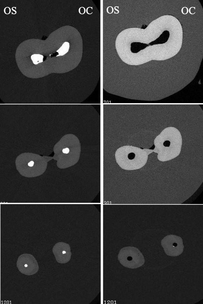
Fig. 2.

Cross section slices taken from raw micro-CT data at level slices 701 (top) 901 (middle), and 1201 (bottom) of both pre- and post- retreatment scans. Cross sections on the left side show pre- retreatment images and cross sections on the left show images of post-retreatment.

Canal straightening for OC and OS groups was 13.93% (±11.93%) and 32.80% (±38.11%), respectively, which means the OS file resulted in more straightening than the OC file, but without statistically significant difference between groups (P > 0.05). The presence of large standard deviation in canal straightening results can be explained by the different canal configuration to start with which cannot be standardized. Mean canal transportation measurements (μm) are summarized in Table 2. Retreatment with the OS group showed significantly more apical transportation than the OC group (P = 0.047). The mean canal transportation for OS was higher than OC at the coronal and middle thirds but without a statistically significant difference (P > 0.05). The mean static voxels and percentages of un-instrumented voxels in both OC and OS groups are summarized in Table 2. The OS group presented more un-instrumented surface area than the OC group, which means the OS group touched less surface area than the OC group. However, there was no statistically significant difference between both groups (P > 0.05).

Table 2.This Table shows mean canal transportation determined for the coronal, middle, and apical third after retreatment with OC and OS files. It also shows mean score of static voxel and mean percentage of un-instrumented canal surface area of root canals after retreatment with OC and OS files and mean score of the percentage in debris reduction of both groups’ reduction.
Variable One Curve n = 10 One Shape n = 10 P-value
Mean canal transportation Coronal third 29.2600 ± 19.40940 33.5400 ± 22.700 0.656
Middle third 31.8000 ± 32.00757 35.7400 ± 33.71641 0.792
Apical third 33.5600 ± 21.64328 86.2600 ± 70.98346 0.047
Un-instrumented Static voxels 23500.20 ± 12179.27 21027.90 ± 8476.27 0.605
Un-instrumented canal surface area (%) 40.32 ± 19.94 41.28 ± 19.41 0.915
Percentage reduction of debris 96.97 ± 1.47 95.82 ± 2.78 0.263

The mean volume of accumulated hard tissue debris (AHTD) before and after retreatment was calculated. There are no statistically significant differences in the experimental groups before or after retreatment (P
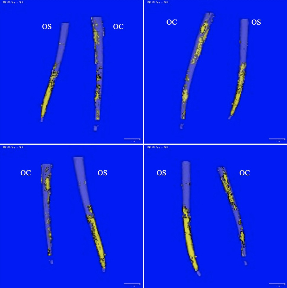
 > 0.05), meaning there is a similarity between the two groups. Reductions in the AHTD were seen in both groups. OC group showed a 95%–99% reduction with a mean of 96.97%. Similarly, OS showed 92%–98% with a mean of 95.82% debris reduction (Table 2). However, there was no statistically significant difference between the groups (P > 0.05). Fig. 3 shows different views of three-dimensional computer constructed images of the root canal system showing the amount of hard tissue debris after retreatment with either OC or OS.

Fig. 3.

Different views of three-dimensional computer constructed images of root canal system showing the amount of hard tissue debris after retreatment with either OC (One Curve) or OS (One Shape) files.

6. Discussion

Recent advances in the metallurgical engineering of NiTi instruments led to the development of various NiTi rotary files that are manufactured by different heat treatment such as max-wire, c-wire, t-wire, controlled memory (CM), R-phase, and M-wire [30, 31, 32, 33]. Different heat-treated files exhibit different phase transformation behavior, which improves fatigue resistance, torsional reissuance, and flexibility [34, 35, 36, 37, 38]. Heat-treated NiTi rotary files are available in two forms, martensitic and austenitic with wide range of motion kinematics, cross section, design, and sizes, which may result in biased shaping ability results.

In the study, we aimed to compare the differential efficacy of two heat-treated rotary files systems which are only different in their metallurgical properties, austenite (OS) and martensite (OC), in the ability to remove gutta-percha from root canals, the time needed for removal, and the morpho geometric changes created during the removal.

The main objective of root canal retreatment is the disinfection of the root canal system which leads to healing of the periapical tissues. Studies have reported that the successful outcome of root canal retreatment ranges from 87 to 97% depending on whether preoperative periapical periodontitis was present or absent [39]. One of the factors that affect the outcome in cases of endodontic retreatment is the ability to completely remove the previous filling material from the root canal [40, 41].

Micro-computed tomography (μ-CT) has been suggested for assessing the tomography of canal walls, as it is a non-destructive technique, which provides accurate three-dimensional reproduction of the tooth without destructive sectioning of the specimens or loss of the root material during sectioning [42]. It also allows superior preoperative and postoperative evaluation of the morphological changes in the root canal trajectory for better assessment of root curvatures and canal transportation [43].

Micro CT analysis was used in our study to evaluate the removal of failing materials as well as to evaluate the morphometric changes created in the root canals after the use of two different rotary systems, One Curve, and One Shape. None of the previous retreatment studies show that it is possible to remove 100% of the filling materials from the root canals [5, 40, 44, 45, 46]. However, in this study, both systems were able to remove around 97% of filling material compared to 75–81% in a previous study in which ProTaper rotary files were used with solvent for retreatment [5]. The mean time to remove gutta-percha ranges from 262 seconds by OS to 308 seconds by OC compared to 540–720 seconds in a study by Mittal 2014 and 412–532 seconds in a study by Giuliani, 2008 [5, 47]. These results come in agreement with previous studies in which the use of rotary instruments resulted in shorter working times [7, 46, 48, 49]. However, other reports show opposing results in which root canal retreatment was achieved using hand instruments compared to rotary instruments [40, 50].

In other parts of this study, the morpho geometric changes in the root canals were observed. The shaping ability of rotary files can be affected by several factors, such as instrument design (cross-section, taper, size, and the number of flutes), motion kinematics (reciprocation, adaptive rotation, continuous rotation, or oscillation), and the number of instruments used [43, 51, 52, 53]. In this study, all these variables were standardized except for file metallurgy between OC (martensitic) and OS (austenitic). Martensitic files such as OC are characterized by enhanced flexibility and controlled memory. However, OS files with its austenitic crystal arrangement tend to be harder and stiffer [32]. This difference in flexibility could be the reason why OS files were able to remove gutta-percha from the root canals in a statistically significant shorter time than OC files. Pretreatment parameters such as volume, area, and SMI did not show significant differences. However, OC shows a statistically significant increase in SMI which was not shown in a study in which canals were not filled with gutta-percha [53]. This means that OC files resulted in more round canals then OS files, which can be explained by the increased flexibility of OC files [32]. When it comes to canal straightening and apical transportation, OS showed a significant increase compared to OC, which comes in agreement with a previous report [53]. This can also be explained by the decreased flexibility of OS file and absence of controlled memory feature [32]. We can assume that the thermomechanical treatment of the files exerted its effect on the behavior of the files inside the root canal especially when it comes to canal straightening and apical transportation. On the other hand, in a study by Almeida et al. [54], there was no statistically significant difference between the heat-treated K3FX files and the superplastic conventional K3 in terms of canal transportation using the same methodology for quantification. However, superior results were found in terms of canal transportation with the use of ProTaper Gold compared to conventional ProTaper Universal [55].

Both OS and OC files showed a significant reduction in accumulated hard tissue debris with no statistically significant difference between both files. This indicates that both files can be used during root canal retreatment for gutta-percha removal.

Limitations of the study are the use of only one type of sealer and only one technique of obturation. Future studies could focus on the retreatment ability to remove bioceramic sealers from root canal walls.

7. Conclusions

Both OS and OC files were unable to completely remove gutta-percha 100% from the root canal with a significantly shorter time using OS for retreatment. OS showed a significant increase in canal straightening and apical transportation, while OC files touched more canals walls. According to our results, both files can be used for root canal retreatment, however OC files should be used in curved and narrow canals and OS should be used in more straight and wide canals.

8. Author contributions

LA—Draft preparation, writing, review, and editing. WR—Formal analysis, methodology, and investigation. MA-H—Writing, review, and editing. HA—Data curation. YA—Resources. KB—Project administration and validation.

9. Ethics approval and consent to participate

The study is in accordance with the guidelines of the Ethical Review Committee, Faculty of Dentistry, King Abdulaziz University, Jeddah 21589, Saudi Arabia.

10. Acknowledgment

Thanks to all the peer reviewers for their opinions and suggestions.

11. Funding

This research was funded by The Deanship of Scientific Research (DSR), King Abdulaziz University, Jeddah, Saudi Arabia, grant number G-1347-165-1440.

12. Conflict of interest

The authors declare no conflict of interest.

References
[1]
Brantley W, Svec T, Iijima M, Powers J, Grentzer T. Differential Scanning Calorimetric Studies of Nickel Titanium Rotary Endodontic Instruments. Journal of Endodontics. 2002; 28: 567–572.
[2]
Braz Fernandes FM, Oliveira JP, Machado A, Schell N. XRD Study of NiTi Endodontic Files Using Synchrotron Radiation. Journal of Materials Engineering and Performance. 2014; 23: 2477–2481.
[3]
Fernandes FMB, Alves AR, Machado A, Oliveira JP. Effect of heat treatments on Ni-Ti endodontic files. CiêNcia & Tecnologia Dos Materiais. 2017; 29: e15–e18.
[4]
Zanza A, Seracchiani M, Di Nardo D, Reda R, Gambarini G, Testarelli L. A Paradigm Shift for Torsional Stiffness of nickel-titanium Rotary Instruments: a Finite Element Analysis. Journal of Endodontics. 2021; 47: 1149–1156.
[5]
Mittal N, Jain J. Spiral computed tomography assessment of the efficacy of different rotary versus hand retreatment system. Journal of Conservative Dentistry. 2014; 17: 8–12.
[6]
Clark D, Khademi J. Modern Molar Endodontic Access and Directed Dentin Conservation. Dental Clinics of North America. 2010; 54: 249–273.
[7]
Hulsmann M, Bluhm V. Efficacy, cleaning ability and safety of different rotary NiTi instruments in root canal retreatment. International Endodontic Journal. 2004; 37: 468–476.
[8]
Hülsmann M, Stotz S. Efficacy, cleaning ability and safety of different devices for gutta-percha removal in root canal retreatment. International Endodontic Journal. 1997; 30: 227–233.
[9]
Rios MDA, Villela AM, Cunha RS, Velasco RC, De Martin AS, Kato AS, et al. Efficacy of 2 reciprocating systems compared with a rotary retreatment system for gutta-percha removal. Journal of Endodontics. 2014; 40: 543–546.
[10]
Alapati SB, Brantley WA, Iijima M, Clark WAT, Kovarik L, Buie C, et al. Metallurgical characterization of a new nickel-titanium wire for rotary endodontic instruments. Journal of Endodontics. 2009; 35: 1589–1593.
[11]
Barrieshi-Nusair KM. Gutta-percha retreatment: effectiveness of nickel-titanium rotary instruments versus stainless steel hand files. Journal of Endodontics. 2002; 28: 454–456.
[12]
Takahashi CM, Cunha RS, de Martin AS, Fontana CE, Silveira CFM, da Silveira Bueno CE. In vitro evaluation of the effectiveness of ProTaper universal rotary retreatment system for gutta-percha removal with or without a solvent. Journal of Endodontics. 2009; 35: 1580–1583.
[13]
Huang X, Ling J, Wei X, Gu L. Quantitative evaluation of debris extruded apically by using ProTaper Universal Tulsa rotary system in endodontic retreatment. Journal of Endodontics. 2007; 33: 1102–1105.
[14]
Pawar AM, Thakur B, Metzger Z, Kfir A, Pawar M. The efficacy of the Self-Adjusting File versus WaveOne in removal of root filling residue that remains in oval canals after the use of ProTaper retreatment files: a cone-beam computed tomography study. Journal of Conservative Dentistry. 2016; 19: 72–76.
[15]
Voet KC, Wu M, Wesselink PR, Shemesh H. Removal of gutta-percha from root canals using the self-adjusting file. Journal of Endodontics. 2012; 38: 1004–1006.
[16]
de Souza PF, Goncalves LCO, Marques AAF, Junior ECS, Garcia LDFR, de Carvalho FMA. Root canal retreatment using reciprocating and continuous rotary nickel-titanium instruments. European Journal of Dentistry. 2015; 9: 234–239.
[17]
Fruchi LDC, Ordinola-Zapata R, Cavenago BC, Hungaro Duarte MA, Bueno CEDS, De Martin AS. Efficacy of reciprocating instruments for removing filling material in curved canals obturated with a single-cone technique: a micro-computed tomographic analysis. Journal of Endodontics. 2016; 40: 1000–1004.
[18]
Monguilhott Crozeta B, Damião de Sousa-Neto M, Bianchi Leoni G, Francisco Mazzi-Chaves J, Terezinha Corrêa Silva-Sousa Y, Baratto-Filho F. A micro-computed tomography assessment of the efficacy of rotary and reciprocating techniques for filling material removal in root canal retreatment. Clinical Oral Investigations. 2016; 20: 2235–2240.
[19]
Koçak MM, Koçak S, Türker SA, Sağlam BC. Cleaning efficacy of reciprocal and rotary systems in the removal of root canal filling material. Journal of Conservative Dentistry. 2016; 19: 184–188.
[20]
Kiefner P, Ban M, De-Deus G. Is the reciprocating movement per se able to improve the cyclic fatigue resistance of instruments? International Endodontic Journal. 2014; 47: 430–436.
[21]
Saber SEDM, Nagy MM, Schäfer E. Comparative evaluation of the shaping ability of ProTaper next, iRaCe and Hyflex CM rotary NiTi files in severely curved root canals. International Endodontic Journal. 2015; 48: 131–136.
[22]
Gutmann JL, Gao Y. Alteration in the inherent metallic and surface properties of nickel-titanium root canal instruments to enhance performance, durability and safety: a focused review. International Endodontic Journal. 2012; 45: 113–128.
[23]
Bürklein S, Benten S, Schäfer E. Shaping ability of different single-file systems in severely curved root canals of extracted teeth. International Endodontic Journal. 2013; 46: 590–597.
[24]
Dhingra A, Ruhal N, Miglani A. Evaluation of Single File Systems Reciproc, Oneshape, and WaveOne using Cone Beam Computed Tomography -an in Vitro Study. Journal of Clinical and Diagnostic Research. 2015; 9: ZC30–ZC34.
[25]
P. Sharma, M. Goel, S. Verma, G. Sachdeva, N. Sharma, and V. Kumar, Entering A New Era in Endodontics with Revolutionary Single File Systems: A Comprehensive Review. EC Dental Science. 2017; 5.3: 1100–1122.
[26]
Yılmaz K, Özyürek T, Uslu G. Comparision of Cyclic Fatigue Resistance of One Curve, Hyflex EDM, WaveOne Gold and Reciproc Blue nickel-titanium Rotary Files at Intra-canal Temperature. Cumhuriyet Dental Journal. 2019; 22: 42–47.
[27]
Bürklein S, Börjes L, Schäfer E. Comparison of preparation of curved root canals with Hyflex CM and Revo-S rotary nickel-titanium instruments. International Endodontic Journal. 2014; 47: 470–476.
[28]
Ganesh A, Venkateshbabu N, John A, Deenadhayalan G, Kandaswamy D. A comparative assessment of fracture resistance of endodontically treated and re-treated teeth: an in vitro study. Journal of Conservative Dentistry. 2014; 17: 61–64.
[29]
Serafin M, De Biasi M, Franco V, Angerame D. In vitro comparison of cyclic fatigue resistance of two rotary single-file endodontic systems: OneCurve versus OneShape. Odontology. 2019; 107: 196–201.
[30]
Gambarini G, Gerosa R, De Luca M, Garala M, Testarelli L. Mechanical properties of a new and improved nickel-titanium alloy for endodontic use: an evaluation of file flexibility. Oral Surgery, Oral Medicine, Oral Pathology, Oral Radiology, and Endodontics. 2008; 105: 798–800.
[31]
Johnson E, Lloyd A, Kuttler S, Namerow K. Comparison between a novel nickel-titanium alloy and 508 nitinol on the cyclic fatigue life of ProFile 25/.04 rotary instruments. Journal of Endodontics. 2008; 34: 1406–1409.
[32]
Shen Y, Zhou H, Zheng Y, Peng B, Haapasalo M. Current challenges and concepts of the thermomechanical treatment of nickel-titanium instruments. Journal of Endodontics. 2013; 39: 163–172.
[33]
Elnaghy AM, Elsaka SE. Cyclic Fatigue Resistance of One Curve, 2Shape, ProFile Vortex, Vortex Blue, and RaCe nickel-titanium Rotary Instruments in Single and Double Curvature Canals. Journal of Endodontics. 2018; 44: 1725–1730.
[34]
Hieawy A, Haapasalo M, Zhou H, Wang Z, Shen Y. Phase Transformation Behavior and Resistance to Bending and Cyclic Fatigue of ProTaper Gold and ProTaper Universal Instruments. Journal of Endodontics. 2015; 41: 1134–1138.
[35]
De-Deus G, Leal Vieira VT, Nogueira da Silva EJ, Lopes H, Elias CN, Moreira EJ. Bending resistance and dynamic and static cyclic fatigue life of Reciproc and WaveOne large instruments. Journal of Endodontics. 2014; 40: 575–579.
[36]
Gao Y, Gutmann JL, Wilkinson K, Maxwell R, Ammon D. Evaluation of the impact of raw materials on the fatigue and mechanical properties of ProFile Vortex rotary instruments. Journal of Endodontics. 2012; 38: 398–401.
[37]
Shen Y, Qian W, Abtin H, Gao Y, Haapasalo M. Fatigue testing of controlled memory wire nickel-titanium rotary instruments. Journal of Endodontics. 2011; 37: 997–1001.
[38]
Silva EJNL, Vieira VTL, Belladonna FG, Zuolo ADS, Antunes HDS, Cavalcante DM, et al. Cyclic and Torsional Fatigue Resistance of XP-endo Shaper and TRUShape Instruments. Journal of Endodontics. 2018; 44: 168–172.
[39]
Farzaneh M, Abitbol S, Friedman S. Treatment outcome in endodontics: the Toronto study. Phases i and II: Orthograde retreatment. Journal of Endodontics. 2004; 30: 627–633.
[40]
Imura N, Kato AS, Hata GI, Uemura M, Toda T, Weine F. A comparison of the relative efficacies of four hand and rotary instrumentation techniques during endodontic retreatment. International Endodontic Journal. 2000; 33: 361–366.
[41]
Ruddle CJ. Nonsurgical retreatment. Journal of Endodontics. 2004; 30: 827–845.
[42]
Denna J, Shafie LA, Alsofi L, Al-Habib M, AlShwaimi E. AlShwaimi, Efficacy of the Rotary Instrument XP-Endo Finisher in the Removal of Calcium Hydroxide Intracanal Medicament in Combination with Different Irrigation Techniques: A Microtomographic Study. Materials. 2020; 13: 2222.
[43]
Alsofi L, Al Harbi M, Stauber M, Balto K. Analysis of the Morpho-Geometrical Changes of the Root Canal System Produced by TF Adaptive vs. BioRace: A Micro-Computed Tomography Study. Materials. 2021; 14: 531.
[44]
Taşdemir T, Yildirim T, Çelik D. Comparative Study of Removal of Current Endodontic Fillings. Journal of Endodontics. 2008; 34: 326–329.
[45]
Friedman S, Mor C. The success of endodontic therapy–healing and functionality. Journal of the California Dental Association. 2004; 32: 493–503.
[46]
Saad AY, Al-Hadlaq SM, Al-Katheeri NH. Efficacy of Two Rotary NiTi Instruments in the Removal of Gutta-Percha during Root Canal Retreatment. Journal of Endodontics. 2007; 33: 38–41.
[47]
Giuliani V, Cocchetti R, Pagavino G. Efficacy of ProTaper universal retreatment files in removing filling materials during root canal retreatment. Journal of Endodontics. 2008; 34: 1381–1384.
[48]
Schirrmeister JF, Wrbas K, Meyer KM, Altenburger MJ, Hellwig E. Efficacy of different rotary instruments for gutta-percha removal in root canal retreatment. Journal of Endodontics. 2006; 32: 469–472.
[49]
West J. Progressive taper technology: rationale and clinical technique for the new ProTaper universal system. Dentistry Today. 2006; 25: 66–69.
[50]
Gergi R, Sabbagh C. Effectiveness of two nickel-titanium rotary instruments and a hand file for removing gutta-percha in severely curved root canals during retreatment: an ex vivo study. International Endodontic Journal. 2007; 40: 532–537.
[51]
Marceliano-Alves MFV, Sousa-Neto MD, Fidel SR, Steier L, Robinson JP, Pécora JD, et al. Shaping ability of single-file reciprocating and heat-treated multifile rotary systems: a micro-CT study. International Endodontic Journal. 2015; 48: 1129–1136.
[52]
Yamamura B, Cox TC, Heddaya B, Flake NM, Johnson JD, Paranjpe A. Comparing canal transportation and centering ability of endosequence and vortex rotary files by using micro-computed tomography. Journal of Endodontics. 2012; 38: 1121–1125.
[53]
Saberi N, Patel S, Mannocci F. Comparison of centring ability and transportation between four nickel titanium instrumentation techniques by micro-computed tomography. International Endodontic Journal. 2017; 50: 595–603.
[54]
de Almeida BC, Ormiga F, de Araújo MCP, Lopes RT, Lima ICB, dos Santos BC, et al. Influence of Heat Treatment of nickel-titanium Rotary Endodontic Instruments on Apical Preparation: a Micro-Computed Tomographic Study. Journal of Endodontics. 2015; 41: 2031–2035.
[55]
Gagliardi J, Versiani MA, de Sousa-Neto MD, Plazas-Garzon A, Basrani B. Evaluation of the Shaping Characteristics of ProTaper Gold, ProTaper next, and ProTaper Universal in Curved Canals. Journal of Endodontics. 2015; 41: 1718–1724.
Share
Back to top