IMR Press / FBL / Volume 29 / Issue 2 / DOI: 10.31083/j.fbl2902054
Open Access Review
Regulation of Sirtuins in Sepsis-Induced Myocardial Damage: The Underlying Mechanisms for Cardioprotection
Zuowei Pei1,2,*,†Wei Yao3,†Shuo Wang3Yaoxin Wu2
Show Less
1 Department of Cardiology, Central Hospital of Dalian University of Technology, 116033 Dalian, Liaoning, China
2 Faculty of Medicine, Dalian University of Technology, 116024 Dalian, Liaoning, China
3 Department of Internal Medicine, Affiliated Zhong Shan Hospital of Dalian University, 116001 Dalian, Liaoning, China
*Correspondence: pzw_dl@163.com (Zuowei Pei)
These authors contributed equally.
Front. Biosci. (Landmark Ed) 2024, 29(2), 54; https://doi.org/10.31083/j.fbl2902054
Submitted: 20 June 2023 | Revised: 18 September 2023 | Accepted: 27 September 2023 | Published: 4 February 2024
Copyright: © 2024 The Author(s). Published by IMR Press.
This is an open access article under the CC BY 4.0 license.
Abstract

Sepsis is defined as “a life-threatening organ dysfunction caused by a dysregulated host response to infection”. Although the treatment of sepsis has evolved rapidly in the last few years, the morbidity and mortality of sepsis in clinical treatment are still climbing. Sirtuins (SIRTs) are a highly conserved family of histone deacetylation involved in energy metabolism. There are many mechanisms of sepsis-induced myocardial damage, and more and more evidence show that SIRTs play a vital role in the occurrence and development of sepsis-induced myocardial damage, including the regulation of sepsis inflammation, oxidative stress and metabolic signals. This review describes our understanding of the molecular mechanisms and pathophysiology of sepsis-induced myocardial damage, with a focus on disrupted SIRTs regulation. In addition, this review also describes the research status of related therapeutic drugs, so as to provide reference for the treatment of sepsis.

Keywords
sirtuins
sepsis
myocardial damage
mitochondria
inflammation
1. Introduction

Sepsis is a life-threatening disease that involves multiple organs of the human body. It is an important cause of global health burden due to its high mortality, long hospital stays and high cost. In recent years, many people have explored the pathophysiology and mechanism of sepsis, involved the interaction of multiple factors, such as the immune system, coagulation cascade, gut microbiota, neuroendocrine system, energy metabolism, and so on, which has led to different degrees of understanding of sepsis [1, 2]. However, the overall situation of sepsis is still uncontrollable. In addition, sepsis survivors have an increased risk of death or decreased health-related quality of life even after hospital discharge [3]. Therefore, sepsis is still one of the most important problems we need to solve. Studies have shown that sepsis inhibits energy metabolism of the cardiac, reduces ATP production, and causes myocardial damage [4]. Myocardial damage is one of the main complications of sepsis, and cardiac dysfunction can predict a poor clinical prognosis.

Sirtuins (SIRTs) are histone deacetylases III, divided into groups I to IV (SIRT1-3, SIRT4, SIRT5, and SIRT6/7). SIRTs regulates a variety of physiological functions ranging from energy metabolism to stress response and also exhibits significant antioxidant activity, mainly due to its deacetylation and activation of antioxidant enzymes [5]. A growing number of evidences support the critical role of the highly conserved nicotinamide adenine dinucleotide (NAD+)-dependent deacetylases of SIRTs in directing the sepsis process [6, 7]. Thus, this review primarily focuses on the progress of potential molecular mechanism, pathophysiology underlying myocardial dysfunction, and therapeutic targets for SIRTs in sepsis.

2. The Molecular Mechanism of Sepsis-Induced Myocardial Damage
2.1 Extramitochondrial Mechanisms

Inflammation, oxidative stress, autophagy, and cardiomyocyte apoptosis ultimately contribute to cardiac dysfunction in sepsis [8, 9]. Large amounts of reactive oxygen species (ROS) and reactive nitrogen species are accumulated. When the oxidation/antioxidation becomes unbalanced, excessive ROS production can cause damage to the cardiac, as well as damage the structure of cardiomyocytes and promote apoptosis directly [10]. In addition, excessive ROS production during sepsis can exacerbate myocardial inflammation, attenuates the endothelium-dependent vasodilatory response and exacerbates microcirculatory disturbances [11].

During the acute infection stage of sepsis, some monocytes are activated and produce a variety of cytokines such as tumor necrosis factor-α (TNF-α) and interleukin-1β (IL-1β). These cytokines trigger an inflammatory cascade, which in turn leads to apoptosis and cytotoxicity of cardiomyocytes and endothelial cells [12]. Sepsis-induced myocardial damage occurs when macrophages infiltrate the heart. Nucleotide-binding and oligomerization domain-like receptor protein 3 (NLRP3)-mediated inflammatory responses occur in macrophages, releasing multiple pro-inflammatory factors and causing accumulation of ROS in cardiomyocytes, leading to cardiac dysfunction. The NLRP3 inflammasome activates caspase-1, produce IL-18, and IL-1β, and triggers inflammation and focal death [13, 14]. Recent studies have provided intriguing evidence suggesting that SIRTs may indeed influence NLRP3 activation. These studies have shown that SIRT3, in particular, can deacetylate and inhibit the activity of the nuclear factor kappa-B (NF-κB) pathway, a major regulator of NLRP3 gene expression [15, 16]. By suppressing NF-κB signaling, SIRT3 may indirectly downregulate NLRP3 inflammasome activation, potentially mitigating the excessive inflammation observed in sepsis.

Several studies have reported that aberrant host responses mediated by Toll-like receptors (TLR) contribute to the pathogenesis of sepsis [17, 18, 19]. TLR is expressed in immune and other cells, including cardiomyocytes, and interacts with different pathogen-associated molecular patterns (PAMPs), leading to activation of NF-κB and subsequent formation of pro-inflammatory cytokines [20]. TNF-α is a cause of cardiovascular changes associated with infectious shock, including myocardial dysfunction [21]. TLR7 as pattern recognition receptors were found in the intranuclear soma membrane [22]. The expression of TLR7 is upregulated in cardiomyocytes in response to sepsis, and TLR7 activation further activates the Cyclic Adenosine monophosphate (cAMP)-protein kinase A (PKA) pathway in the sarcoplasmic reticulum [19, 22]. Activated TLR7 narrow moves into the Golgi apparatus and endosomes, and enhances the myeloid differentiation primary response 88 (MyD88)/interleukin-1 receptor associated kinase1 (IRAK)/NF-κB cascade, ultimately leading to inflammation [23]. Furthermore, TLR7 regulates Ca2+ release through activation of the adenosine (cAMP)-protein kinase A (PKA)-phospholamban (PLN) pathway and protects against sepsis-induced myocardial damage [19].

Activation of the TLR4 signaling pathway may directly contribute to cardiomyocyte dysfunction. Besides, sepsis directly impairs cardiac function by binding to its receptor TLR4 through its binding protein CD14, which produces inflammatory factors such as TNF-α, IL-1β, and IL-18 [24]. Invasive bacteria or other external stimuli first trigger innate immunity, which then induces TLR4 expression through activation of NF-κB transcription, leading to the production of various inflammatory mediators such as cytokines, chemokines, and antimicrobial peptides [25]. Other cell wall components of pathogenic microorganisms (e.g., lipoproteins) are released and recognized by pattern recognition receptors (e.g., 2, 5, 9), leading to inflammation and dysfunction of cardiomyocytes [26, 27]. Sepsis activates Janus Kinase 2 (JAK2)/transcription 6 (STAT6) phosphorylation, which is accompanied by increased levels of oxidative stress, apoptosis and inflammatory responses in cardiomyocytes thereby causing cardiomyocyte damage [12].

MiR-146a is typically considered a negative regulator of the TLR4 and NF-κB signaling pathways. It acts to inhibit the expression and activity of TLR4 and NF-κB, thus exerting an anti-inflammatory effect [28]. NF-κB rapidly translocate from the cytoplasm to the nucleus, initiating transcription of target genes and releasing downstream inflammatory factors, including TNF-α and IL-6. Then TNF-α and IL-6 contribute to the myocardial dysfunction observed in sepsis by inhibiting Ca2+ transport, regulating nitric oxide pathways, degrading key contractile proteins, affecting mitochondrial function, and activating intracellular signaling to inhibit the myocardium [29, 30, 31, 32, 33].

Sepsis triggers endoplasmic reticulum stress, leading to activation of calmodulin-dependent protein kinase kinase beta (CaMKKβ) and adenosine 5-monophosphate-activated protein kinase (AMPK), inhibiting the mTOR signaling pathway and ultimately leading to excessive autophagy. Meanwhile, sepsis promoted early endosome formation to favor autophagosome formation, while inhibiting late endosome formation to delay autophagic lysosome formation [34].

2.2 Mitochondrial Mechanisms

Mitochondria, as energy suppliers to cardiomyocytes, contribute to the pathophysiology of myocardial dysfunction in sepsis [35]. Mitochondrial dysfunction associated with septic cardiomyopathy consists of the following: changes in mitochondrial structure (swelling, internal vesicle formation, and cristae abnormalities), mitochondrial DNA damage, elevated mitochondrial permeability transition, and inhibition of cytochrome C oxidase activity [36, 37, 38, 39]. Sepsis causes mitochondrial damage through multiple mechanisms [40, 41]. Damaged mitochondria promote cardiomyocyte apoptosis, which in turn exacerbates sepsis-induced myocardial damage. Biogenesis of damaged or defective mitochondria is associated with accumulation of useless or dysfunctional mitochondria with reduced mitochondrial potential or increased ROS [42]. These damaged mitochondria subsequently interfere with cardiomyocyte metabolism and induce mitochondrial apoptosis or necrosis via activation of 1-Methyl-4-phenyl-1,2,3,6-tetrahydropyridine (mPTP) [43, 44].

Mitochondrial damage and dysfunction are highly associated with sepsis-induced myocardial damage [45, 46]. Mitochondria are the main source of intracellular ROS in the myocardium during sepsis. Sepsis damages myocardial mitochondria by disrupting membrane integrity, which in turn increases ROS production and oxidative stress [47]. Increased ROS production leads to oxidative stress that impairs mitochondrial biosynthesis, division/fusion homeostasis, and mitochondrial autophagy, thereby promoting apoptosis, hypertrophy, and remodeling, which have been widely reported in sepsis-induced cardiac, renal, and pulmonary damage [48]. The expression of Gasdermin D (GSDMD) functional fragment is up-regulated in the heart tissue of septic WT mice, accompanied by reduced cardiac function and myocardial injury. GSDMD leads to mitochondrial dysfunction and ROS overproduction by enriching mitochondria, which in turn regulates the activation of NLRP3 inflammasome, leading to myocardial injury, pyroptosis, and cell death in sepsis. GSDMD enrichment in the mitochondrial membrane triggers ROS production and may be partially involved in NLRP3 activation [49, 50]. NLPR3-deficient mice show improved cardiac systolic and diastolic function and increased survival from sepsis [51]. GSDMD plays a role in sepsis plays an important role in the pathophysiology of sepsis-induced myocardial damage [50].

In addition, inhibition of the NLRP3/IL-1β axis prevents systolic and diastolic dysfunction in sepsis [51]. Because IL-1β activates NF-κB through its receptor, this leads to decrease deformability, and decreased cardiomyocyte contraction and diastole. In turn, it leads to septic cardiomyopathy with myocardial atrophy, cardiac systolic and diastolic dysfunction and increased expression of pro-inflammatory cytokines of the cardiac [51]. Overall, inhibitors of NLRP3 inflammasomes, IL-1β, interleukin- 1 receptor antagonist (IL-1RA), and NF-κB are used to prevent sepsis-induced myocardial damage.

We summarized the current mechanism of myocardial damage caused by sepsis, and then we started from the damage mechanism to explore whether SIRTs can inhibit these damages and thus achieve protective effects. The molecular mechanisms of mitochondria associated with myocardial damage caused by sepsis are summarized in Fig. 1.
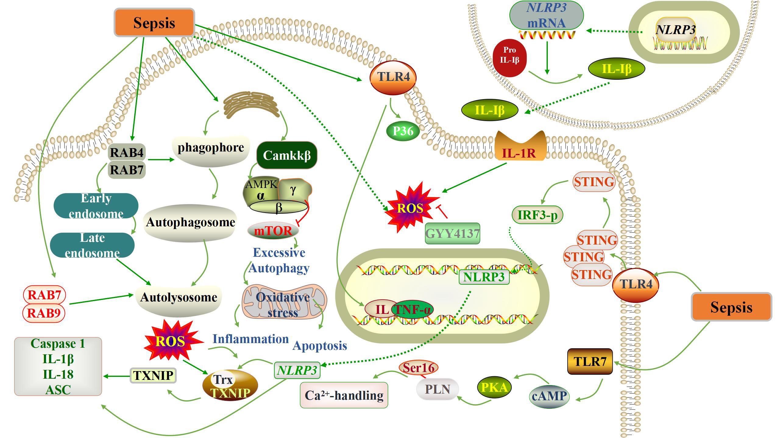
Fig. 1.

Schematic representation of the pathogenesis of sepsis-induced myocardial damage. Sepsis triggers a complex cascade of reactions, including the activation of oxidative stress, the occurrence of autophagy, apoptosis and systemic inflammatory response. IL, interleukin; AMPK, adenosine 5-monophosphate-activated protein kinase; ROS, reactive oxygen species; NLRP3, Nucleotide-binding and oligomerization domainlike receptor protein 3; TLR, Toll-like receptors; TNF-α, tumor necrosis factor-α; PLN, phospholamban; PKA, protein kinase A; cAMP, Cyclic Adenosine monophosphate; IRF3-p, Type-I interferons regulatory factor 3- Phosphorylated; Trx, thioredoxin; TXNIP, Thioredoxin-interacting protein; mTOR, mammalian target of rapamycin; ASC, apoptosis-associated speckle-like protein; RAB, Ras-related proteins in brain; GYY4137, hydrogen sulfide donor.

3. The Relationship Between Sirtuins and Sepsis
3.1 SIRT1

SIRT1 is a conserved NAD+-dependent protein deacetylase that exerts anti-inflammatory, antioxidant, and anti-aging effects by regulating gene transcription, chromosome stability, and target protein activity through deacetylation [52], which is reported to be involved in sepsis-induced myocardial damage.

Clinical studies have found that SIRT1 can still be clinically relevant as a rapid measurable prognostic biomarker for patients with sepsis in the clinical setting [53]. SIRT1 expression is downregulated in the hyper-inflammatory response to sepsis, and SIRT1 activation counteracts sepsis-induced myocardial damage [54, 55, 56]. Besides, SIRT1 played a negative role in regulating the activation of NLRP3 inflammasome [57, 58]. NLRP3 interacts with apoptosis-associated speckle-like protein (ASC) and promotes its own cleavage and activation through ASC recruitment of caspase1 precursor monomers. Thus, activated caspase 1 stimulates the cleavage of pro-IL-1β and pro-IL-18 into mature IL-1β and IL-18, which triggers an inflammatory response [59, 60]. Resveratrol (RESV), plant antioxidant, has been shown to reduce myocardial damage in sepsis by activating SIRT1 signaling to reduce neutrophil accumulation, TNF-α expression, and cardiomyocyte apoptosis [61]. In studies of acute renal damage caused by sepsis, SIRT1 mitigates acute renal damage by promoting autophagy mediated by deacetylation of Beclin1. However, this mechanism has not been validated in the cardiac and therefore needs to be further explored [7].

Besides, injection of a cell-permeable Tat-Beclin-1 peptide to activate autophagy improved cardiac function, attenuated inflammation, and rescued the phenotypes caused by Beclin-1 deficiency in sepsis-challenged mice, this suggests that Beclin-1 protects against cardiac damage during sepsis [62]. miR-181a is one of the MicroRNAs that represses gene expression at the post-transcriptional level by binding to target mRNAs [63]. MiR-181 levels were significantly increased in the presence of enhanced inflammatory response and apoptosis. MiR-181 binds directly to its target genes and inhibits SIRT1 expression. MiR-181a inhibition leads to upregulation of SIRT1 expression and downregulation of pro-inflammatory cytokines and apoptosis. Inhibition of miR-181a attenuates sepsis-induced inflammation and apoptosis by activating the Nuclear factor erythroid2-related factor 2 (Nrf2) pathway while inhibiting the NF-κB pathway, achieved through the targeting of SIRT1 [63]. Thus, miR-181 plays a key role in inflammation during sepsis and could be a potential therapeutic target for patients with sepsis. Serum lactate has been recognized as a biomarker of sepsis prognosis, and elevated serum lactate levels are positively correlated with sepsis mortality [64]. Clinical studies have shown that circulating high mobility group box-1 (HMGB1) levels are significantly elevated and positively correlate with the severity of sepsis and mortality [65, 66]. Lactate can strongly inhibit the gene expression of SIRT1 [67]. SIRT1 acetylation promotes HMGB1 release and is essential for sepsis pathogenesis [68]. This process is closely linked to the pathogenic role of lactate in sepsis, as lactate can inhibit SIRT1’s nuclear translocation and its function in deacetylation [7].

Restoring mitochondrial quality control mechanisms by activating mitochondrial biosynthesis or reducing mitochondrial division is a promising approach to improve organ dysfunction in sepsis [69, 70]. SIRT1/peroxisome proliferator-activated receptor-gamma co-activator-1alpha (PGC-1α) network enhances the regulation of cardiac mitochondrial activity and plays a protective role in sepsis-induced myocardial injury [71, 72]. Melatonin upregulates SIRT1 and autophagy, protecting from sepsis in relation to mitochondrial mechanism [73].

3.2 SIRT2

SIRT2, far less studied than SIRT1, is mainly located in the cytoplasm, but under certain conditions, it also enters the nucleus and performs the corresponding functions [74]. SIRT2 is a nutrient sensor in adipose and immune cells, like SIRT1, increased caloric restriction lead to a high nutrient and high fat diet decreases SIRT2 expression in white adipose tissue of mice [75, 76]. In obese mice with sepsis, elevated levels of SIRT2 protein instead prolonged low inflammation; SIRT2 is the most abundant SIRTs in adipose tissue [77]. Specifically, there are some studies found that through direct deacetylation of NF-κB and p65. SIRT2 expression in obesity with sepsis was reduced in the high inflammatory phase and increased in the low inflammatory phase [78].

3.3 SIRT3

SIRT3 is an NAD+-dependent histone deacetylase with functions in the regulation of mitochondrial oxidative stress, bioenergetics, anti-senescence, anti-fibrosis, and anti-inflammation [79, 80, 81, 82]. In the cardiac, SIRT3 is highly expressed and is required for the maintenance of cardiomyocyte energy and cardiac contractility [83]. SIRT3 located mainly in mitochondria, represents the major protein deacetylase in mitochondria and detoxifies ROS and increases mitochondrial ATP regeneration by activating manganese superoxide dismutase (MnSOD) and various energy metabolizing enzymes [83, 84, 85, 86]. The activation of SIRT3 is currently considered as a potential therapeutic strategy for the treatment of septic cardiomyopathy [87]. It was demonstrated that SIRT3 expression was significantly reduced in myocardial tissue from mice with sepsis-induced cardiac damage, and that restoration of SIRT3 expression prevented sepsis-induced cardiac damage [88]. Antioxidant and anti-inflammatory effects on the SIRT3 have also been demonstrated in sepsis-induced cardiac damage [89].

Mitochondrial dysfunction impairs the contractility of cardiomyocytes and even triggers cardiomyocyte death [90]. Annexin A1 (ANXA1), a glucocorticoid-regulated protein that is widely expressed in human tissues and cells [91]. ANXA1 promotes p53 deacetylation in sepsis-treated H9C2 cells by enhancing SIRT3 expression thereby reducing cardiomyocyte death [92]. Impaired SIRT3 activity may mediate cardiac dysfunction in endotoxemia by promoting calpain-mediated disruption of ATP synthesis, suggesting SIRT3 activation as a potential therapeutic strategy for the treatment of septic cardiomyopathy [87]. Tubeimoside I (TBM) prevents sepsis-induced endothelial dysfunction by reducing oxidative stress and apoptosis through SIRT3. TBM is a promising new therapeutic agent for sepsis-induced endothelial dysfunction [89]. Polydatin-mediated SIRT3 activation preserves mitochondrial function through mitochondrial superoxide dismutase 2 (SOD2) and Cyclophilin D (CypD) deacetylation, thereby reversing sepsis-induced endothelial hyperpermeability [93]. In addition, the role of SIRT3-CypD signaling in barrier protection was identified. SIRT 3-mediated deacetylation inhibited CypD activity, thereby inhibiting mPTP opening and ΔΨm reduction [94]. It was shown that reduced CypD hyperacetylation and interaction with SIRT3 in response to sepsis attack resulted in a reduction in mPTP opening and subsequent ΔΨm [93]. Estrogen related receptors (ERRs) can bind to the SIRT3 promoter and induce transcription of SIRT3 [95]. Tubeimoside I may protect against sepsisinduced cardiac dysfunction (SICD) by binding to and activating ERRα, thereby activating ERRα to promote SIRT3 transcription to reduce inflammation, oxidative stress and apoptosis [89].

SIRT3 deficiency leads to hyperacetylation of key enzymes in the cardiac trichloroacetic acid (TCA) cycle, which subsequently shifts myocardial metabolism to anaerobic glycolysis and subsequently promotes lactate and nicotinamide adenine dinucleotide (NADH) production, ultimately exacerbating cardiac function after sepsis [88]. SIRT3 overexpression increases AMPK activity and improves mitochondrial biosynthesis, which maintains mitochondrial function and reduces sepsis-associated cardiomyocyte damage [90]. These studies suggest that targeting SIRT3 may provide a potential new target to maintain normal cardiac performance after sepsis. However, the precise mechanisms of protection still need further investigation.

3.4 SIRT5

Unlike SIRT1 and SIRT3 deacetylases, SIRT5 has relatively weak deacetylation, but shows stronger desuccinylation activity, mainly removing succinyl, malonyl and glutaryl groups from lysine residues of mitochondria and peroxisome metabolic enzymes [96]. For example, SIRT5 has been reported to be able to descuccinylate pyruvate dehydrogenase 1 (PDHA1) and succinate dehydrogenase (SHDB), and play a role in regulating the activity of these protein [97]. As a member of SIRTs, SIRT5 has been proved to be located in mitochondria and cytosol [98]. As far as its cell biological function is concerned, SIRT5 can promote ammonia detoxification, regulate glycolysis, TCA cycle and electron transfer chain. In addition, SIRT5 can also promote fatty acid β oxidation and ketone body production [99]. Studies have shown that SIRT5 deficiency can promote enteritis, acute lung damage and ischemia-reperfusion damage [96].

In sepsis, SIRT5 has also attracted great attention. Studies have shown that in sepsis, SIRT5 can enhance the innate inflammatory response of macrophages or endotoxin-tolerant macrophages by promoting the acetylation of p65 and activating NF-κB pathway. This process is completed by competing with SIRT2 and blocking the deacetylation of SIRT 2 to p65 [100]. In an experimental study on burn sepsis model in mice, the inhibition of SIRT5 expression reduced the deacetylation of PDHA1, restored the activity of pyruvate dehydrogenase, promoted the polarization of macrophage M2, and alleviated burn sepsis in mice [101]. Another study using renal tubular cells (RTSCs) showed that in sepsis, the expression level of phosphorylation-AMPK (p-AMPK) decreased, the structure of mitochondria was destroyed, and the content of ATP decreased, while AMPK agonists could alleviate septic acute renal damage (SAKI), and this process was mediated by SIRT5. Knockout of SIRT5 will significantly aggravate SAKI. On the contrary, up-regulation of SIRT5 expression can reduce mitochondrial dysfunction of renal tubular epithelial cells (RTECs) and reduce SAKI by enhancing AMPK phosphorylation [102]. In a word, SIRT5 may play a regulatory role in the pathogenesis of sepsis by regulating cell metabolism, apoptosis and oxidative stress.

3.5 SIRT6

SIRT6 is another protein that depends on NAD+ for its deacetylation activity. It regulates gene expression by removing acetyl groups from histones and transcription factors. Its biological functions include deacetylation, defatty-acylation, and ADP-ribosylation, which are involved in DNA repair, gene expression, and telomere maintenance, ultimately regulating lifespan and controlling aging [103]. In addition, SIRT6 is also related to energy metabolism, aging, obesity, inflammation, cancer and so on [104].

As far as sepsis is concerned, SIRT6 can reduce the inflammatory reaction of human venous endothelial cells (HUVECs) induced by sepsis by positively regulating the expression of Nrf2 and activating anti-inflammatory and antioxidant enzymes regulated by Nrf2, which suggests that SIRT6 is a possible target for preventing lung damage induced by sepsis [105]. Further research shows that the overexpression of SIRT6 in renal proximal convoluted tubule epithelial cells will alleviate SAKI by activating autophagy, which promotes AMPK activation, inhibits mTOR signaling pathway, promotes the polarization of macrophage M2 in autophagy-dependent and non-autophagy-dependent ways, and then plays a role in alleviating SAKI [104]. In another experiment of damage and apoptosis of renal tubular epithelial cells induced by sepsis, it was found that SIRT6 can activate nuclear factor erythroid-2-related factor 2-antioxidant response element (Nrf2)/ARE signal, alleviate renal dysfunction induced by sepsis, and reduce apoptosis and oxidative stress of renal tubular epithelial cells [106].

Because SIRT6 can promote autophagy, studies show that the overexpression of SIRT6 can alleviate sepsis-induced apoptosis, and further induce autophagy to play a protective role in renal epithelial cell damage caused by sepsis [107]. On the one hand, the up-regulated expression of SIRT6 can promote autophagy, on the other hand, it can protect renal tubular epithelial cells from oxidative stress, inflammatory reaction and apoptosis, and then play a protective role in sepsis-induced renal damage [108]. Another study on the treatment of sepsis shows that the activation of SIRT6 can inhibit the apoptosis of myocardial cells and the expression of inflammatory factors by promoting autophagy, and then inhibit the myocardial dysfunction induced by sepsis [109]. Besides promoting autophagy, SIRT6 plays an important role in regulating glucose metabolism in vivo, and the decrease of SIRT6 activity will reverse glycolysis block related to monocytes caused by endotoxin, which is a process that leads to immune metabolism paralysis of monocytes in human and mice sepsis [110]. This shows that SIRT6 mainly promotes autophagy, reduces oxidative stress and alleviates sepsis mediated by inflammatory factors.

3.6 SIRT7

SIRT7 is also a deacetylase belonging to SIRT family. Many studies have shown that SIRT7 can regulate protein deposition, endoplasmic reticulum stress, mitochondrial protein folding and mitochondrial metabolism [111]. SIRT7 is mainly located in nucleoli [112], which combines with ribosomal RNA gene and participates in the transcription process of ribosomal DNA during mitosis. One of its important functions is to regulate chromatin remodeling [113]. SIRT7 can exert deacetylation activity at DNA damage sites, and show other catalytic activities for protein involved in DNA damage and repair [111, 112]. Although there are few studies on the role of SIRT7 in sepsis at present, a study on the mechanism of resveratrol found that the activation of SIRT7 can reduce inflammatory factors and play a positive role in the treatment and protection of sepsis and its cardiac damage [114]. Another study shows that liver damage will be caused during the pathogenesis of sepsis, and the activation of endoplasmic reticulum stress (ERS) pathway will cause apoptosis of hepatocytes. SIRT7 can inhibit glucose-r\regulated protein 78 (GRP78) in ERS pathway to inhibit apoptosis of hepatocytes, which provides clues for the treatment of liver damage caused by sepsis in the future [115]. Due to its unique biological function, SIRTs reduces sepsis by deacetylating several histone and non-histone proteins. Fig. 2 summarizes the role of these SIRTs in cells and their mechanisms of action in sepsis.
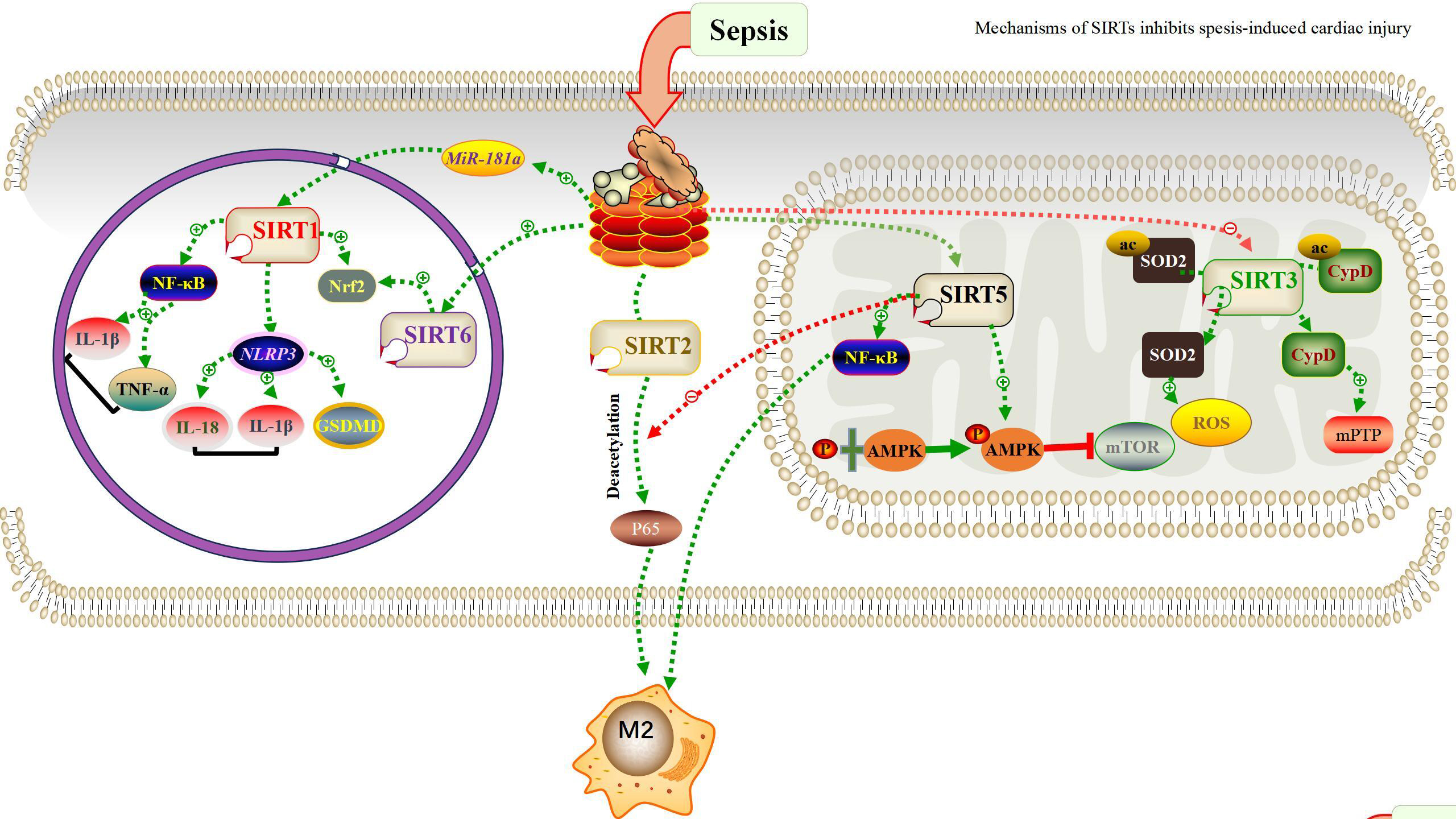
Fig. 2.

Schematic representation of SIRTs modulates the inhibition of sepsis through intracellular mechanisms. SIRTs is dispersed in subcellular compartments of cells. Each SIRTs has its own unique targets that define its biological activity, as described in the review. SIRT, Sirtuin; NF-κB, nuclear factor kappa-B; GSDMD, Gasdermin D; Nrf2, Nuclear factor erythroid2-related factor 2; SOD2, superoxide dismutase 2; mPTP, 1-Methyl-4-phenyl-1,2,3,6-tetrahydropyridine; CypD, Cyclophilin D; ac, acetylation; M2, Macrophages 2; MiR-181a, miRNAs-181a; p65, reLA.

4. Application of Therapeutic Targets of SIRTs in Sepsis

As mentioned above, SIRTs play an important role in the development of sepsis. Therefore, it is extremely important to find a cure for sepsis with SIRTs as the target. On the one hand, the up-regulation of SIRT1 induces deacetylation of NF-κB, and then reduces inflammatory cytokines. On the other hand, it upregulates the deacetylation of SOD2 induced by SIRT3, reduces oxidative stress, improves autophagy and protects mitochondrial function [116]. Syringaresinol is a natural extract with anti-inflammatory properties, which improves sepsis-induced myocardial damage through SIRT1/NLRP3/GSDMD pathway [117]. Resveratrol alleviates sepsis-induced myocardial damage by inhibiting ferroptosis through up-regulation of SIRT1/Nrf2 signaling pathway [118]. Melatonin, an important hormone that regulates sleep, regulates apoptosis and autophagy by activating SIRT1 in mice, and protects mice from septicaemia induced cardiac dysfunction [73]. Ganoderma lucidum polysaccharides regulates inflammation, apoptosis and proliferation by activating SIRT1 [119]. Rosmarinic acid alleviates sepsis-induced cardiac damage by activating SIRT1, thereby alleviating mitochondrial damage [120]. Ttrimetazidine has a protective effect on sepsis-induced myocardial damage and apoptosis, and is associated with inhibition of macrophage proinflammatory through normalizing the SIRT1/AMP signal pathway [121]. A study using renal tubular epithelial cells showed that SIRT3 was activated and p-AMPK was also activated after 2-deoxy-D-glucose (2-DG) treatment, thus promoting autophagy and reducing cell apoptosis [122]. Tubeimoside I supplementation can significantly relieve sepsis by reversing SIRT3 expression [89]. Dietary eicosapentaenoic acid protects mitochondrial integrity and protects sepsis-induced cardiac damage through increased expression of SIRT3 [123]. Experiments have proved that after SIRT1 is activated by resveratrol, Beclin1 is induced to deacetylate, and then autophagy is promoted and it plays a protective role in sepsis-induced acute renal damage [7]. By detecting the expression level of SIRT2 mRNA in patients with sepsis and septic shock, it was found that the expression level of SIRT2 mRNA in patients with sepsis and septic shock decreased, which indicated that SIRT2 could be used as a potential biomarker for diagnosis on the one hand and a therapeutic target on the other [124]. Other studies have shown that SIRT2 is a potential therapeutic target for ethanol-induced sepsis [125]. It is further found that obesity will increase the incidence of sepsis patients, and the treatment with SIRT2 inhibitor adenylate kinase (AK7) can reverse autophagy disorder and improve the autophagy clearance rate of free fatty acid tolerant cells in endotoxin tolerance period [126, 127], which indicates that SIRT2 is a potential target in the treatment of sepsis. In acute renal damage induced by sepsis, SIRT5 knockout significantly aggravated acute renal damage, indicating that SIRT5 plays an important role in the treatment of sepsis [102]. The activation of SIRT6 can inhibit the apoptosis of myocardial cells and the expression of inflammatory factors by promoting autophagy, and then inhibit the myocardial dysfunction induced by sepsis, which shows that SIRT6 also plays an important role in alleviating the damage of sepsis [109]. Different from other SIRTs, SIRT7 has advantages in the treatment of liver damage caused by sepsis. SIRT7 can inhibit GRP78 in ERS pathway to inhibit hepatocyte apoptosis [115]. Generally, SIRTs play a role by inhibiting the production of inflammatory mediators and regulating the function of immune cells, so SIRTs will be a promising target in the treatment of sepsis.

5. Conclusion and Prospect

In summary, SIRTs are involved in important metabolic reactions and play an important role in the development of sepsis. We have witnessed great advances in our understanding of sepsis in the SIRTs involved. However, our exploration of the upstream and downstream pathways of SIRTs is very limited, we will also focus on the SIRTs up-down pathway. We believe that it is feasible to regulate the occurrence and development of sepsis by regulating the level of SIRTs, and the subsequent research on its mechanism and the development of new drugs will open up new avenues for the treatment of sepsis.

Author Contributions

ZP and WY: contributing to conceptualization ideas; formulation and evolution of review aims; contributing to management and coordination responsibility for the research activity planning and execution; revise the draft. SW and YW: contributing to the visualization preparation, creation and presentation of the published work; contributing to write the initial draft. All authors contributed to editorial changes in the manuscript. All authors read and approved the final manuscript. All authors have participated sufficiently in the work and agreed to be accountable for all aspects of the work.

Ethics Approval and Consent to Participate

Not applicable.

Acknowledgment

Not applicable.

Funding

This study was funded by Liaoning Province Applied Basic Research Program [grant number 2023JH2/101300098] and Beijing Jiekai Cardiovascular Health Foundation [grant number BW20220302].

Conflict of Interest

The authors declare no conflict of interest. Zuowei Pei is serving as one of the Guest editors of this journal. Zuowei Pei had no involvement in the peer-review of this article and has no access to information regarding its peer review. Full responsibility for the editorial process for this article was delegated to Graham Pawelec.

References
[1]
van der Poll T, Shankar-Hari M, Wiersinga WJ. The immunology of sepsis. Immunity. 2021; 54: 2450–2464.
[2]
McCall CE, Zhu X, Zabalawi M, Long D, Quinn MA, Yoza BK, et al. Sepsis, pyruvate, and mitochondria energy supply chain shortage. Journal of Leukocyte Biology. 2022; 112: 1509–1514.
[3]
Prescott HC, Osterholzer JJ, Langa KM, Angus DC, Iwashyna TJ. Late mortality after sepsis: propensity matched cohort study. BMJ (Clinical Research Ed.). 2016; 353: i2375.
[4]
Wasyluk W, Nowicka-Stążka P, Zwolak A. Heart Metabolism in Sepsis-Induced Cardiomyopathy-Unusual Metabolic Dysfunction of the Heart. International Journal of Environmental Research and Public Health. 2021; 18: 7598.
[5]
Singh CK, Chhabra G, Ndiaye MA, Garcia-Peterson LM, Mack NJ, Ahmad N. The Role of Sirtuins in Antioxidant and Redox Signaling. Antioxidants & Redox Signaling. 2018; 28: 643–661.
[6]
Liu TF, Vachharajani V, Millet P, Bharadwaj MS, Molina AJ, McCall CE. Sequential actions of SIRT1-RELB-SIRT3 coordinate nuclear-mitochondrial communication during immunometabolic adaptation to acute inflammation and sepsis. The Journal of Biological Chemistry. 2015; 290: 396–408.
[7]
Deng Z, Sun M, Wu J, Fang H, Cai S, An S, et al. SIRT1 attenuates sepsis-induced acute kidney injury via Beclin1 deacetylation-mediated autophagy activation. Cell Death & Disease. 2021; 12: 217.
[8]
Zhao L, Jin L, Luo Y, Wang L, Li Y, Xian S, et al. Shenfu injection attenuates cardiac dysfunction and inhibits apoptosis in septic mice. Annals of Translational Medicine. 2022; 10: 597.
[9]
Zhu H, Zhang L, Jia H, Xu L, Cao Y, Zhai M, et al. Tetrahydrocurcumin improves lipopolysaccharide-induced myocardial dysfunction by inhibiting oxidative stress and inflammation via JNK/ERK signaling pathway regulation. Phytomedicine: International Journal of Phytotherapy and Phytopharmacology. 2022; 104: 154283.
[10]
Vellinga NAR, Boerma EC, Koopmans M, Donati A, Dubin A, Shapiro NI, et al. International study on microcirculatory shock occurrence in acutely ill patients. Critical Care Medicine. 2015; 43: 48–56.
[11]
Joffre J, Hellman J. Oxidative Stress and Endothelial Dysfunction in Sepsis and Acute Inflammation. Antioxidants & Redox Signaling. 2021; 35: 1291–1307.
[12]
Jiang L, Zhang L, Yang J, Shi H, Zhu H, Zhai M, et al. 1-Deoxynojirimycin attenuates septic cardiomyopathy by regulating oxidative stress, apoptosis, and inflammation via the JAK2/STAT6 signaling pathway. Biomedicine & Pharmacotherapy. 2022; 155: 113648.
[13]
Zhou R, Yazdi AS, Menu P, Tschopp J. A role for mitochondria in NLRP3 inflammasome activation. Nature. 2011; 469: 221–225.
[14]
Tschopp J, Schroder K. NLRP3 inflammasome activation: The convergence of multiple signalling pathways on ROS production? Nature Reviews. Immunology. 2010; 10: 210–215.
[15]
Rahim I, Djerdjouri B, Sayed RK, Fernández-Ortiz M, Fernández-Gil B, Hidalgo-Gutiérrez A, et al. Melatonin administration to wild-type mice and nontreated NLRP3 mutant mice share similar inhibition of the inflammatory response during sepsis. Journal of Pineal Research. 2017; 63.
[16]
Lv D, Luo M, Yan J, Yang X, Luo S. Protective Effect of Sirtuin 3 on CLP-Induced Endothelial Dysfunction of Early Sepsis by Inhibiting NF-κB and NLRP3 Signaling Pathways. Inflammation. 2021; 44: 1782–1792.
[17]
Alves-Filho JC, Freitas A, Souto FO, Spiller F, Paula-Neto H, Silva JS, et al. Regulation of chemokine receptor by Toll-like receptor 2 is critical to neutrophil migration and resistance to polymicrobial sepsis. Proceedings of the National Academy of Sciences of the United States of America. 2009; 106: 4018–4023.
[18]
Plitas G, Burt BM, Nguyen HM, Bamboat ZM, DeMatteo RP. Toll-like receptor 9 inhibition reduces mortality in polymicrobial sepsis. The Journal of Experimental Medicine. 2008; 205: 1277–1283.
[19]
Saiyang X, Qingqing W, Man X, Chen L, Min Z, Yun X, et al. Activation of Toll-like receptor 7 provides cardioprotection in septic cardiomyopathy-induced systolic dysfunction. Clinical and Translational Medicine. 2021; 11: e266.
[20]
Akira S, Uematsu S, Takeuchi O. Pathogen recognition and innate immunity. Cell. 2006; 124: 783–801.
[21]
Kumar A, Thota V, Dee L, Olson J, Uretz E, Parrillo JE. Tumor necrosis factor alpha and interleukin 1beta are responsible for in vitro myocardial cell depression induced by human septic shock serum. The Journal of Experimental Medicine. 1996; 183: 949–958.
[22]
Takeuchi O, Akira S. Pattern recognition receptors and inflammation. Cell. 2010; 140: 805–820.
[23]
Majer O, Liu B, Kreuk LSM, Krogan N, Barton GM. UNC93B1 recruits syntenin-1 to dampen TLR7 signalling and prevent autoimmunity. Nature. 2019; 575: 366–370.
[24]
Tan S, Long Z, Hou X, Lin Y, Xu J, You X, et al. H_2 Protects Against Lipopolysaccharide-Induced Cardiac Dysfunction via Blocking TLR4-Mediated Cytokines Expression. Frontiers in Pharmacology. 2019; 10: 865.
[25]
Kawai T, Akira S. Signaling to NF-kappaB by Toll-like receptors. Trends in Molecular Medicine. 2007; 13: 460–469.
[26]
Knuefermann P, Sakata Y, Baker JS, Huang CH, Sekiguchi K, Hardarson HS, et al. Toll-like receptor 2 mediates Staphylococcus aureus-induced myocardial dysfunction and cytokine production in the heart. Circulation. 2004; 110: 3693–3698.
[27]
Lohner R, Schwederski M, Narath C, Klein J, Duerr GD, Torno A, et al. Toll-like receptor 9 promotes cardiac inflammation and heart failure during polymicrobial sepsis. Mediators of Inflammation. 2013; 2013: 261049.
[28]
Taganov KD, Boldin MP, Chang KJ, Baltimore D. NF-kappaB-dependent induction of microRNA miR-146, an inhibitor targeted to signaling proteins of innate immune responses. Proceedings of the National Academy of Sciences of the United States of America. 2006; 103: 12481–12486.
[29]
Zhou Q, Pan X, Wang L, Wang X, Xiong D. The protective role of neuregulin-1: A potential therapy for sepsis-induced cardiomyopathy. European Journal of Pharmacology. 2016; 788: 234–240.
[30]
Park H, Huang X, Lu C, Cairo MS, Zhou X. MicroRNA-146a and microRNA-146b regulate human dendritic cell apoptosis and cytokine production by targeting TRAF6 and IRAK1 proteins. The Journal of Biological Chemistry. 2015; 290: 2831–2841.
[31]
Wei J, Wang J, Zhou Y, Yan S, Li K, Lin H. MicroRNA-146a Contributes to SCI Recovery via Regulating TRAF6 and IRAK1 Expression. BioMed Research International. 2016; 2016: 4013487.
[32]
Jiang W, Kong L, Ni Q, Lu Y, Ding W, Liu G, et al. miR-146a ameliorates liver ischemia/reperfusion injury by suppressing IRAK1 and TRAF6. PLoS ONE. 2014; 9: e101530.
[33]
Hiram R, Rizcallah E, Sirois C, Sirois M, Morin C, Fortin S, et al. Resolvin D1 reverses reactivity and Ca2+ sensitivity induced by ET-1, TNF-α, and IL-6 in the human pulmonary artery. American Journal of Physiology. Heart and Circulatory Physiology. 2014; 307: H1547–H1558.
[34]
Pang J, Peng H, Wang S, Xu X, Xu F, Wang Q, et al. Mitochondrial ALDH2 protects against lipopolysaccharide-induced myocardial contractile dysfunction by suppression of ER stress and autophagy. Biochimica et Biophysica Acta. Molecular Basis of Disease. 2019; 1865: 1627–1641.
[35]
Mantzarlis K, Tsolaki V, Zakynthinos E. Role of Oxidative Stress and Mitochondrial Dysfunction in Sepsis and Potential Therapies. Oxidative Medicine and Cellular Longevity. 2017; 2017: 5985209.
[36]
Suliman HB, Carraway MS, Piantadosi CA. Postlipopolysaccharide oxidative damage of mitochondrial DNA. American Journal of Respiratory and Critical Care Medicine. 2003; 167: 570–579.
[37]
Brealey D, Brand M, Hargreaves I, Heales S, Land J, Smolenski R, et al. Association between mitochondrial dysfunction and severity and outcome of septic shock. Lancet (London, England). 2002; 360: 219–223.
[38]
Martin L, Derwall M, Al Zoubi S, Zechendorf E, Reuter DA, Thiemermann C, et al. The Septic Heart: Current Understanding of Molecular Mechanisms and Clinical Implications. Chest. 2019; 155: 427–437.
[39]
Stanzani G, Duchen MR, Singer M. The role of mitochondria in sepsis-induced cardiomyopathy. Biochimica et Biophysica Acta. Molecular Basis of Disease. 2019; 1865: 759–773.
[40]
Santulli G, Xie W, Reiken SR, Marks AR. Mitochondrial calcium overload is a key determinant in heart failure. Proceedings of the National Academy of Sciences of the United States of America. 2015; 112: 11389–11394.
[41]
Bauer M, Gerlach H, Vogelmann T, Preissing F, Stiefel J, Adam D. Mortality in sepsis and septic shock in Europe, North America and Australia between 2009 and 2019- results from a systematic review and meta-analysis. Critical Care (London, England). 2020; 24: 239.
[42]
Delmotte P, Sieck GC. Endoplasmic Reticulum Stress and Mitochondrial Function in Airway Smooth Muscle. Frontiers in Cell and Developmental Biology. 2020; 7: 374.
[43]
Denton D, Kumar S. Autophagy-dependent cell death. Cell Death and Differentiation. 2019; 26: 605–616.
[44]
Galluzzi L, Vitale I, Aaronson SA, Abrams JM, Adam D, Agostinis P, et al. Molecular mechanisms of cell death: recommendations of the Nomenclature Committee on Cell Death 2018. Cell Death and Differentiation. 2018; 25: 486–541.
[45]
Lin Y, Xu Y, Zhang Z. Sepsis-Induced Myocardial Dysfunction (SIMD): the Pathophysiological Mechanisms and Therapeutic Strategies Targeting Mitochondria. Inflammation. 2020; 43: 1184–1200.
[46]
Jiang T, Peng D, Shi W, Guo J, Huo S, Men L, et al. IL-6/STAT3 Signaling Promotes Cardiac Dysfunction by Upregulating FUNDC1-Dependent Mitochondria-Associated Endoplasmic Reticulum Membranes Formation in Sepsis Mice. Frontiers in Cardiovascular Medicine. 2022; 8: 790612.
[47]
Zang Q, Maass DL, Tsai SJ, Horton JW. Cardiac mitochondrial damage and inflammation responses in sepsis. Surgical Infections. 2007; 8: 41–54.
[48]
Wang R, Xu Y, Fang Y, Wang C, Xue Y, Wang F, et al. Pathogenetic mechanisms of septic cardiomyopathy. Journal of Cellular Physiology. 2022; 237: 49–58.
[49]
Platnich JM, Chung H, Lau A, Sandall CF, Bondzi-Simpson A, Chen HM, et al. Shiga Toxin/Lipopolysaccharide Activates Caspase-4 and Gasdermin D to Trigger Mitochondrial Reactive Oxygen Species Upstream of the NLRP3 Inflammasome. Cell Reports. 2018; 25: 1525–1536.e7.
[50]
Dai S, Ye B, Zhong L, Chen Y, Hong G, Zhao G, et al. GSDMD Mediates LPS-Induced Septic Myocardial Dysfunction by Regulating ROS-dependent NLRP3 Inflammasome Activation. Frontiers in Cell and Developmental Biology. 2021; 9: 779432.
[51]
Busch K, Kny M, Huang N, Klassert TE, Stock M, Hahn A, et al. Inhibition of the NLRP3/IL-1β axis protects against sepsis-induced cardiomyopathy. Journal of Cachexia, Sarcopenia and Muscle. 2021; 12: 1653–1668.
[52]
Meng X, Tan J, Li M, Song S, Miao Y, Zhang Q. Sirt1: Role Under the Condition of Ischemia/Hypoxia. Cellular and Molecular Neurobiology. 2017; 37: 17–28.
[53]
Cheng X, Zhang S, Wen Y, Shi Z. Clinical significance of sirtuin 1 level in sepsis: correlation with disease risk, severity, and mortality risk. Brazilian Journal of Medical and Biological Research. 2020; 54: e10271.
[54]
Han D, Li X, Li S, Su T, Fan L, Fan WS, et al. Reduced silent information regulator 1 signaling exacerbates sepsis-induced myocardial injury and mitigates the protective effect of a liver X receptor agonist. Free Radical Biology & Medicine. 2017; 113: 291–303.
[55]
Hong G, Zheng D, Zhang L, Ni R, Wang G, Fan GC, et al. Administration of nicotinamide riboside prevents oxidative stress and organ injury in sepsis. Free Radical Biology & Medicine. 2018; 123: 125–137.
[56]
Smith LM, Yoza BK, Hoth JJ, McCall CE, Vachharajani V. SIRT1 Mediates Septic Cardiomyopathy in a Murine Model of Polymicrobial Sepsis. Shock (Augusta, Ga.). 2020; 54: 96–101.
[57]
Peng Z, Zhang W, Qiao J, He B. Melatonin attenuates airway inflammation via SIRT1 dependent inhibition of NLRP3 inflammasome and IL-1β in rats with COPD. International Immunopharmacology. 2018; 62: 23–28.
[58]
Guo T, Jiang ZB, Tong ZY, Zhou Y, Chai XP, Xiao XZ. Shikonin Ameliorates LPS-Induced Cardiac Dysfunction by SIRT1-Dependent Inhibition of NLRP3 Inflammasome. Frontiers in Physiology. 2020; 11: 570441.
[59]
Gong T, Yang Y, Jin T, Jiang W, Zhou R. Orchestration of NLRP3 Inflammasome Activation by Ion Fluxes. Trends in Immunology. 2018; 39: 393–406.
[60]
Katsnelson MA, Lozada-Soto KM, Russo HM, Miller BA, Dubyak GR. NLRP3 inflammasome signaling is activated by low-level lysosome disruption but inhibited by extensive lysosome disruption: roles for K+ efflux and Ca2+ influx. American Journal of Physiology. Cell Physiology. 2016; 311: C83–C100.
[61]
An R, Zhao L, Xu J, Xi C, Li H, Shen G, et al. Resveratrol alleviates sepsis induced myocardial injury in rats by suppressing neutrophil accumulation, the induction of TNF α and myocardial apoptosis via activation of Sirt1. Molecular Medicine Reports. 2016; 14: 5297–5303.
[62]
Sun Y, Yao X, Zhang QJ, Zhu M, Liu ZP, Ci B, et al. Beclin-1-Dependent Autophagy Protects the Heart During Sepsis. Circulation. 2018; 138: 2247–2262.
[63]
Wu Z, Chen J, Zhao W, Zhuo CH, Chen Q. Inhibition of miR-181a attenuates sepsis-induced inflammation and apoptosis by activating Nrf2 and inhibiting NF-κB pathways via targeting SIRT1. The Kaohsiung Journal of Medical Sciences. 2021; 37: 200–207.
[64]
Broder G, Weil MH. Excess lactate: an index of reversibility of shock in human patients. Science (New York, N.Y.). 1964; 143: 1457–1459.
[65]
Sundén-Cullberg J, Norrby-Teglund A, Rouhiainen A, Rauvala H, Herman G, Tracey KJ, et al. Persistent elevation of high mobility group box-1 protein (HMGB1) in patients with severe sepsis and septic shock. Critical Care Medicine. 2005; 33: 564–573.
[66]
Karlsson S, Pettilä V, Tenhunen J, Laru-Sompa R, Hynninen M, Ruokonen E. HMGB1 as a predictor of organ dysfunction and outcome in patients with severe sepsis. Intensive Care Medicine. 2008; 34: 1046–1053.
[67]
Yang K, Fan M, Wang X, Xu J, Wang Y, Tu F, et al. Lactate promotes macrophage HMGB1 lactylation, acetylation, and exosomal release in polymicrobial sepsis. Cell Death and Differentiation. 2022; 29: 133–146.
[68]
Kim YM, Park EJ, Kim HJ, Chang KC. Sirt1 S-nitrosylation induces acetylation of HMGB1 in LPS-activated RAW264.7 cells and endotoxemic mice. Biochemical and Biophysical Research Communications. 2018; 501: 73–79.
[69]
Hernandez-Resendiz S, Prunier F, Girao H, Dorn G, Hausenloy DJ, EU-CARDIOPROTECTION COST Action (CA16225). Targeting mitochondrial fusion and fission proteins for cardioprotection. Journal of Cellular and Molecular Medicine. 2020; 24: 6571–6585.
[70]
Lightowlers RN, Chrzanowska-Lightowlers ZM, Russell OM. Mitochondrial transplantation-a possible therapeutic for mitochondrial dysfunction?: Mitochondrial transfer is a potential cure for many diseases but proof of efficacy and safety is still lacking. EMBO Reports. 2020; 21: e50964.
[71]
Mokhtari B, Hamidi M, Badalzadeh R, Mahmoodpoor A. Mitochondrial transplantation protects against sepsis-induced myocardial dysfunction by modulating mitochondrial biogenesis and fission/fusion and inflammatory response. Molecular Biology Reports. 2023; 50: 2147–2158.
[72]
Mohan MS, Aswani SS, Aparna NS, Boban PT, Sudhakaran PR, Saja K. Effect of acute cold exposure on cardiac mitochondrial function: role of sirtuins. Molecular and Cellular Biochemistry. 2023; 478: 2257–2270.
[73]
Zhang WX, He BM, Wu Y, Qiao JF, Peng ZY. Melatonin protects against sepsis-induced cardiac dysfunction by regulating apoptosis and autophagy via activation of SIRT1 in mice. Life Sciences. 2019; 217: 8–15.
[74]
North BJ, Marshall BL, Borra MT, Denu JM, Verdin E. The human Sir2 ortholog, SIRT2, is an NAD+-dependent tubulin deacetylase. Molecular Cell. 2003; 11: 437–444.
[75]
Wang F, Nguyen M, Qin FXF, Tong Q. SIRT2 deacetylates FOXO3a in response to oxidative stress and caloric restriction. Aging Cell. 2007; 6: 505–514.
[76]
Krishnan J, Danzer C, Simka T, Ukropec J, Walter KM, Kumpf S, et al. Dietary obesity-associated Hif1α activation in adipocytes restricts fatty acid oxidation and energy expenditure via suppression of the Sirt2-NAD+ system. Genes & Development. 2012; 26: 259–270.
[77]
Wang X, Buechler NL, Martin A, Wells J, Yoza B, McCall CE, et al. Sirtuin-2 Regulates Sepsis Inflammation in ob/ob Mice. PLoS ONE. 2016; 11: e0160431.
[78]
Wang X, Buechler NL, Woodruff AG, Long DL, Zabalawi M, Yoza BK, et al. Sirtuins and Immuno-Metabolism of Sepsis. International Journal of Molecular Sciences. 2018; 19: 2738.
[79]
Li R, Xin T, Li D, Wang C, Zhu H, Zhou H. Therapeutic effect of Sirtuin 3 on ameliorating nonalcoholic fatty liver disease: The role of the ERK-CREB pathway and Bnip3-mediated mitophagy. Redox Biology. 2018; 18: 229–243.
[80]
Xiang X, Huang J, Song S, Wang Y, Zeng Y, Wu S, et al. 17β-estradiol inhibits H2O2-induced senescence in HUVEC cells through upregulating SIRT3 expression and promoting autophagy. Biogerontology. 2020; 21: 549–557.
[81]
Gao J, Zhang K, Wang Y, Guo R, Liu H, Jia C, et al. A machine learning-driven study indicates emodin improves cardiac hypertrophy by modulation of mitochondrial SIRT3 signaling. Pharmacological Research. 2020; 155: 104739.
[82]
Dikalova AE, Pandey A, Xiao L, Arslanbaeva L, Sidorova T, Lopez MG, et al. Mitochondrial Deacetylase Sirt3 Reduces Vascular Dysfunction and Hypertension While Sirt3 Depletion in Essential Hypertension Is Linked to Vascular Inflammation and Oxidative Stress. Circulation Research. 2020; 126: 439–452.
[83]
Koentges C, Pfeil K, Schnick T, Wiese S, Dahlbock R, Cimolai MC, et al. SIRT3 deficiency impairs mitochondrial and contractile function in the heart. Basic Research in Cardiology. 2015; 110: 36.
[84]
Cooper HM, Spelbrink JN. The human SIRT3 protein deacetylase is exclusively mitochondrial. The Biochemical Journal. 2008; 411: 279–285.
[85]
Sundaresan NR, Gupta M, Kim G, Rajamohan SB, Isbatan A, Gupta MP. Sirt3 blocks the cardiac hypertrophic response by augmenting Foxo3a-dependent antioxidant defense mechanisms in mice. The Journal of Clinical Investigation. 2009; 119: 2758–2771.
[86]
Murugasamy K, Munjal A, Sundaresan NR. Emerging Roles of SIRT3 in Cardiac Metabolism. Frontiers in Cardiovascular Medicine. 2022; 9: 850340.
[87]
Koentges C, Cimolai MC, Pfeil K, Wolf D, Marchini T, Tarkhnishvili A, et al. Impaired SIRT3 activity mediates cardiac dysfunction in endotoxemia by calpain-dependent disruption of ATP synthesis. Journal of Molecular and Cellular Cardiology. 2019; 133: 138–147.
[88]
Xu Y, Zhang S, Rong J, Lin Y, Du L, Wang Y, et al. Sirt3 is a novel target to treat sepsis induced myocardial dysfunction by acetylated modulation of critical enzymes within cardiac tricarboxylic acid cycle. Pharmacological Research. 2020; 159: 104887.
[89]
Cheng Z, Lv D, Luo M, Wang R, Guo Y, Yang X, et al. Tubeimoside I protects against sepsis-induced cardiac dysfunction via SIRT3. European Journal of Pharmacology. 2021; 905: 174186.
[90]
Xin T, Lu C. SirT3 activates AMPK-related mitochondrial biogenesis and ameliorates sepsis-induced myocardial injury. Aging. 2020; 12: 16224–16237.
[91]
Purvis GSD, Solito E, Thiemermann C. Annexin-A1: Therapeutic Potential in Microvascular Disease. Frontiers in Immunology. 2019; 10: 938.
[92]
Qin S, Ren Y, Feng B, Wang X, Liu J, Zheng J, et al. ANXA1sp Protects against Sepsis-Induced Myocardial Injury by Inhibiting Ferroptosis-Induced Cardiomyocyte Death via SIRT3-Mediated p53 Deacetylation. Mediators of Inflammation. 2023; 2023: 6638929.
[93]
Wu J, Deng Z, Sun M, Zhang W, Yang Y, Zeng Z, et al. Polydatin protects against lipopolysaccharide-induced endothelial barrier disruption via SIRT3 activation. Laboratory Investigation; a Journal of Technical Methods and Pathology. 2020; 100: 643–656.
[94]
Hafner AV, Dai J, Gomes AP, Xiao CY, Palmeira CM, Rosenzweig A, et al. Regulation of the mPTP by SIRT3-mediated deacetylation of CypD at lysine 166 suppresses age-related cardiac hypertrophy. Aging. 2010; 2: 914–923.
[95]
Giralt A, Hondares E, Villena JA, Ribas F, Díaz-Delfín J, Giralt M, et al. Peroxisome proliferator-activated receptor-gamma coactivator-1alpha controls transcription of the Sirt3 gene, an essential component of the thermogenic brown adipocyte phenotype. The Journal of Biological Chemistry. 2011; 286: 16958–16966.
[96]
Heinonen T, Ciarlo E, Le Roy D, Roger T. Impact of the Dual Deletion of the Mitochondrial Sirtuins SIRT3 and SIRT5 on Anti-microbial Host Defenses. Frontiers in Immunology. 2019; 10: 2341.
[97]
Yihan L, Xiaojing W, Ao L, Chuanjie Z, Haofei W, Yan S, et al. SIRT5 functions as a tumor suppressor in renal cell carcinoma by reversing the Warburg effect. Journal of Translational Medicine. 2021; 19: 521.
[98]
Hu T, Shukla SK, Vernucci E, He C, Wang D, King RJ, et al. Metabolic Rewiring by Loss of Sirt5 Promotes Kras-Induced Pancreatic Cancer Progression. Gastroenterology. 2021; 161: 1584–1600.
[99]
Wang Y, Chen H, Zha X. Overview of SIRT5 as a potential therapeutic target: Structure, function and inhibitors. European Journal of Medicinal Chemistry. 2022; 236: 114363.
[100]
Qin K, Han C, Zhang H, Li T, Li N, Cao X. NAD+ dependent deacetylase Sirtuin 5 rescues the innate inflammatory response of endotoxin tolerant macrophages by promoting acetylation of p65. Journal of Autoimmunity. 2017; 81: 120–129.
[101]
Zhu Y, Chen X, Lu Y, Xia L, Fan S, Huang Q, et al. Glutamine mitigates murine burn sepsis by supporting macrophage M2 polarization through repressing the SIRT5-mediated desuccinylation of pyruvate dehydrogenase. Burns & Trauma. 2022; 10: tkac041.
[102]
Wang T, Lin B, Qiu W, Yu B, Li J, An S, et al. ADENOSINE MONOPHOSPHATE-ACTIVATED PROTEIN KINASE PHOSPHORYLATION MEDIATED BY SIRTUIN 5 ALLEVIATES SEPTIC ACUTE KIDNEY INJURY. Shock (Augusta, Ga.). 2023; 59: 477–485.
[103]
Liu G, Chen H, Liu H, Zhang W, Zhou J. Emerging roles of SIRT6 in human diseases and its modulators. Medicinal Research Reviews. 2021; 41: 1089–1137.
[104]
Wang QL, Yang L, Liu ZL, Peng Y, Gao M, Deng LT, et al. Sirtuin 6 regulates macrophage polarization to alleviate sepsis-induced acute respiratory distress syndrome via dual mechanisms dependent on and independent of autophagy. Cytotherapy. 2022; 24: 149–160.
[105]
Qin Y, Cao L, Hu L. Sirtuin 6 mitigated LPS-induced human umbilical vein endothelial cells inflammatory responses through modulating nuclear factor erythroid 2-related factor 2. Journal of Cellular Biochemistry, 2019; 120: 11305–11317.
[106]
Gao F, Qian M, Liu G, Ao W, Dai D, Yin C. USP10 alleviates sepsis-induced acute kidney injury by regulating Sirt6-mediated Nrf2/ARE signaling pathway. Journal of Inflammation (London, England). 2021; 18: 25.
[107]
Zhang Y, Wang L, Meng L, Cao G, Wu Y. Sirtuin 6 overexpression relieves sepsis-induced acute kidney injury by promoting autophagy. Cell Cycle (Georgetown, Tex.). 2019; 18: 425–436.
[108]
Li Z, Zhou L, Du Y, Li H, Feng L, Li X, et al. Polydatin Attenuates Cisplatin-Induced Acute Kidney Injury via SIRT6-Mediated Autophagy Activation. Oxidative Medicine and Cellular Longevity. 2022; 2022: 9035547.
[109]
Yuan X, Chen G, Guo D, Xu L, Gu Y. Polydatin Alleviates Septic Myocardial Injury by Promoting SIRT6-Mediated Autophagy. Inflammation. 2020; 43: 785–795.
[110]
Long D, Wu H, Tsang AW, Poole LB, Yoza BK, Wang X, et al. The Oxidative State of Cysteine Thiol 144 Regulates the SIRT6 Glucose Homeostat. Scientific Reports. 2017; 7: 11005.
[111]
Tang M, Tang H, Tu B, Zhu WG. SIRT7: a sentinel of genome stability. Open Biology. 2021; 11: 210047.
[112]
Tang X, Li G, Shi L, Su F, Qian M, Liu Z, et al. Combined intermittent fasting and ERK inhibition enhance the anti-tumor effects of chemotherapy via the GSK3β-SIRT7 axis. Nature Communications. 2021; 12: 5058.
[113]
He X, Li Y, Chen Q, Zheng L, Lou J, Lin C, et al. O-GlcNAcylation and stablization of SIRT7 promote pancreatic cancer progression by blocking the SIRT7-REGγ interaction. Cell Death and Differentiation. 2022; 29: 1970–1981.
[114]
Sharma M, Mohapatra J, Wagh A, Patel HM, Pandey D, Kadam S, et al. Involvement of TACE in colon inflammation: a novel mechanism of regulation via SIRT-1 activation. Cytokine. 2014; 66: 30–39.
[115]
Kny M, Fielitz J. Hidden Agenda - The Involvement of Endoplasmic Reticulum Stress and Unfolded Protein Response in Inflammation-Induced Muscle Wasting. Frontiers in Immunology. 2022; 13: 878755.
[116]
Xu S, Li L, Wu J, An S, Fang H, Han Y, et al. Melatonin Attenuates Sepsis-Induced Small-Intestine Injury by Upregulating SIRT3-Mediated Oxidative-Stress Inhibition, Mitochondrial Protection, and Autophagy Induction. Frontiers in Immunology. 2021; 12: 625627.
[117]
Wei A, Liu J, Li D, Lu Y, Yang L, Zhuo Y, et al. Syringaresinol attenuates sepsis-induced cardiac dysfunction by inhibiting inflammation and pyroptosis in mice. European Journal of Pharmacology. 2021; 913: 174644.
[118]
Zeng Y, Cao G, Lin L, Zhang Y, Luo X, Ma X, et al. Resveratrol Attenuates Sepsis-Induced Cardiomyopathy in Rats through Anti-Ferroptosis via the Sirt1/Nrf2 Pathway. Journal of Investigative Surgery: the Official Journal of the Academy of Surgical Research. 2023; 36: 2157521.
[119]
Xu ZH, Su X, Yang G, Qin T, Liu Y. Ganoderma lucidum polysaccharides protect against sepsis-induced cardiac dysfunction by activating SIRT1. The Journal of Pharmacy and Pharmacology. 2022; 74: 124–130.
[120]
Peng K, Yang F, Qiu C, Yang Y, Lan C. Rosmarinic acid protects against lipopolysaccharide-induced cardiac dysfunction via activating Sirt1/PGC-1α pathway to alleviate mitochondrial impairment. Clinical and Experimental Pharmacology and Physiology. 2023; 50: 218–227.
[121]
Chen J, Lai J, Yang L, Ruan G, Chaugai S, Ning Q, et al. Trimetazidine prevents macrophage-mediated septic myocardial dysfunction via activation of the histone deacetylase sirtuin 1. British Journal of Pharmacology. 2016; 173: 545–561.
[122]
Tan C, Gu J, Li T, Chen H, Liu K, Liu M, et al. Inhibition of aerobic glycolysis alleviates sepsis induced acute kidney injury by promoting lactate/Sirtuin 3/AMPK regulated autophagy. International Journal of Molecular Medicine. 2021; 47: 19.
[123]
Leger T, Azarnoush K, Traoré A, Cassagnes L, Rigaudière JP, Jouve C, et al. Antioxidant and Cardioprotective Effects of EPA on Early Low-Severity Sepsis through UCP3 and SIRT3 Upholding of the Mitochondrial Redox Potential. Oxidative Medicine and Cellular Longevity. 2019; 2019: 9710352.
[124]
Xu H, Yu X, Wang B, Zhang H, Li J, Gao H, et al. The clinical significance of the SIRT2 expression level in the early stage of sepsis patients. Annals of Palliative Medicine. 2020; 9: 1413–1419.
[125]
Gandhirajan A, Roychowdhury S, Kibler C, Bauer SR, Nagy LE, Vachharajani V. Ethanol Exposure Attenuates Immune Response in Sepsis via Sirtuin 2 Expression. Alcoholism, Clinical and Experimental Research. 2021; 45: 338–350.
[126]
Wang X, Buechler NL, Long DL, Furdui CM, Yoza BK, McCall CE, et al. Cysteine thiol oxidation on SIRT2 regulates inflammation in obese mice with sepsis. Inflammation. 2019; 42: 156–169.
[127]
Roychowdhury S, Gandhirajan A, Kibler C, Wang X, Vachharajani V. Sirtuin 2 Dysregulates Autophagy in High-Fat-Exposed Immune-Tolerant Macrophages. Cells. 2021; 10: 731.

Publisher’s Note: IMR Press stays neutral with regard to jurisdictional claims in published maps and institutional affiliations.

Share
Back to top