IMR Press / FBL / Volume 29 / Issue 2 / DOI: 10.31083/j.fbl2902088
Open Access Original Research
The Use of Human Platelet Lysate as a Coating Substance for Adipose-Derived Stem Cell Expansion
Show Less
1 Department of Clinical Microscopy, Faculty of Medical Technology, Mahidol University, 73170 Nakhon Pathom, Thailand
2 Department of Surgery, Phramongkutklao Hospital and Phramongkutklao College of Medicine, 10400 Bangkok, Thailand
3 Department of Biological Engineering Program, Faculty of Engineering, King Mongkut’s University of Technology Thonburi, 10140 Bangkok, Thailand
4 Department of Clinical Microbiology anẠpplied Technology, Faculty of Medical Technology, Mahidol University, 73170 Nakhon Pathom, Thailand
*Correspondence: witchayapon.kam@mahidol.ac.th (Witchayapon Kamprom); aungkura.jer@mahidol.ac.th (Aungkura Supokawej)
Front. Biosci. (Landmark Ed) 2024, 29(2), 88; https://doi.org/10.31083/j.fbl2902088
Submitted: 7 November 2023 | Revised: 3 December 2023 | Accepted: 22 December 2023 | Published: 23 February 2024
Copyright: © 2024 The Author(s). Published by IMR Press.
This is an open access article under the CC BY 4.0 license.
Abstract

Background: Large-scale production of mesenchymal stromal cells is essential for sufficient therapeutic doses in regenerative medicine. However, long-term cultivation encounters limited cell growth and cellular aging. Therefore, an alternative cell culture approach that promotes proliferation and attenuates cell senescence is required. Human platelet lysate (HPL) is a potent supplement for in vitro cell expansion. Applying HPL as a coating material can potentially improve mesenchymal stromal cell cultures. Method: To examine the capacity of HPL, it was used to pre-coat a tissue culture plate for in vitro adipose-derived mesenchymal stromal cell expansion. Alterations in biological features of adipose-derived stem cells (ADSCs) were investigated, including cell adhesion assays, cell proliferation, population doubling time, and cellular senescence. Results: ADSCs cultured on HPL-coated plates significantly increased cell adhesion rate, shortened population doubling time, and stimulated cell growth. The senescent cells were significantly decreased in ADSCs cultured in an HPL-coated plate, and the expression levels of senescence-associated genes, including p16, p21, and p53, were downregulated. Furthermore, Western blotting analysis revealed that HPL was enriched with fibronectin and vitronectin, essential cell adhesive proteins. Conclusions: HPL was effectively used as a coating material for ADSC expansions. Cellular cultivation on the HPL coating is an alternative approach for producing mesenchymal stromal cells.

Keywords
mesenchymal stromal cell
adipose-derived stem cell
human platelet lysate
coating substance
cell senescence
1. Introduction

Mesenchymal stromal cells (MSCs) offer great promise as advanced therapy medicinal products in regenerative medicine to repair or replace damaged tissues and organs [1]. MSCs gain marked therapeutic properties, such as high proliferation ability, multilineage differentiation potential, and secretion of various biological components, especially anti-inflammatory and immunomodulatory molecules [2, 3]. MSCs have been successfully isolated from several sources, including bone marrow and gestational and adipose tissues [4]. According to the International Society for Cell and Gene Therapy (ISCT) criteria, MSC characteristics are defined by their differentiation potential and the expression of a set of stem cell markers [5, 6]. MSCs express CD73, CD90, and CD105 on the surface, yet they lack the expression of hematopoietic cell surface markers: CD45, CD34, CD14, CD19, and HLA-DR. In addition, MSCs can also differentiate into multi-lineage cells, such as chondrocytes, osteocytes, and adipocytes.

Most clinical applications use extensive MSC numbers to achieve effective outcomes; thus, large-scale expansion of MSCs in vitro is required to reach adequate therapeutic MSC doses [7]. Although MSCs expand with ease as conventional monolayer cultures, serially passaged MSCs begin to form alterations in their biological features and functions. After serial cell passaging, MSCs decrease in replication rate, alter their morphology, and express distinct transcriptome profiles [8]. In addition, late-passaged cells usually undergo senescence [9, 10]. These limitations make optimizing the MSC culture system necessary to enhance cell expansion and maintain cell functionalities.

MSCs are adherent cells that attach to the extracellular matrix or an artificial material of the cell culture surface. Generally, the polystyrene surface of cell culture plasticware is treated with corona discharge. This process generates a high-energy oxygen ion that allows the culture surface to become hydrophilic and negatively charged [11, 12]. In addition, the culture surface can be coated by various biological materials, including extracellular matrix and adhesion proteins, such as collagen, fibronectin, vitronectin, and laminin [13, 14, 15]. Therefore, surface modification possibly improves cell adhesion, enhances cell proliferation, and regulates cell behavior.

At present, many studies have revealed the efficiency of using human platelet lysates (HPLs) as supplements for expanding MSC growth [16, 17, 18]: HPLs were superior to fetal bovine serum (FBS) at enhancing higher growth rates [16], cellular viability [17], and differentiation potential [18]. HPL is derived from human platelets using repeated freeze and thawing cycles to disrupt the cell membrane. Increasing evidence has shown that HPL contains several growth factors and adhesion molecules supporting cellular adhesion and cell proliferation [19, 20]. Substituting FBS with xeno-free HPL can be an alternative nutrient for cell culture and is also suitable for cell-based therapies. The enrichment of several adhesion molecules in the HPL, including fibronectin, vitronectin, and collagen, has been reported [21]. Therefore, applying HPL to coat surfaces in cell culture may establish an appropriate environment for cell expansion [22].

Taken together, cultivating MSCs on HPL-coated surfaces may improve cell expansion, augment cell adhesion, and decrease cellular senescence in long-term cultures. This study aimed to modify cell culture surfaces to enrich adipose-derived stem cells (ADSCs) expansion in vitro, using HPL as a biological coating material. Changes in ADSC biological features such as cell adhesion, proliferation, and replicative senescence were determined. Moreover, the presence of key adhesive molecules in HPL samples, especially fibronectin and vitronectin, was identified by Western blotting analysis. The finding of this study could be applied to cell expansion during the manufacturing of ADSCs, which promotes cost savings by reducing the use of additional adhesion molecules or HPL volumes.

2. Materials and Methods
2.1 Isolation and Culture of Adipose-Derived Stem Cells

After receiving written informed consent, adipose tissue samples were harvested from healthy subjects using lipoaspiration. The Ethical Committee approved the procedure for Human Research on the Mahidol University Central Institutional Review Board in accordance with the Declaration of Helsinki (MU-CIRB 2018/202.1411). The lipoaspirate was washed with pre-warmed in phosphate-buffered saline (PBS) to remove blood and oil, followed by digestion in 0.025% type I collagenase (Worthington Biochemical Corporation, Lakewood, NJ, USA) at 37 °C for one hour with shaking. To separate the stromal vascular fraction (SVF) containing ADSCs, the lipoaspirate was centrifuged at 300 ×g for 5 minutes. The cell pellet was resuspended in complete stromal medium containing Dulbecco’s Modified Eagle Medium (DMEM-LG; #31600034, Gibco, Carlsbad, CA, USA), 10% (v/v) fetal bovine serum (Sigma-Aldrich, St. Louis, MO, USA), 1% penicillin–streptomycin (#15140122, Gibco, Carlsbad, CA, USA), and 1% GlutaMAX (#35050061, Gibco, Carlsbad, CA, USA). The cells were plated in tissue culture vessels at a density of 103 cells/cm2 and incubated at 37 °C in 5% CO2 for 48 hours. After that, the non-adherent cells were removed while the adherent cells were further maintained at 37 °C in 5% CO2until reaching 80–90% confluency. ADSCs were harvested by trypsinization using 0.25% trypsin–EDTA (#25200072, Gibco, Carlsbad, CA, USA) for cell expansion and cryopreservation. ADSCs were characterized according to the MSC characteristics using a previously published protocol [23]. Briefly, the expression of positive MSC markers (CD73, CD90, and CD105) and negative markers (CD34 and CD45) in ADSCs were investigated by flow cytometry analysis. For osteogenic and adipogenic differentiation, ADSCs were cultured in either an osteogenic induction medium or an adipogenic induction medium for 14–21 days. Osteoblast characteristics were assessed by Alizarin Red S staining. Adipocyte differentiation was determined by Oil Red O staining.

2.2 Human Platelet Lysate Preparation

The expired leukocyte-poor platelet concentrates (LPPCs) received from the blood bank were used as a source for human platelet lysate (HPL) preparation. The study was approved by the Ethical Committee for Human Research on the Mahidol University Central Institutional Review Board in accordance with the Declaration of Helsinki (MU-CIRB 2020/180.2307). HPLs were prepared using the freeze and thawing technique. Briefly, frozen LPPC was thawed in a 37 °C water bath for 90 minutes with shaking every 15 minutes. Then, LPPC was frozen at –80 °C for 24 hours. The freeze–thawing process was repeated to lyse the platelets completely. For fibrin depletion, sterile 2 mM CaCl2H2O was added to the platelet lysate and incubated at 37 °C for 120 minutes. The HPL was centrifuged at 4000 ×g and 4 °C for 30 minutes, then debris was removed by filtration through a 0.45µm membrane filter. The HPL protein concentration was quantified by Bradford assay and equaled 6408.61 ± 1783.39 µg/mL.

2.3 Surface Modification

To investigate the effects of HPL as a coating material for in vitro ADSCs cultures, the experiments were divided into two groups: ADSCs cultured on an uncoated surface and those cultured on an HPL-coated surface. For the HPL-coated group, a 35 mm polystyrene tissue culture dish was pre-coated with 1 mL HPL and incubated at 37 °C in 5% CO2 overnight. After incubation, the excess HPL was removed completely before the cell suspension was introduced.

2.4 Cell Adhesion Assay

ADSCs at passage 5 were seeded at 1 × 10⁵ cells/well in a 24-well plate coated with HPL in the complete stromal medium containing DMEM-LG (#31600034, Gibco, Carlsbad, CA, USA), 10% (v/v) fetal bovine serum (Sigma-Aldrich, St. Louis, MO, USA), 1% penicillin–streptomycin (#15140122, Gibco, Carlsbad, CA, USA), and 1% GlutaMAX (#35050061, Gibco, Carlsbad, CA, USA). ADSCs cultured on the uncoated surface served as the control. The cells were allowed to attach to the surface at 37 °C in a humidified atmosphere containing 5% CO2 for 12 hours. After which, the non-adherent cells were carefully removed and discarded. To determine the cell adhesion number, the adherent cells were washed twice with PBS and trypsinization using 0.25% trypsin–EDTA (#25200072, Gibco, Carlsbad, CA, USA). Cells were counted manually using a hemocytometer. The cell adhesion rate was calculated and presented as the cell adhesion percentage.

2.5 Proliferative Activity

To assess the effects of the HPL-coated surface on cell growth, ADSCs were seeded at 1 × 10⁵ cells in a 35 mm polystyrene tissue culture dish coated with HPL in the complete stromal medium containing DMEM-LG (#31600034, Gibco, Carlsbad, CA, USA), 10% (v/v) fetal bovine serum (Sigma-Aldrich, St. Louis, MO, USA), 1% penicillin–streptomycin (#15140122, Gibco, Carlsbad, CA, USA), and 1% GlutaMAX (#35050061, Gibco, Carlsbad, CA, USA). Cells were grown until a confluency of nearly 90% before being detached through trypsinization. The trypan blue exclusion test was employed to evaluate the number of live and dead cells in each culture. The proliferative activity of the ADSCs was determined by the population doubling time (PDT), which was calculated using the following formula. PDT = CT/PDN, where culture time (CT) is the time of culture (hours). The population doubling number (PDN) was calculated using the formula: PDN = [logNH – logNI]/log2—where NI is the cell seeding number and NH is the harvested cell number. The PDT of the cells cultured on the uncoated surface was set as the control group.

To determine the growth curve of the ADSCs cultured on the HPL-coated surface, the ADSCs were seeded at 1.5 × 10⁴ cells/well in a 24-well plate and cultured for 7 days at 37 °C in a humidified atmosphere containing 5% CO2. The culture medium was divided into two conditions: complete stromal medium supplemented with 10% FBS and stromal medium supplemented with 10% HPL. After incubating for 7 days, cells were detached by treatment with 0.25% trypsin–EDTA (#25200072, Gibco, Carlsbad, CA, USA). The total numbers of cells in each day were manually counted using a hemocytometer and used to plot the growth curve.

2.6 Senescence-Associated β-Galactosidase Staining

For the cellular senescence assay and senescence-associated gene expression, 1 × 10⁵ ADSCs at passage 2 (P2) were seeded in HPL-coated plates containing complete stromal medium and cultivated at 37 °C in 5% CO2 until the cells reached passage 5 (P5) and 10 (P10), respectively. ADSCs were maintained in HPL-coated plates throughout the cell culture period. Meanwhile, ADSCs at P2 were cultured in plates with uncoated surfaces until P5 and P10 served as the respective control groups. The biological activities of the ADSCs were investigated using the cellular senescence assay and senescence-associated gene expression of individual conditions.

To assess whether the HPL-coated surface affected cellular senescence, the senescence-associated β-galactosidase (SA-β-gal) activity was detected by staining with the senescence cell staining kit (#9860, Cell Signaling Technology, Danvers, MA, USA). Briefly, 3 × 10⁴ ADSCs at passages 5 and 10 were seeded in HPL-pre-coated 35 mm polystyrene dishes and cultured at 37 °C in a humidified atmosphere containing 5% CO2. After the cultures reached 60% confluency, the cells were washed with PBS and fixed at room temperature for 15 minutes. Then, the cells were stained with SA-β-gal staining solution in a dry incubator at 37 °C for 16–18 hours. The development of a blue color, which represented senescent positive cells, was observed using an inverted microscope (Olympus, Shinjuku, Tokyo, Japan). The number of positives was counted from a total of 103 cells and presented as the percentage of the SA-β-gal activity.

2.7 RNA Extraction and Quantitative Real-Time Polymerase Chain Reaction

ADSCs were cultured in individual conditions until passages 5 and 10, as described previously, were harvested for RNA extraction. RNA was collected using TRIzol reagent (#15596026, Invitrogen, Waltham, MA, USA) and extracted through the phenol–chloroform procedure. The RNA concentration was assessed using a nanodrop spectrometer (Thermo Scientific, Waltham, MA, USA). The cDNA was synthesized from 1 µg of RNA using iScript reverse transcription (#170-8841, Bio-Rad Laboratories, CA, USA). The p16, p21, and p53 mRNA levels were investigated using quantitative real-time PCR. Each cDNA sample was mixed with the PCR master mix, containing forward and reverse primers of the target genes (Table 1), nuclease-free water, and SYBR FAST qPCR master mix (#KK4600, KAPA Biosystem, Wilmington, MA, USA). The expression levels of the genes of interest were normalized to glyceraldehyde-3-phosphate dehydrogenase (GAPDH), an internal reference control. The difference in transcript levels of the senescence-associated genes was calculated using the comparative CT method (2-ΔΔCt) and presented as a relative gene expression to the control group.

Table 1.Oligonucleotide primers.
Gene name Forward primer sequences Reverse primer sequences
GAPDH 5-CAACTACATGGTTTACATGTTCCAA-3 5-CAGCCTTCTCCATGGTGGT-3
p16 5-TGAGGGTTTTCGTGGTTCAC-3 5-TGGTCTTCTAGGAAGCGGC-3
p21 5-GATGAGTTGGGAGGAGGCAG-3 5-CTGAGAGTCTCCAGGTCCAC-3
p53 5-ATGATTTGATGCTGTCCCCG-3 5-CAAGAAGCCCAGACGGAAAC-3
2.8 Western Blotting Analysis

For the Western blotting analysis, an equal amount of protein (20 µg) was separated on a 7% polyacrylamide gel and transferred to an immobilon P transfer membrane (Merck Millipore, Burlington, MA, USA) using the Mini-Protean© system (Bio-Rad Laboratories, Feldkirchen, Germany). The membranes were incubated in 5% skimmed milk (Merck, Darmstadt, Germany) for 2 hours to block any non-specific binding sites, followed by incubation with anti-fibronectin (Ab268020) and anti-vitronectin (Ab45139) antibodies (Abcam, Cambridge, UK) at 4 °C overnight. β-actin (MAB1501, Merck, Germany) was used as a loading control to normalize protein expressions. The membranes were incubated with the required secondary antibodies, such as anti-mouse IgG-HRP (#7074P2) and anti-rabbit IgG-HRP (#7076S) (Cell Signaling Technology, Danvers, MA, USA) at room temperature for 2 hours, followed by incubation with an ECL Prime Western Blotting Detection reagent (#RPN2232, GE Healthcare, Buckinghamshire, UK). The signal was detected and analyzed using the ChemiDoc™ MP imaging system (Bio-Rad Laboratories, Los Angeles, CA, USA).

2.9 Statistical Analysis

All data are presented as mean ± standard deviation (SD) from at least three individual experiments. Statistical comparisons of the observed data were assessed using the unpaired Student’s t-test of GraphPad Prism 9.0 software, (GraphPad Software, San Diego, CA, USA). A p-value less than 0.05 was determined as statistical significance.

3. Results
3.1 Characteristics of Adipose-Derived Stem Cells

In vitro cultivation of adipose-derived stem cells (ADSCs) exhibited a spindle-shaped morphology with plastic adherent capacity (Fig. 1A). Multi-lineage differentiation properties of the ADSCs were evaluated by culturing the cells in osteogenic induction medium and adipogenic induction medium. The results showed that ADSCs could differentiate toward an osteoblast and adipocyte, as shown by the positive staining for Alizarin Red S (Fig. 1B) and Oil Red O staining (Fig. 1C), respectively. Immunophenotyping demonstrated that more than 95% of the ADSCs expressed mesenchymal stem cell surface markers, including CD105, CD90, and CD73. However, the cells rarely expressed both CD34 and CD45 hematopoietic markers (Fig. 1D).
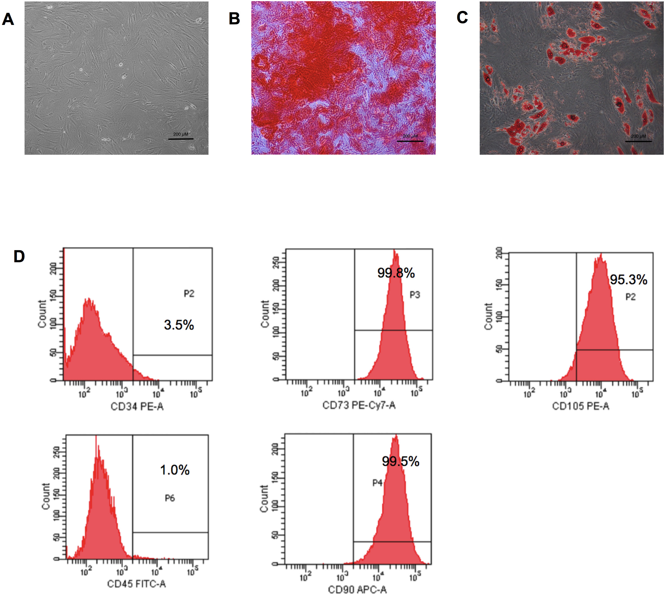
Fig. 1.

Adipose-derived stem cell characteristics. (A) Adipose-derived stem cells (ADSCs) displayed a fibroblast-like morphology. (B) Osteogenic differentiation of ADSCs after 21 days of differentiating—positive for Alizarin Red S staining. (C) Adipogenic differentiation of ADSCs after 14 days—positive for Oil Red O staining (scale bar = 200 µm). (D) Immunophenotyping of ADSCs demonstrated expressions of CD90, CD105, and CD73. PE-A, phycoerythrin; PE-Cy7-A, phycoerythrin-Cyanine7; FITC-A, fluorescein isothiocyanate; APC-A, allophycocyanin.

3.2 Effect of HPL Coating on ADSC Adhesion

A cell adhesion assay was performed to investigate the alteration in the biological features of ADSCs after being cultured on an HPL-pre-coated surface. The ability of ADSCs to attach to HPL-coated surfaces was investigated after being incubated for 12 hours. As shown in Fig. 2A, ADSCs cultured on HPL-coated surfaces had significantly higher cell adhesion rates than those cultured on the uncoated surfaces (96.49%
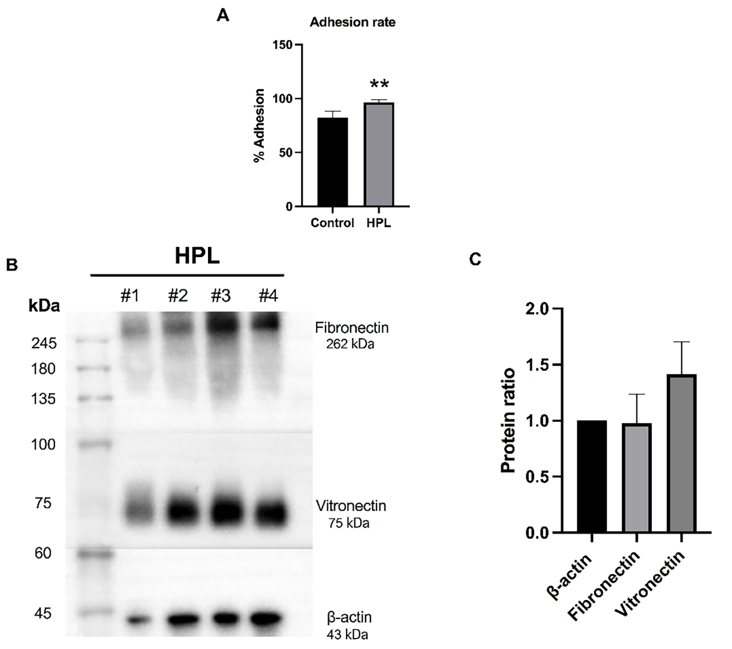
 ± 2.63% vs. 82.03% ± 6.12%, p < 0.01). Moreover, the roles of HPL in promoting ADSC adhesion were comparable with those cultured on fibronectin and vitronectin coatings. The adhesion rates of the ADSCs significantly increased when cultured on HPL (98.32% ± 6.90%), fibronectin (92.55% ± 5.12%), and vitronectin-coated (89.34% ± 9.55%) surfaces compared to the uncoated control (80.52% ± 9.74) (p < 0.05) (Supplementary Fig. 1). Therefore, HPLs possibly contain adhesive molecules that regulate cell behavior. Additionally, Western blotting analysis revealed that the HPLs were enriched with the cell adhesive proteins fibronectin and vitronectin (Fig. 2B). The semi-quantitative levels of fibronectin and vitronectin expressions were 0.976 and 1.416, respectively, relative to β-actin (Fig. 2C). Collectively, HPL facilitated ADSC attachment partly due to the presence of these cell adhesion molecules.

Fig. 2.

Effects of human platelet lysate (HPL) coating on cell adhesion and the expression of adhesive molecules. (A) The ADSC cell adhesion percentage when cultured on HPL (HPL group) for 12 hours compared to ADSCs cultured on an uncoated surface (control group). Data are presented as the mean ± standard deviation (SD) from four individual experiments. **p < 0.01 as analyzed by unpaired t-test. (B) The presence of adhesive molecules, fibronectin, and vitronectin, from four individual HPL samples, as detected by Western blotting. (C) Representative quantification of fibronectin and vitronectin levels relative to β-actin. Data are presented as the mean ± SD from four individual HPL samples.

3.3 Effect of HPL Coating on ADSC Proliferation

To investigate the effect of HPL coating on the growth rate, the ADSC population doubling times (PDTs) when cultured in the presence or absence of HPL were evaluated. Analysis of the PDTs revealed that HPL coating significantly decreased the PDT of ADSCs to 44.17
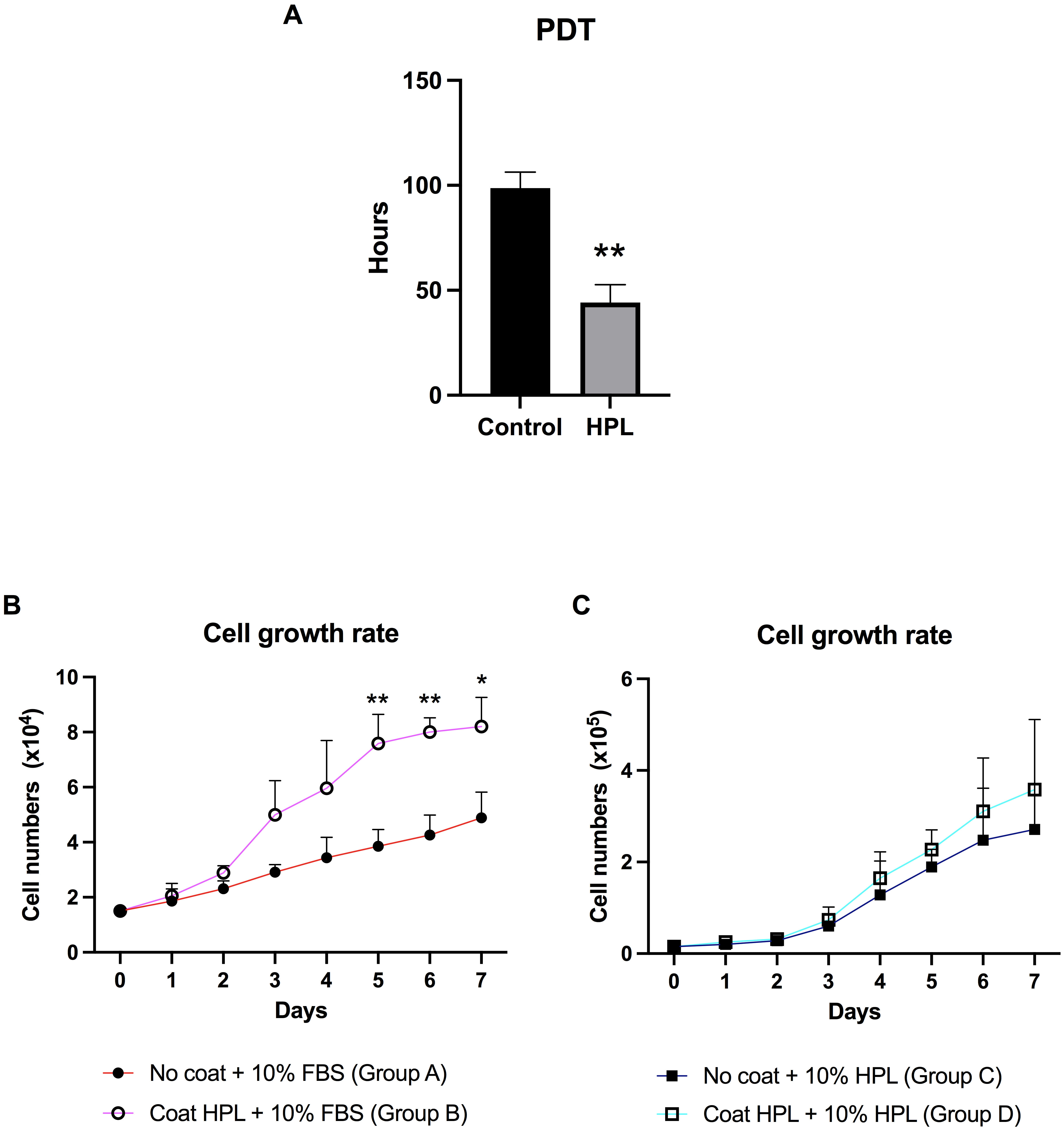
 ± 8.49 hours, whereas the ADSC control group PDT was 98.65 ± 7.64 hours (Fig. 3A). Furthermore, the ADSC PDT was significantly reduced when cultured in the HPL, fibronectin, and vitronectin coating substances (Supplementary Fig. 2). A marked decline in PDT was observed in cells grown on the HPL coating compared with the other coating substances. Thus, HPL coating increases robust ADSC growth by shortening the PDT. In addition, the population growth curve of ADSCs cultured with or without HPL coating for 7 days was examined by counting the cells at a specific time point. On day 1, ADSCs cultured on HPL-coated surfaces in stromal culture media containing 10% FBS (group B) were in the lag phase. After that, the cells gradually increased over time and reached the highest levels on day 7 (Fig. 3B). The number of cells in the group B condition was significantly higher than those grown on uncoated surfaces (group A) at days 5–7. We investigated whether substituting FBS with HPL and culturing on an HPL coating would further enhance ADSC expansion. The results demonstrated that the growth curve pattern of ADSCs cultured on HPL-coated surfaces in a stromal medium containing 10% HPL (group D) was similar to those cultured in 10% HPL supplements on the uncoated surface (group C). On days 0–2, the cell numbers began to gradually increase before proliferating to the highest levels on day 7 (Fig. 3C). Although there was no difference in the proliferation rates between the two groups, the results suggested that HPL contains growth factors necessary for supporting ADSC expansion in vitro.

Fig. 3.

Population doubling time (PDT) and growth curve of ADSCs cultured on HPL coating. (A) The population doubling time (PDT) of ADSCs derived from the HPL coating group compared to the control group. (B) The growth curve of ADSCs was maintained on an HPL-coated surface (group B) for 7 days, compared to an uncoated surface (group A). Cells in both groups were supplemented with stromal medium and 10% FBS (fetal bovine serum). (C) The growth curve of ADSCs was maintained on an HPL-coated surface (group D) compared to those cultured on an uncoated surface (group C), although both had FBS substituted for 10% HPL. Data are presented as mean ± SD (n = 3). *p < 0.05 and **p < 0.01 vs. the control group.

3.4 Effect of HPL Coating on Cellular Senescence in ADSC Cultures

To determine whether HPL coating affects ADSC senescence rates after serial passage, senescent cells were visualized using senescence-associated beta-galactosidase (SA
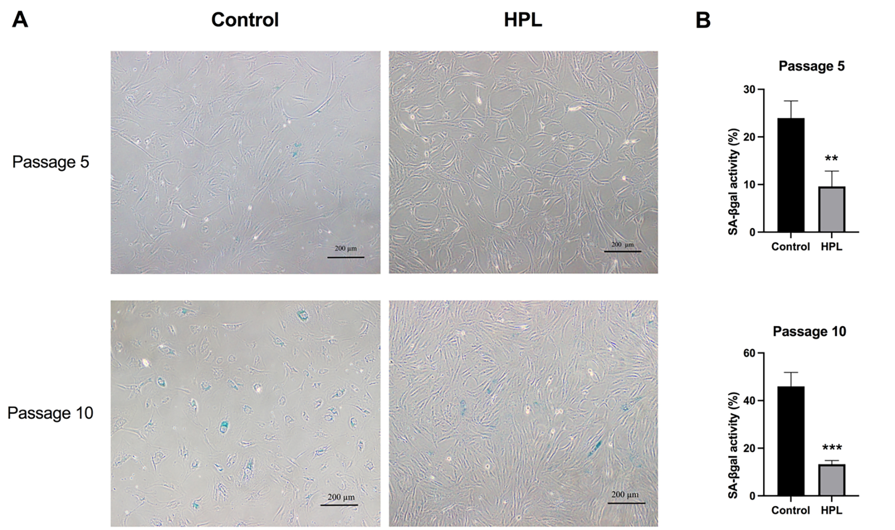
-β-gal) staining. ADSCs were continually maintained in the presence or absence of HPL coating from P2 to P5 and P2 to P10. The results showed that the morphologies of the ADSCs cultured on HPL-coated surfaces differed from those cultured in the control group. Without HPL coating, ADSCs were large and flat in appearance and contained more granules than those cultured on HPL-coated surfaces (Fig. 4A). The SA-β-gal staining demonstrated that the number of senescent ADSCs cultured in the control group was higher than in the HPL-coating group (Fig. 4A,B). The number of senescent cells was significantly decreased in the HPL-coated surface group for ADSCs cultured from P2 to P5 (9.61 ± 3.23%) compared to the control group (23.97 ± 3.59%). Similarly, the number of senescent ADSCs at P10 was significantly lower in the HPL-coating group (13.28 ± 1.52%) compared to the control group (45.97 ± 5.83%) (Fig. 4B). The results indicated that HPL coating could protect the ADSCs from undergoing senescence during in vitro expansion over a long period of culturing.

Fig. 4.

Effects of HPL coating on ADSCs senescence after serial passages. (A) Positive β-galactosidase stained ADSCs cultured with or without HPL coating—depicted in blue (scale bar = 200 µm). (B) The number of senescent ADSCs at P5 and P10 were counted and calculated as percentages of β-galactosidase activity compared to the control. Data are presented as mean ± SD (n = 3). **p < 0.01 and ***p < 0.001 vs. the control group.

3.5 HPL Coating Downregulates Senescence-Associated Gene Expressions during ADSC Cultivation

The previous results demonstrated a decrease in the number of senescent ADSCs maintained on the HPL-coated surface. Thus, to clarify whether HPL coating influenced the activation of the senescence signaling pathway during cellular aging, the transcription levels of p16, p21, and p53 were investigated in P5 and P10 ADSCs. Gene expression analysis showed that the p16, p21, and p53 mRNA levels were significantly downregulated in ADSCs cultured on HPL-coated plates for five passages compared to the control group (0.501
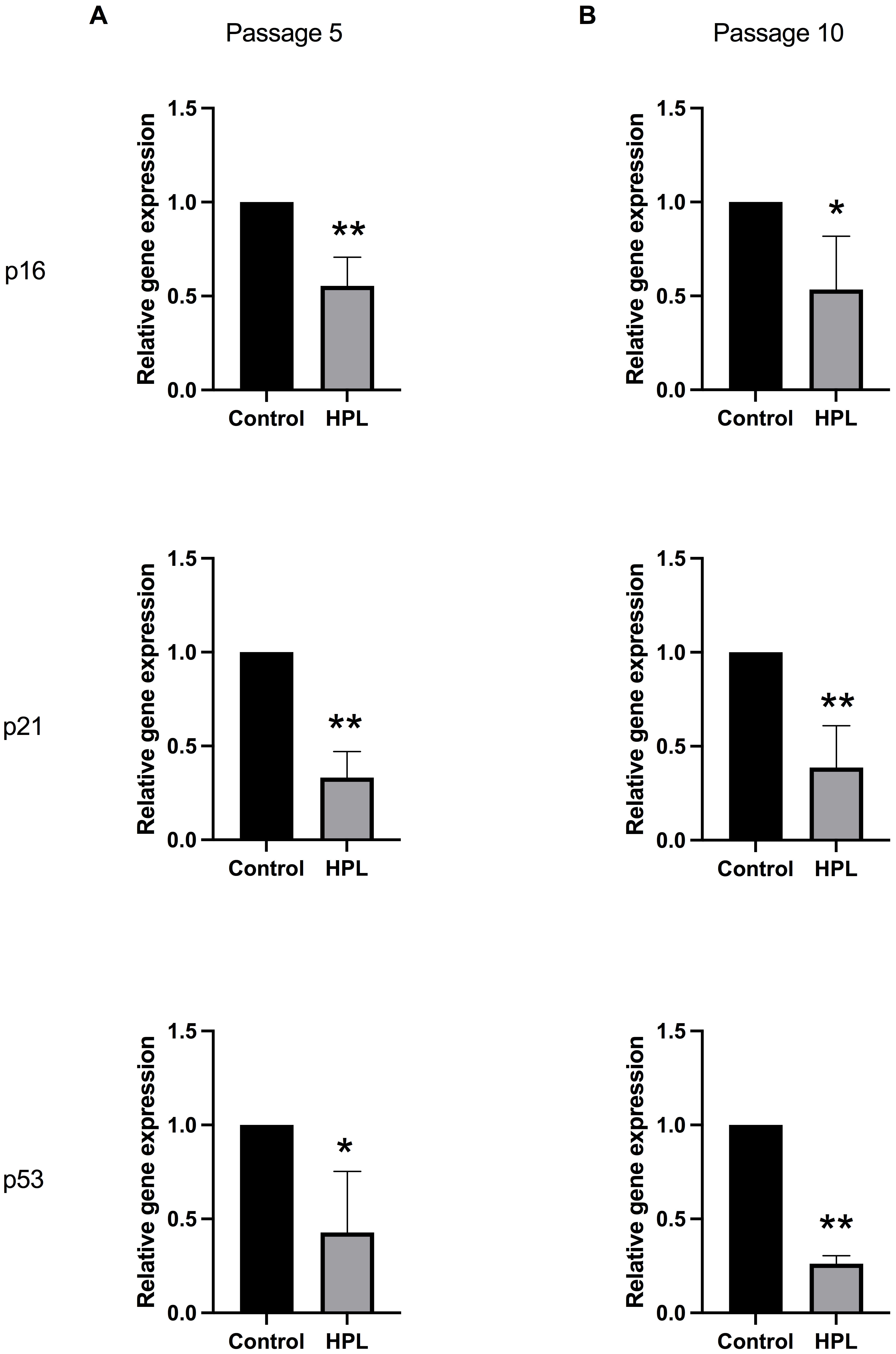
 ± 0.153, 0.333 ± 0.138 and 0.428 ± 0.325-fold of the control, respectively) (Fig. 5A). In addition, the p16, p21, and p53 transcription levels were significantly decreased in ADSCs maintained on HPL-coated surfaces for 10 passages compared with the control group (0.674 ± 0.285, 0.486 ± 0.223, 0.253 ± 0.042-fold of the control, respectively) (Fig. 5B). The results suggested that HPL coating downregulates the activation of the p16, p21, and p53 signaling pathway in ADSCs during in vitro expansion.

Fig. 5.

Alteration of senescent-associated gene expressions in ADSCs cultured on HPL coating. (A) Passage 5 and (B) passage 10 populations maintained on HPL-coated surfaces. Data are presented as mean ± SD from four individual experiments. *p < 0.05 and **p < 0.01 vs. the control group.

4. Discussion

This study demonstrated the role of HPL as a coating material in supporting in vitro ADSC expansion. Changes in the biological features of ADSCs, including cell adhesion, cell proliferation, and cellular senescence, were investigated at different periods of ADSC growth on pre-coated HPL plates. HPL supplements have beneficial effects on MSC growth; however, the cells had difficulty attaching to the plastic surface of tissue culture vessels. According to a recent study, MSCs cultured in FBS medium interacted more with the tissue culture vessel than those cultured in HPL [24]. The MSCs cultured in FBS had a significantly higher amount of focal adhesion than those cultured in the HPL additive.

In cell-based therapy, expanding ADSCs holds significance in cell manufacturing procedures. An alternative approach to enhancing growth during this process involves surface coating with adhesion molecules. Notably, utilizing HPL coating has emerged as a strategic method for MSC expansion [25], one which closely mimics the natural cellular environment through the enrichment of adhesion molecules [19, 20]. Interestingly, the findings in our recent study clearly showed that HPL coating could promote the adhesion of ADSCs. Western blot analysis of the HPL samples indicated that they possess key adhesive molecules, including fibronectin and vitronectin. This finding agrees with those in prior studies, which showed that HPL has a biological richness and contains various growth factors, cytokines, and extracellular matrix proteins that can positively influence cell behavior, which is crucial for cell growth and differentiation [26, 27, 28]. Moreover, recent studies that thoroughly characterized protein contents of HPL samples following proteomic analysis revealed a remarkable richness in multiple growth factors and adhesion molecules [19, 20]. Adhesive molecules, such as laminin, vitronectin, and fibronectin, have been shown to play roles in improving cellular adhesion and promoting colony formation [29, 30]. Thus, it seems possible that the increase in the cell adhesion ability of ADSCs cultured on HPL-coated plates is partly due to the presence of key adhesive molecules in the HPL samples, including fibronectin and vitronectin.

Evidence supporting the substitution of FBS with HPL supplements could enhance MSC proliferation [16, 31] and facilitate MSC differentiation [18]. We further investigated whether minimal amounts of HPL that remained as a coating substance could promote ADSC growth. Our finding first reported that the PDT of ADSCs cultured on HPL-coated plates was reduced, which led to robust increases in ADSC growth. Likewise, the growth curve of the ADSCs cultured in HPL-coated plates demonstrated a significant increase in ADSC numbers [32]. HPL as a coating material successfully enhanced in vitro ADSC expansion. Moreover, adding HPL supplements to HPL-coated plates slightly increased ADSC numbers. However, no significant differences were found. HPL is enriched with an abundance of growth factors, such as insulin-like growth factor 1 (IGF-1), platelet-derived growth factor (PDGF), transforming growth factor-beta (TGF-β), and vascular endothelial growth factor (VEGF) [19, 33]; therefore, the addition of high concentrations of HPL supplements possibly concealed the effects of the HPL coating. Additionally, a study was conducted that indicated the use of HPL as a standalone supplement was ineffective. However, cell adhesion and proliferation were improved for cells grown on laminin-coated surfaces and cultured with the HPL supplement. This enhancement can potentially reduce the necessity for HPL supplementation for specific cell culture applications [34]. In this investigation, applying an HPL coating holds promise in potentially lowering the required HPL volume from 10% to 5%. This improvement reduces the supplement volume required during the expansion of ADSCs, promoting cost-effective practices throughout the culture process and resulting in overall cost reductions.

Serial cell expansions and prolonged cell culture times usually lead to cellular aging [35]. However, cellular senescence is induced by repetitive passaging and high glucose levels [36]. The alterations in cellular behaviors during senescence can be demonstrated by morphological changes and increased senescence-associated β-galactosidase (SA-β-gal) expressions [37, 38]. Indeed, this study clearly demonstrated that HPL coating delayed the senescence of ADSCs after long-term cell expansion. SA-β-gal positive cells were initially found in early-passage ADSCs (passage 5) and were massively increased in cells of high passages (passage 10). In addition, the ADSC morphologies altered from spindle-shaped cells to cells of enlarged sizes, especially after a high number of passages. This finding is comparable to prior studies that reported a change in cell morphologies following serial passaging and cellular aging [39, 40]. Cultivating ADSCs on HPL-coated surfaces diminished cells preceding senescence even at late passages (passage 10). The appearance of high passage ADSCs maintained on HPL-coated surfaces displayed fibroblast-like morphologies similar to early-passage ADSCs. The results indicated that HPL coating effectively reduced the number of senescent ADSCs and maintained ADSC morphologies in long-term cultures. These results support the previous observation that small amounts of HPL being supplemented (2–5% HPL) into the culture medium could attenuate MSC senescence and rejuvenate aged MSCs [40]. The p53/p21/p16 pathway plays a major role in regulating the cell cycle and cellular senescence, whereby activating the p53/p21/p16 pathway leads to cell cycle exit [41, 42, 43]. Our results demonstrated that the transcript levels of senescence-associated genes, including p53, p21, and p16, in ADSCs cultured on HPL-coated plates were significantly downregulated. Collectively, the results implied that HPL coating altered the expression profiles of senescence-associated genes in ADSCs, alleviating cellular senescence during serial passaging. These findings align with a previous study showing that p21 expression was reduced in ADSCs cultured in a medium containing HPL [44].

5. Conclusions

Our experiments demonstrated the beneficial effects of HPL coating on ADSC expansion. Culturing ADSCs using an HPL-coating system resulted in increased cell adhesion, enhanced cell proliferation, and reduced cellular senescence. This study has also identified the presence of key adhesive molecules in HPL. This study strengthens the idea that cell culture surface modification by HPL coating would be an additional method for expanding in vitro ADSC growth. Minimal HPL volumes are required to improve ADSC growth during long periods of in vitro cell culture.

Abbreviations

ADSCs, adipose-derived stem cells; MSCs, mesenchymal stromal cells; FBS, fetal bovine serum; HPL, human platelet lysate; PDT, population doubling time.

Availability of Data and Materials

All data generated and/or analyzed in this study are included in the published article and supplementary information.

Author Contributions

WK and AS designed and conceptualized the research study. PT, NC and SH performed the research. CP performed sample collection and the data analysis. PT, SH, and KN participated in collecting and analyzing the data. WK and AS supervised the experiments and analyzed the data. PT, WK and AS wrote and edited the manuscript. AS made significant revisions and proofread the manuscript. All authors contributed to editorial changes in the manuscript. All authors read and approved the final manuscript. All authors have participated sufficiently in the work and agreed to be accountable for all aspects of the work.

Ethics Approval and Consent to Participate

This study was approved by ethical committee for human research of the Mahidol University Central Institutional Review Board in accordance with the Declaration of Helsinki (MU-CIRB 2018/202.1411 and MU-CIRB 2020/180.2307).

Acknowledgment

We would like to express our sincere gratitude to our colleagues for their advice on the article. Thanks to all those who helped us during the writing of this manuscript.

Funding

This research project is supported by Mahidol University (NDFR 30/2564).

Conflict of Interest

The authors declare no conflict of interest.

References
[1]
Pittenger MF, Discher DE, Péault BM, Phinney DG, Hare JM, Caplan AI. Mesenchymal stem cell perspective: cell biology to clinical progress. NPJ Regenerative Medicine. 2019; 4: 22.
[2]
Bacakova L, Zarubova J, Travnickova M, Musilkova J, Pajorova J, Slepicka P, et al. Stem cells: their source, potency and use in regenerative therapies with focus on adipose-derived stem cells - a review. Biotechnology Advances. 2018; 36: 1111–1126.
[3]
Müller L, Tunger A, Wobus M, von Bonin M, Towers R, Bornhäuser M, et al. Immunomodulatory Properties of Mesenchymal Stromal Cells: An Update. Frontiers in Cell and Developmental Biology. 2021; 9: 637725.
[4]
Mushahary D, Spittler A, Kasper C, Weber V, Charwat V. Isolation, cultivation, and characterization of human mesenchymal stem cells. Cytometry. Part A. 2018; 93: 19–31.
[5]
Dominici M, Le Blanc K, Mueller I, Slaper-Cortenbach I, Marini F, Krause D, et al. Minimal criteria for defining multipotent mesenchymal stromal cells. The International Society for Cellular Therapy position statement. Cytotherapy. 2006; 8: 315–317.
[6]
Viswanathan S, Shi Y, Galipeau J, Krampera M, Leblanc K, Martin I, et al. Mesenchymal stem versus stromal cells: International Society for Cell & Gene Therapy (ISCT®) Mesenchymal Stromal Cell committee position statement on nomenclature. Cytotherapy. 2019; 21: 1019–1024.
[7]
Kabat M, Bobkov I, Kumar S, Grumet M. Trends in mesenchymal stem cell clinical trials 2004-2018: Is efficacy optimal in a narrow dose range? Stem Cells Translational Medicine. 2020; 9: 17–27.
[8]
Yang YHK, Ogando CR, Wang See C, Chang TY, Barabino GA. Changes in phenotype and differentiation potential of human mesenchymal stem cells aging in vitro. Stem Cell Research & Therapy. 2018; 9: 131.
[9]
Wagner W, Horn P, Castoldi M, Diehlmann A, Bork S, Saffrich R, et al. Replicative senescence of mesenchymal stem cells: a continuous and organized process. PLoS ONE. 2008; 3: e2213.
[10]
Yu Y, Park YS, Kim HS, Kim HY, Jin YM, Jung SC, et al. Characterization of long-term in vitro culture-related alterations of human tonsil-derived mesenchymal stem cells: role for CCN1 in replicative senescence-associated increase in osteogenic differentiation. Journal of Anatomy. 2014; 225: 510–518.
[11]
Curtis AS, Forrester JV, McInnes C, Lawrie F. Adhesion of cells to polystyrene surfaces. The Journal of Cell Biology. 1983; 97: 1500–1506.
[12]
Lerman MJ, Lembong J, Muramoto S, Gillen G, Fisher JP. The Evolution of Polystyrene as a Cell Culture Material. Tissue Engineering. Part B, Reviews. 2018; 24: 359–372.
[13]
Cai S, Wu C, Yang W, Liang W, Yu H, Liu L. Recent advance in surface modification for regulating cell adhesion and behaviors. Nanotechnology Reviews. 2020; 9: 971–989.
[14]
Cooke MJ, Phillips SR, Shah DSH, Athey D, Lakey JH, Przyborski SA. Enhanced cell attachment using a novel cell culture surface presenting functional domains from extracellular matrix proteins. Cytotechnology. 2008; 56: 71–79.
[15]
Li X, Yao J, Yang X, Tian W, Liu L. Surface modification with fibronectin or collagen to improve the cell adhesion. Applied Surface Science. 2008; 255: 459–461.
[16]
Guiotto M, Raffoul W, Hart AM, Riehle MO, di Summa PG. Human platelet lysate to substitute fetal bovine serum in hMSC expansion for translational applications: a systematic review. Journal of Translational Medicine. 2020; 18: 351.
[17]
Escobar CH, Chaparro O. Xeno-Free Extraction, Culture, and Cryopreservation of Human Adipose-Derived Mesenchymal Stem Cells. Stem Cells Translational Medicine. 2016; 5: 358–365.
[18]
Gao Y, Ku NJ, Sung TC, Higuchi A, Hung CS, Lee HHC, et al. The effect of human platelet lysate on the differentiation ability of human adipose-derived stem cells cultured on ECM-coated surfaces. Journal of Materials Chemistry. B. 2019; 7: 7110–7119.
[19]
Viau S, Lagrange A, Chabrand L, Lorant J, Charrier M, Rouger K, et al. A highly standardized and characterized human platelet lysate for efficient and reproducible expansion of human bone marrow mesenchymal stromal cells. Cytotherapy. 2019; 21: 738–754.
[20]
Widyaningrum R, Burnouf T, Nebie O, Delila L, Wang TJ. A purified human platelet pellet lysate rich in neurotrophic factors and antioxidants repairs and protects corneal endothelial cells from oxidative stress. Biomedicine & Pharmacotherapy. 2021; 142: 112046.
[21]
Cheng NC, Tu YK, Lee NH, Young TH. Influence of Human Platelet Lysate on Extracellular Matrix Deposition and Cellular Characteristics in Adipose-Derived Stem Cell Sheets. Frontiers in Cell and Developmental Biology. 2020; 8: 558354.
[22]
Cimino M, Gonçalves RM, Barrias CC, Martins MCL. Xeno-Free Strategies for Safe Human Mesenchymal Stem/Stromal Cell Expansion: Supplements and Coatings. Stem Cells International. 2017; 2017: 6597815.
[23]
Phetfong J, Tawonsawatruk T, Seenprachawong K, Srisarin A, Isarankura-Na-Ayudhya C, Supokawej A. Re-using blood products as an alternative supplement in the optimisation of clinical-grade adipose-derived mesenchymal stem cell culture. Bone & Joint Research. 2017; 6: 414–422.
[24]
Fernandez-Rebollo E, Mentrup B, Ebert R, Franzen J, Abagnale G, Sieben T, et al. Human Platelet Lysate versus Fetal Calf Serum: These Supplements Do Not Select for Different Mesenchymal Stromal Cells. Scientific Reports. 2017; 7: 5132.
[25]
Loubière C, Sion C, De Isla N, Reppel L, Guedon E, Chevalot I, et al. Impact of the type of microcarrier and agitation modes on the expansion performances of mesenchymal stem cells derived from umbilical cord. Biotechnology Progress. 2019; 35: 6.
[26]
Liu S, Yang W, Li Y, Sun C. Fetal bovine serum, an important factor affecting the reproducibility of cell experiments. Scientific Reports. 2023; 13: 1942.
[27]
Liau LL, Hassan MNFB, Tang YL, Ng MH, Law JX. Feasibility of Human Platelet Lysate as an Alternative to Foetal Bovine Serum for In Vitro Expansion of Chondrocytes. International Journal of Molecular Sciences. 2021; 22: 1269.
[28]
Vasanthan P, Gnanasegaran N, Govindasamy V, Abdullah AN, Jayaraman P, Ronald VS, et al. Comparison of fetal bovine serum and human platelet lysate in cultivation and differentiation of dental pulp stem cells into hepatic lineage cells. Biochemical Engineering Journal. 2014; 88: 142–153.
[29]
Pattappa G, Reischl F, Jahns J, Schewior R, Lang S, Zellner J, et al. Fibronectin Adherent Cell Populations Derived From Avascular and Vascular Regions of the Meniscus Have Enhanced Clonogenicity and Differentiation Potential Under Physioxia. Frontiers in Bioengineering and Biotechnology. 2022; 9: 789621.
[30]
Zhu XY, Chen YH, Zhang T, Liu SJ, Bai XY, Huang XY, et al. Improvement of human embryonic stem cell-derived retinal pigment epithelium cell adhesion, maturation, and function through coating with truncated recombinant human vitronectin. International Journal of Ophthalmology. 2021; 14: 1160–1167.
[31]
Bieback K, Fernandez-Muñoz B, Pati S, Schäfer R. Gaps in the knowledge of human platelet lysate as a cell culture supplement for cell therapy: a joint publication from the AABB and the International Society for Cell & Gene Therapy. Cytotherapy. 2019; 21: 911–924.
[32]
Oliveira SM, Santo VE, Gomes ME, Reis RL, Mano JF. Layer-by-layer assembled cell instructive nanocoatings containing platelet lysate. Biomaterials. 2015; 48: 56–65.
[33]
Shanskii YD, Sergeeva NS, Sviridova IK, Kirakozov MS, Kirsanova VA, Akhmedova SA, et al. Human platelet lysate as a promising growth-stimulating additive for culturing of stem cells and other cell types. Bulletin of Experimental Biology and Medicine. 2013; 156: 146–151.
[34]
Guiotto M, Raffoul W, Hart AM, Riehle MO, di Summa PG. Human Platelet Lysate Acts Synergistically With Laminin to Improve the Neurotrophic Effect of Human Adipose-Derived Stem Cells on Primary Neurons in vitro. Frontiers in Bioengineering and Biotechnology. 2021; 9: 658176.
[35]
Gruber HE, Somayaji S, Riley F, Hoelscher GL, Norton HJ, Ingram J, et al. Human adipose-derived mesenchymal stem cells: serial passaging, doubling time and cell senescence. Biotechnic & Histochemistry. 2012; 87: 303–311.
[36]
Aswamenakul K, Klabklai P, Pannengpetch S, Tawonsawatruk T, Isarankura-Na-Ayudhya C, Roytrakul S, et al. Proteomic study of in vitro osteogenic differentiation of mesenchymal stem cells in high glucose condition. Molecular Biology Reports. 2020; 47: 7505–7516.
[37]
McHugh D, Gil J. Senescence and aging: Causes, consequences, and therapeutic avenues. The Journal of Cell Biology. 2018; 217: 65–77.
[38]
Mohamad Kamal NS, Safuan S, Shamsuddin S, Foroozandeh P. Aging of the cells: Insight into cellular senescence and detection Methods. European Journal of Cell Biology. 2020; 99: 151108.
[39]
Fraile M, Eiro N, Costa LA, Martín A, Vizoso FJ. Aging and Mesenchymal Stem Cells: Basic Concepts, Challenges and Strategies. Biology. 2022; 11: 1678.
[40]
Griffiths S, Baraniak PR, Copland IB, Nerem RM, McDevitt TC. Human platelet lysate stimulates high-passage and senescent human multipotent mesenchymal stromal cell growth and rejuvenation in vitro. Cytotherapy. 2013; 15: 1469–1483.
[41]
Di Micco R, Krizhanovsky V, Baker D, d’Adda di Fagagna F. Cellular senescence in ageing: from mechanisms to therapeutic opportunities. Nature Reviews. Molecular Cell Biology. 2021; 22: 75–95.
[42]
Kumari R, Jat P. Mechanisms of Cellular Senescence: Cell Cycle Arrest and Senescence Associated Secretory Phenotype. Frontiers in Cell and Developmental Biology. 2021; 9: 645593.
[43]
Roger L, Tomas F, Gire V. Mechanisms and Regulation of Cellular Senescence. International Journal of Molecular Sciences. 2021; 22: 13173.
[44]
Søndergaard RH, Follin B, Lund LD, Juhl M, Ekblond A, Kastrup J, et al. Senescence and quiescence in adipose-derived stromal cells: Effects of human platelet lysate, fetal bovine serum and hypoxia. Cytotherapy. 2017; 19: 95–106.

Publisher’s Note: IMR Press stays neutral with regard to jurisdictional claims in published maps and institutional affiliations.

Share
Back to top