IMR Press / FBL / Volume 29 / Issue 4 / DOI: 10.31083/j.fbl2904145
Open Access Review
Progenitor Cell Function and Cardiovascular Remodelling Induced by SGLT2 Inhibitors
Show Less
1 Department of Cardiothoracic Surgery, Democritus University of Thrace – University General Hospital, 68100 Alexeandroupolis, Greece
2 Second Propaedeutic Department of Internal Medicine, Hippokration General Hospital, Aristotle University of Thessaloniki, 54642 Thessaloniki, Greece
3 Department of Adult and Congenital Cardiac Surgery, Mitera Hospital, 15123 Marousi, Greece
4 Department of Anesthesia, St. Michael's Hospital, University of Toronto, Toronto, ON M5B 1W8, Canada
*Correspondence: theodorastougiannos@gmail.com (Theodora M. Stougiannou)
Front. Biosci. (Landmark Ed) 2024, 29(4), 145; https://doi.org/10.31083/j.fbl2904145
Submitted: 10 December 2023 | Revised: 18 February 2024 | Accepted: 28 February 2024 | Published: 9 April 2024
Copyright: © 2024 The Author(s). Published by IMR Press.
This is an open access article under the CC BY 4.0 license.
Abstract

Sodium-glucose cotransporters 2 (SGLT2) are high-capacity, low-affinity transporters, expressed mainly in the early portion of the proximal renal tube, mediating up to 90% of renal glucose uptake, while SGLT1 receptors are found mainly in the small intestine, facilitating glucose absorption. SGLT2 inhibitors (SGLT2i) originally emerged as agents for the treatment of type 2 diabetes mellitus; however, they soon demonstrated remarkable cardio- and renoprotective actions that led to their licensed use for the treatment of heart failure and chronic kidney disease, regardless of the diabetic status. Cardiovascular remodelling represents an umbrella term that encompasses changes that occur in the cardiovascular system, from the molecular and cellular level, to tissue and organs after local injury, chronic stress, or pressure. SGLT modulation has been shown to positively affect many of these molecular and cellular changes observed during pathological remodelling. Among the different pathophysiological mechanisms that contribute to adverse remodelling, various stem and progenitor cells have been shown to be involved, through alterations in their number or function. Recent studies have examined the effects of SGLT2i on stem and progenitor cell populations and more specifically on endothelial progenitor cells (EPCs). Although some found no significant effect, others showed that SGLT2i can modulate the morphology and function of EPCs. These preliminary observations of the effect of SGLT2i on EPCs may be responsible for some of the beneficial effects of gliflozins on pathological remodelling and, by extension, on cardiovascular disease. The purpose of this narrative review is to critically discuss recent evidence on the cardioprotective effects of SGLT2is, in the context of cardiac remodelling.

Keywords
cardiac remodelling
heart failure
endothelial progenitor
hemopoietic stem cells
1. Introduction

Cardiac remodelling is a complex biological process, characterized by a number of changes in molecular and cellular tissue architecture, in response to long-term stress, such as pressure or volume overload, tissue inflammation, or local ischemia due to myocardial infarction; gross tissue function will eventually change as well, to accommodate these new conditions. Remodelling may be a physiological adaptation, for example, during exercise or pregnancy, or it may be pathological, for example following chronic pressure overload or ischemic injury. A number of different mechanisms have been discovered to contribute to this process, from alterations in cellular and subcellular organization, to metabolic derangements, inflammation, and even stem and progenitor cell dysfunction [1, 2].

Eventually, the resulting cardiac dysfunction can lead to heart failure (HF) [1], often described as a clinical syndrome [3]; in fact, in 2019, the Heart Failure Association (HFA) ATLAS Project estimated the median prevalence of HF to be 17 per 1000 persons, while in North America the prevalence is projected to increase from 2.4% (2012) to an estimated 3.0% in 2030 (United States). On the other hand, incidence is relatively stable and has been shown to be decreasing, at least in the developed world [4].

Risk for HF may increase due to various conditions, including hypertension and coronary artery disease; type 2 diabetes mellitus (T2DM) in particular, confers an increased risk of HF-related morbidity and mortality, along with an increased risk of occurrence, highlighting the need for prompt and appropriate management [5]. Not only do therapies for both HF and diabetes mellitus act synergistically, but treatment of one disorder can positively affect the outcomes in the other [6]. Among the various pharmaceutical compounds developed for the treatment of T2DM, sodium-glucose cotransporters 2 inhibitors (SGLT2is) have emerged as a viable option, due to the low risk of hypoglycemia as well as the weight-lowering effects [7]. They were first studied more than 20 years ago, when their potential for inhibition of the kidney SGLT transporter was evaluated [8]. Ever since, their effectiveness in the treatment of hyperglycemia [9], mitigation of kidney damage [10] and alleviation of cardiovascular risk has been confirmed [5]. SGLT inhibition has been shown to exert protective effects, not only on kidney function, but also on the heart and vasculature [11]. Recent research on SGLT2is has revealed a variety of mechanisms, targeting several of the derangements observed in pathological remodelling [5].

Although there has been extensive research into the mechanisms that could contribute to pathological remodelling, evidence for stem and progenitor cell involvement is relatively limited. In more detail, dysfunctional or even decreased numbers of progenitor populations have been shown to be involved in the process [12, 13]. Investigation into the relationship between SGLT modulation and progenitor cell function has been a relatively recent pursuit, with few studies examining this association yet, often yielding conflicting results [14]. Thus, the purpose of this narrative review is to gather and critically discuss recent evidence on the cardioprotective effects of gliflozins, in the context of cardiac remodelling, with an emphasis on pathological remodeling.

2. SGLT Receptors

SGLTs are a large family of membrane cotransporters found on the plasma membrane of various cells that, in addition to glucose, transport various other molecules, such as vitamins, amino acids, and ions across the apical cellular membrane. In general, there are 6 SGLT isoforms, of which SGLT1 and SGLT2 have been the most studied. SGLT1 is most frequently found in the small intestine and is responsible for glucose and galactose absorption. The first SGLT protein transporter to be discovered [7], SGLT1 is encoded by the SLC5A1 gene and consists of 664 amino acids. It functions primarily by facilitating the simultaneous transport of 2 Na+ ions along with one glucose or galactose molecule within enterocytes and epithelial cells, as well as within cells of the S2 and S3 regions of the proximal convoluted tubule (PCT) of the kidney. In addition, they have been further identified in the heart, lungs, liver, skeletal muscles, and pancreatic α cells [7, 15, 16]. SGLT2, encoded by SLC5A2, consists of a 672 amino acid structure and is expressed predominantly within cells of the S1 and S2 segments of the PCT. Since 90% of the glucose within the glomerular filtrate is absorbed in the PCT, it is evident that SGLT2 receptors play an important role in this process. SGLT2 receptors have also been identified in the mammary glands, testes, lungs, liver, skeletal muscles and nervous system [7, 16, 17].

3. Cardiovascular Remodelling

The series of structural and functional changes observed in cardiac remodelling assist to the better adjustment of the heart to new conditions, leading to either physiological or pathological adaptation; these may be caused by a variety of different factors which are summarized in Table 1 [18, 19, 20, 21, 22, 23, 24, 25, 26]. Eventually, left ventricle (LV) pressure, radius, and wall thickness are altered, so that the developed wall stress remains within an appropriate range to maintain the cardiac output (CO) [19].

Table 1.Causes of physiological and pathological cardiac remodelling.
Physiological remodelling Normal growth (neonatal/postnatal period)
Normal aging
Exercise
Pregnancy
Pathological remodelling Pathological aging-related processes
Inflammation
Infiltrative conditions (cardiac amyloidosis, sarcoidosis, hemochromatosis)
Autoimmune conditions (RA, SLE, PM/DM, pSS, SSc)
Ischemia
DCM
Endocrine conditions (for example acromegaly, Cushing’s Syndrome, hyperthyroidism/hypothyroidism, hyperaldosteronism)
HOCM
Chronic mechanical stress
Various drugs/toxins (for example cocaine, amphetamines, isoprenaline, anticancer drugs – doxorubicin, and ethanol, leading to cardiomyopathy)

RA, Rheumatoid Arthritis; SLE, Systemic lupus erythematosus; PM, polymyositis; DM, dermatomyositis; pSS, primary Sjögren’s Syndrome; SSc, Systemic sclerosis; HOCM, Hypertrophic Cardiomyopathy; DCM, Diabetic Cardiomyopathy.

3.1 Signalling Pathways Implicated in Physiological and Pathological Cardiac Remodelling

Changes occurring with physiological remodelling adaptations include alterations in cell morphology, protein expression, and signalling pathways [27]. These frequently result in cardiac hypertrophy through an increase in cardiomyocyte size [19]; a number of signalling pathways are implicated in physiological cardiac hypertrophy, such as mammalian target of rapamycin (mTOR) and phosphoinositide-3-kinase/protein kinase B (PI3K/Akt), leading to increased Akt levels and thus increased cardiomyocyte size [28]. In fact, studies in mice have shown higher levels of PI3K and Akt when cardiomyocytes are subjected to increased pressures (hypertrophy signals), in turn causing an increase in size [29]. The role of Akt in normal growth has been exhibited in Akt-deficient mice; insulin-like growth factor (IGF) or exercise had no effect, which further proved that IGF acts through Akt to induce physiological cardiomyocyte hypertrophy [30]. IGF/Akt is inhibited by SIRT6, proving that SIRT6 is capable of attenuating cardiomyocyte hypertrophy; indeed, SIRT6 has been found in reduced quantities in dysfunctional cardiac tissue and failing hearts, while SIRT6 deficient mice appeared to be unaffected by hypertrophic stimuli in general [31]. Akt may also become activated by mTOR [19], and also lead to cardiomyocyte hypertrophy, experimentally shown through pharmacological mTOR inhibition by rapamycin [32].

Hypertrophy may also be mediated by the expression of certain myosin and sarcoplasmic reticulum (SR) genes, α-MyHC, β-MyHC, and SERCA respectively, controlled through thyroid hormone (TH) receptors; though α-MyHC and SERCA expression seems to increase in response to stimulation of the TH receptor (hyperthyroid cell state), β-MyHC expression seems to decrease. On the other hand, pathological hypertrophy is associated with decreased stimulation of the TH receptor (hypothyroid cell state), where only β-MyHC expression increases instead, as a result of fetal programming [33, 34]; overexpression of certain miRNAs including miR – 132, has also been observed, in turn leading to FoxO3 repression, and calcineurin/NFAT signalling pathway activation [35]. In essence, it seems that thyroid hormone receptors (TR) are downregulated in cases of pathological hypertrophy, and up-regulated in cases of physiological hypertrophy [36] (Table 2).

Table 2.Signaling pathways implicated in physiological and pathological remodelling.
Cardiac remodelling Signalling mechanisms
Physiological Insulin, PI3K/Akt signalling pathway; Akt interacts with PI3K, affecting L-type calcium channels (LTCC)-Akt inhibited by SIRT6.
mTOR/Akt pathway.
TRα, TRβ receptor expression and stimulation; TRβ1 receptor expression upregulated, causing increased expression of α-MyHC and SERCA (hyperthyroid cell state).
Pathological CM thyroid receptors downregulated (hypothyroid cell state), (pathological aging-induced cardiac remodelling an additional cause); α-MyHC, SERCA downregulated, β-MyHC upregulated.
miR-132 overexpression, FoxO3 (Transcription factor-TF) repression and activation of the calcineurin/NFAT signalling pathway, induction of pathological cardiac hypertrophy, impairment of autophagy.

PI3K, phosphoinositide-3-kinase; Akt, Protein Kinase B; LTCC, L-type Calcium Channel; SIRT6, Sirtuin 6; mTOR, mammalian target of rapamycin; TRα, Thyroid receptor α; TRβ, Thyroid receptor β; α-MyHC, α-Myosin heavy chain; β-MyHC, β-Myosin heavy chain; SERCA, Sarcoendoplasmic reticulum Calcium ATPase; CM, cardiomyocyte; miR, micro ribonucleic acid; FoxO3, Forkhead transcription factor O subfamily member 3a; NFAT, Nuclear factor of activated T-cells.

3.2 Pathological Cardiac Remodelling

Pathological cardiac remodelling is characterized by a series of molecular and cellular changes, related to alterations in cells, subcellular organelles, intracellular and extracellular protein expression summarized in Table 3, as well as changes in various intracellular processes, including metabolism, reactive oxygen species (ROS) and Ca2+ handling, inflammatory and neurohumoral pathways summarized in Table 4, which will in turn affect microscopic as well as macroscopic tissue architecture, and eventually function [27, 37].

Table 3.Summary of structural changes implicated in pathological cardiac remodelling.
Structure Alteration Additional information
Cardiomyocytes (CM) Hypertrophy Increased protein expression (biomechanical stress, neurohumoral activation, fibroblast/myofibroblast growth-factor secretion).
Progressive CM lengthening, rearrangement within the myocardial matrix (‘cell slippage’), cell death and replacement by fibrous matrix.
Cell death Apoptosis, autophagy, pyroptosis, necroptosis.
Mitochondria IF mitochondria IF mitochondria smaller and rounder, increased numbers, increased recruitment of immature mitochondria.
Sarcoplasmic reticulum (SR) Disordered Structure Changes non-specific, not easily quantified.
Cell Membrane T-tubule Reduced surface area and density, loss of some T-tubules.
Intracellular proteins Myofibrillar proteins Increase in absolute numbers, and length, depending on orientation of added sarcomeres (if added in parallel or in series respectively).
Remodelling due to pressure increases causes increase in myofibril number, while remodelling due to volume increase causes increase in myofibril length.
ICD proteins ICDs exhibit a smaller surface area, along with the presence of vacuoles.
Desmosomes Increased expression, toxic desmin oligomers.
Extracellular proteins Collagen Increased collagen deposition (Type I collagen, type III collagen), mediated by fibroblasts, myofibroblasts, CMs, vascular cells, various immune system cells (macrophages, lymphocytes).
Induced by TGF-β1/SMAD3 signaling through Angiotensin II; TGF-β1/SMAD3/Stat3 regulated by EphrinB2 and myofibroblast activation.
ECM collagen disruption by metalloproteinase activation.

CM, cardiomyocyte; ICD, intercalated disk; TGF-β1, Transforming Growth Factor beta 1; SMAD3, Suppressor of Mothers against Decapentaplegic 3; Stat3, signal transducer and activator of transcription 3; EphrinB2, Eph family receptor interacting protein B2; ECM, extracellular matrix; IF, intrafibrillar.

Table 4.Summary of changes in processes implicated in pathological cardiac remodelling.
Process Alteration Causes/Results
Metabolism Switch towards glycolysis Increased aspartate production from glucose, low oxygen tension and HIF-1a production also induce switch to glycolysis (cell hypertrophy).
Preferential use of ketone bodies (AcAc, DβOHB) for energy production; this adaptation ensures fuel provision, cardioprotection (vasodilation, reduction in total peripheral vascular resistance, reduction in atherosclerosis through niacin signalling, reduction in oxidative stress).
Variations in FA metabolism Increase, decrease, as well as no change in the rate of FA use as substrate; variation in study results due to different stages of remodelling and heart failure studied.
Diminished FA metabolism in later stages of heart failure, may correlate with cardiac function (ejection fraction-EF).
ROS ROS increase Produced by NADPH oxidases (NOX2, NOX4) (NOX2 activation after myocardial infarction), xanthine oxidoreductase, nitric oxide synthase (NOS), by-product of the mitochondrial electron transport chain (ETC).
Increase in Na+ retention in heart failure, leads to increase in rate of Ca2+ exit from mitochondria, decrease in NADPH dehydrogenase activity, increase ROS production during activity.
ROS from ischemia-reperfusion injury (due to ADP depletion in the early stages, and oxygen reintroduction later on).
Effects: lipid peroxidation, damage in excitation-contraction coupling proteins, mitochondrial damage, increase in fibroblast (CM hypetrophy through β1 integrin) and metalloproteinase activity, CM hypertrophy and death, inflammation (NF-κβ activation causing increase in TNF-α, Bax and TGF-b1).
TGF-β1/ROS feedback loop: TGF-β1 may increase production of ROS from mitochondria, through NADPH oxidases (PI3K, MAPK or Rhoa/ROCK pathway), and through anti-oxidant system downregulation. This leads to an increase in ROS within cytoplasm and mitochondria, further upregulating TGF-β1 production.
TGF-β1: Increased fibroblast activation and fibrosis, inflammation, potentiation of ROS damage inflicted upon blood vessels (affecting VSMCs and ECs).
Inflammation Inflammatory pathway activation Associated with various stages of atherosclerosis (atherogenesis, atherosclerosis and plaque rupture, myocardial infarction).
NLRP3 inflammasome formation, resident macrophages and monocytes activation, production of IL-Ib, TNF-α, IL-6, IL-18, TGF-β1signaling pathway activation (cardiac fibrosis).
Epicardial fat deposits secrete factors (IL-1b, IL-6, TNF-α, adiponectin, a1-antichymotrypsin, p53, MMP14, NA) harmful to myocardial tissue, contributing to local inflammation, as well as fibrosis.
Ca2+ Cycling Ca2+ binding protein activity Increased Ca2+ uptake by the SR due to increased SERCA2a activity, increase in uncoupled RyR receptors, decrease in Ca2+ binding protein activity (calmodulin, calsequestrin).
Effects: reduction in rate of Ca2+ decrease during cardiac action potential (reduced contractility), Ca2+ release at non-coupled sites (due to increases in uncoupled RyR receptors), arrhythmias.
Na+ Regulation NKA, phospholemman Intracellular Na+ concentration lower than extracellular Na+ concentration (NKA).
Regulated through phospholemman (inhibits its activity); inhibition of phospholemman mediated through phosphorylation by PKA and PKC.
‘Energy-starvation hypothesis’, pathological changes in metabolic processes usually precede changes in contractile function, eventually leading to hypertrophy and heart failure; preferential use of ATP derived from glycolysis for NKA function might explain Na+ accumulation.
Intracellular Na+ elevation in the early phases of cardiac remodelling due to NKA inhibition attributed to decreased NKA subunit expression (hypoxia-induced HIF-1a expression in ischemic myocardial tissue, HIF-1a-mediated ANP and BNP, glutathionylation of NKA subunits, NO accumulation, PI3K/Rac1/NADPH oxidase signalling pathway).
SNS Nerve density and neurotransmitter release Increased excitability, further increase in neurotransmitter levels (NA) due to circadian rhythm disruption.
RAAS Increased aldosterone-mediated signaling Aldosterone, through AT1R receptor binding, affects vascular tone and blood volume, may induce fibrotic and hypertrophic changes, increase ROS production, induce inflammation.

AcAc, acetoacetate; DβOHB, D-β-hydroxybutyrate; VSMC, Vascular smooth muscle cells; EC, Endothelial cell; HIF-1a, Hypoxia inducible factor-1a; FA, fatty acid; EF, ejection fraction; NOX, NADPH oxidase; NOS, nitric oxide synthase; ETC, electron transport chain; NADPH, Nicotinamide Adenine Dinucleotide Phosphate Hydrogen; ROS, reactive oxygen species; ADP, adenosine diphosphate; CM, cardiomyocyte; NF-κβ, Nuclear Factor kappa-light-chain enhancer of activated B cells; TNF-α, Tumor necrosis factor alpha; Bax, Bcl2-associated X protein; TGF-β1, transforming growth factor betta; NLRP3, NLR family pyrin domain containing 3; IL-Ib, interleukin Ib; IL-6, interleukin-6; IL-18, interleukin-18; SR, sarcoendoplasmic reticulum; SERCA2a, Sarcoendoplasmic reticulum calcium transport ATPase 2a; RyR, ryanodine receptor; NA, noradrenaline; AT1R, angiotensin 1 receptor; NKA, Na+/K+ ATPase; SNS, Sympathetic nervous system; RAAS, Renin angiotensin aldosterone system; MMP14, matrix metalloproteinase 14; PKA, Protein kinase A; PKC, Protein kinase C; ANP, Atrial natriuretic peptide; BNP, Brain natriuretic peptide; NO, Nitric oxide; PI3K, Phosphoinositide 3 kinase; Rac1, Ras-related C3 botulinum toxin substrate 1.

3.2.1 Cellular Changes

Most changes during cardiac remodelling will eventually contribute to increased cardiomyocyte size and cellular death. Cardiomyocyte hypertrophy is often the result of increased protein synthesis and addition of sarcomeres, while it may be attributed to many processes including biomechanical stress, neurohormonal stimulation, and fibroblast, or myofibroblast-derived growth factors [38]. A mechanism described for this process includes progressive lengthening of cardiomyocytes, rearrangement within the myocardial matrix (‘cell slippage’), cell death, and fibrous matrix replacement [39]. Forms of cell death observed in cases of cardiac remodelling include programmed cell death or apoptosis [40], pyroptosis, a form of apoptosis caused by inflammatory processes [41], autophagy, programmed necrotic cardiomyocyte death [39], and regulated necrosis, also known as necroptosis, often observed after myocardial ischemia [42].

3.2.2 Subcellular Organelles

During cardiac remodelling a series of alterations in cellular organelles, including mitochondria, the cellular membrane (sarcolemma), and the sarcoplasmic reticulum (SR) have been described. Mitochondria often display abnormal localization, changes in total intracellular mass, or increases in immature forms [43, 44]. Of the various mitochondrial subgroups, only mitochondria located around myofilaments (intrafibrillar-IF) appear to exhibit changes in shape and size, being smaller and rounder than normal, with increased numbers possibly due to increased recruitment of immature forms [43], with the overall area occupied by mitochondria increased as well [45]. The sarcoplasmic reticulum (SR) also seems to exhibit structural changes, with affected cells showing more disordered SR in sheep models of HF, although these changes do not seem to be specific, or easily quantified [45]. Changes in cellular membrane morphology appear to occur as well, particularly in areas where cellular invaginations along the Z-line area of the sarcomere (T-tubules) make contact with the SR [46]. These modifications include reduced surface area and density [47], as well as loss of some T-tubule formations entirely, at least in age-related remodelling [48].

3.2.3 Protein Structure and Expression

Myofibrillar protein structure is also affected. Depending on the orientation of added sarcomeres, if added in parallel or in series, an increase in absolute numbers, and length have been documented. Remodelling due to elevated pressure usually promotes a proliferation in myofibril number, while remodelling due to volume increase usually leads to an increase in myofibril length [49, 50]. Fetal myosin isoforms may also persist in a greater number in cases of pathological remodelling [1]. Proteins comprising intercalated disks (ICDs) may be affected as well [51], with a higher percentage of ICDs showing a smaller surface area, a mechanism possibly responsible for arrhythmogenicity and conduction disturbances; presence of vacuoles with an increase in associated protein expression may also be observed. Furthermore, desmosome protein expression seems to upregulated, with desmin forming toxic oligomers [52, 53].

The extracellular matrix (ECM) also undergoes changes [54], including both accumulation and disruption of the normal collagen network, due to metalloproteinase activation [1]. Collagen accumulation (fibrosis) usually occurs as a result of tissue injury after myocardial infarction (MI), inflammation or chronic pressure overload, perpetuated by fibroblasts, myofibroblasts, cardiomyocytes, vascular cells, and various immune system cells (macrophages, lymphocytes) [54]. Increased collagen deposition, which can be of type I or III [20], can be induced by many different signalling pathways, including TGF-β1/SMAD3 signalling (induced by Angiotensin II), and it can be associated with inflammation [55]. The interaction between TGF-β1/SMAD3 and Stat3 seems to be important in cardiac fibrosis and is regulated by EphrinB2 and myofibroblast activation. EphrinB2 appears to be found in high concentrations in patients with HF, while elimination of the EphrinB2 gene in mice seems to decrease cardiac fibrosis following myocardial tissue injury [56].

3.2.4 Metabolism

The heart usually relies on oxidative phosphorylation for about 95% of its needs in ATP, of which about 6 kg seem to be needed on daily basis by the myocardium [57]. Glycolysis provides the remaining energy requirements; a number of substrates are utilized for this purpose, including fatty acids (FA), glucose, lactate, as well as ketone bodies, such as acetoacetate (AcAc) and D-β-hydroxybutyrate (DβOHB) [58, 59]. During pathological remodelling, there seems to be a switch toward glucose use, with an associated increase in aspartate production, which favors cardiomyocyte hypertrophy [60]. Low oxygen tension can shift metabolism toward glycolysis, with subsequent activation of hypoxia-inducible factor 1a (HIF-1a), with its persistent activation promoting hypertrophy [61]. Regarding fatty acid (FA) utilization, which seems to be the usual substrate for energy production under physiological conditions [58], several articles have noted either an increase, a decrease, or even no change in the rate of FA use as substrate [62]. This variation in study results can be attributed to the different stages of remodelling and HF studied each time [59], with some authors proposing that diminished FA metabolism appears to be a feature in later stages, correlating with cardiac function (ejection fraction-EF) [63].

It seems that, in general, other alternative fuels seem to be preferred for energy production under pathological conditions, including ketone bodies, as was exhibited in relevant experiments in both murine and human myocardial tissue; in the former, increased expression of the Bdh1 gene was observed, encoding the enzyme for the first step in oxidation of β-hydroxybutyrate (DβOHB), while in the latter, there was upregulation of OXCT1, encoding for the enzyme involved in the rate-limiting step during ketolysis. In other experiments, there seemed to be increased flux of DβOHB products towards the tricarboxylic acid (TCA) cycle; all these results are consistent with increased ketone production and utilization under the conditions of pathological remodelling observed in failing hearts. This adaptation not only seems to preserve fuel provision, but also contributes to various mechanisms of cardioprotection, including vasodilation and reduction in total peripheral vascular resistance [64], reduction in atherosclerosis through niacin receptor signalling [65], and possibly, a reduction in oxidative stress [58, 66, 67].

3.2.5 Reactive Oxygen Species (ROS)

A product of aerobic metabolism, reactive oxygen species (ROS) [68], are commonly produced in cardiomyocytes by enzymes such as NADPH oxidases (NOX2, NOX4), xanthine oxidoreductase, or nitric oxide synthase (NOS), and are usually neutralized by antioxidant enzyme systems, including catalases, glutathione peroxidases (GHSPx) and superoxide dismutases (SOD) [69]. Mechanical stress and increased pressure seem to be a stimulus for increased activation of NOX2, 4 and NOS (NOS decoupling) [70], with NOX2 also activated after myocardial infarction (MI) [71]. ROS are also a by-product of the mitochondrial electron transport chain (ETC), with a decrease in oxidative phosphorylation causing an increase in ROS production [72]. One mechanism that may lead to increased ROS in this setting could be increased Na+ in HF, which accelerates mitochondrial Ca2+ exit, reducing dehydrogenase activity, thus increasing ROS during exersice [73]. ROS may also be produced during ischemia-reperfusion injury [74], due to ADP depletion in its early stages and the introduction of oxygen during reperfusion, along with an increased risk of ROS leak from the ETC and decreased ROS enzyme scavenger activity [72]. Increased ROS production can in turn damage lipids, through lipid peroxidation [68], proteins involved in excitation-contraction coupling and mitochondria, as well as cause an increase in fibroblast and metalloproteinase activity. The former contributes to cardiomyocyte hypertrophy through β1 integrin [75, 76, 77] and cardiomyocyte death [1], thus contributing to cardiac remodelling.

Additional ROS sequelae include inflammation, owing to activation of NF-κβ, and the subsequent production of TNF-α, Bax and TGF-β1 [69]. In turn, TGF-β1 may then increase production of ROS from mitochondria, through activation of NADPH oxidases (Nox) via various signalling pathways (PI3K, MAPK or Rhoa/ROCK pathway), and through downregulation of anti-oxidant systems. This leads to an increase in the overall concentration of ROS within cytoplasm and mitochondria; in some cells, TGF-β1 may also activate the mTOR pathway, increasing oxygen consumption, further contributing to ROS production. ROS produced in this manner upregulate TGF-β1, generating a pathological feedback loop. Eventually, this results in potentiation of pathological processes, including fibroblast activation and as a result, fibrosis [78, 79], as well as inflammation. This further potentiates the damage inflicted upon blood vessels by reactive oxygen species (ROS), compromising not only vascular integrity through effects on vascular smooth muscle (VSMC) [80] and endothelial cells (EC) [81] leading to vascular dysfunction [82], but also contributing to the fibrotic processes and increased cardiomyocyte growth characterizing cardiac remodelling [78, 83, 84, 85].

3.2.6 Inflammation

Inflammation during cardiac remodelling is associated with the various stages of atherosclerotic disease, from early atherogenesis, to plaque rupture and MI, thus playing a role in the remodelling that occurs in ischemic heart disease (IHD) [86]. Various molecules might become a nidus for an inflammatory response, including cholesterol crystals, ROS, ischemia, as well as reperfusion injury [87], promoting the formation of the NLRP3 inflammasome, which will activate pathways within cells of the innate immune system, like resident macrophages and monocytes [88]. Eventually, this will cause the production of IL-Ib and the activation of TNF-α, IL-6 [89] and IL-18 [86, 90]. Furthermore, NLRP3 may also activate the TGF-β1 signalling pathway, contributing to cardiac fibrosis [91].

Epicardial fat has also emerged as a significant nidus for inflammation that may contribute to pathological cardiac remodelling and as a result, dysfunction; epicardial fat deposits possess unique secretomic characteristics, and exhibit both thermogenic functions reminiscent of brown-fat adipose tissue, as well as secrete factors that may be harmful to myocardial tissue, contributing to local inflammation and fibrosis. These factors include pro-inflammatory proteins, such as interleukin-1b (IL-1b), interleukin-6 (IL-6), tumor necrosis factor (TNF-α), adiponectin [92], a1-antichymotrypsin, p53, matrix metalloproteinase 14 (MMP14) [93] and catecholamines (noradrenaline-NA). Epicardial fat also contributes to myocardial injury through increased provision of fatty acids (FA); furthermore, the volume of the epicardial adipose tissue itself seems to be increased as well, exerting mechanical effects [92, 93].

3.2.7 Ca2+ Handling

Ca2+ cycling is important for excitation-contraction coupling, cardiac rhythm, intracellular signaling, and mitochondrial function [94]. Excitation-contraction coupling in particular, is a process dependent on the interplay between different Ca2+ transporters, their localization within the sarcolemma and sarcoplasmic reticulum (SR), as well as the resulting interactions of Ca2+ between troponin, and thick/thin myosin filaments. In short, after a wave of depolarization travels along the sarcolemma and into the invaginations known as transverse T-tubules, opening of L-type calcium channels/dihydropyridine (DHP) receptors occurs, allowing for Ca2+ entry into the cytoplasm. This triggers opening of ryanodine receptors (RyR2) situated within the membrane of the sarcoplasmic reticulum (SR), which facilitate the release of Ca2+ from the SR (though they have also been identified in mitochondria) [95], a process known as Ca2+ induced Ca2+ release. The Ca2+ introduced within the cardiomyocytes is then free to bind troponin-C, part of a protein complex including troponin-I and troponin-T, all bound to actin; a conformational change then occurs, allowing actin to bind the ATPase within the myosin head, which after hydrolysis and release of energy, allows for sliding movement between actin and myosin filaments (contraction). Once the cycle is complete, Ca2+ entry into the cell steadily diminishes, while existing Ca2+ within the cytoplasm is sequestered by sarcoendoplasmic reticulum calcium transport ATPase (SERCA2a) and the mitochondrial calcium uniporter (MCU) into the mitochondria, or transported out of the cell, through NCX (Sodium calcium exchanger), a Na+ exchanger and PMCA (plasma membrane Ca2+ ATPase), both of which facilitate Ca2+ exit from the cell [96, 97]. There is thus little Ca2+ left to bind troponin-C, which may be once again bound by troponin-I, inhibiting the binding site [98].

Ca2+ levels must therefore be tightly regulated, through a variety of Ca2+ handling proteins, described previously, including L-type calcium channels (LTCC), which facilitate Ca2+ entry into the cell, ryanodine receptors (RyR2) aiding in SR Ca2+ release [95], sarcoendoplasmic reticulum calcium transport ATPase (SERCA2a) (Ca2+ sequestration into the SR, during the plateau of the action potential), mitochondrial calcium uniporter (MCU) (Ca2+ sequestration into mitochondria, during the plateau of the action potential) [99], NCX (Sodium calcium exchanger) and PMCA (plasma membrane Ca2+ ATPase) [96, 97]. During adverse cardiovascular remodelling, a decrease in Ca2+ uptake by the SR reduces the rate of Ca2+ decrease during the cardiac action potential, thus affecting contractility [100]; furthermore, preferential use of ATP derived from glycolysis rather than oxidative phosphorylation, for SERCA function, leads to increased SERCA activity, an adaptation usually reflecting the need to maintain adequate Ca2+ handling during this time [101]. Additional alterations include transverse tubule remodelling (transverse – axial tubular system-TATS) [102], or loss of T-tubules entirely, which increases the amount of uncoupled RyR, leading to Ca2+ release at noncoupled sites. This facilitates the occurrence of arrhythmias [103], due to alteration in the generated Ca2+ transients [104]. Finally, there is a decrease of Ca2+ binding protein activity, including calsequestrin and calmodulin [1].

3.2.8 Na+ Regulation

In general, intracellular Na+ concentration within most mammalian myocardial cells is lower than its corresponding extracellular concentration, usually kept at around 4–8 mM; the number seems to differ depending on species, with murine hearts usually exhibiting a higher intracellular Na+concentration, at around 10–20 mM [101]. This is mainly due to the action of the Na+/K+ ATPase (NKA), which in turn creates a Na+ gradient advantageous for the transport of other ions across the sarcolemma, some of which are important for the Na+ upstroke during the cardiac action potential. Cardiomyocyte NKA is regulated through phospholemman, which inhibits its activity. In turn, phospholemman inhibition is mediated through phosphorylation by the protein kinases A (PKA) and C (PKC), allowing for more effective control of Na+ efflux during periods of variable heart rate, including disease or sympathetic stimulation [57].

Based on the ‘energy-starvation hypothesis’, pathological changes in metabolic processes usually precede changes in contractile function during pathological remodelling, eventually leading to hypertrophy and heart failure; preferential use of ATP derived from glycolysis for NKA function might in part explain the accumulation of Na+ within cardiomyocytes in such cases [105]. Additional studies have further examined the relationship between intracellular Na+ elevation and adverse metabolic remodelling during its early phases; some have shown a reduction in cellular respiration within murine cardiomyocytes upon elevation of intracellular Na+ above a certain threshold [106], whilst others have uncovered a connection between intracellular Na+, Ca2+ concentrations and intracellular energetics, revealing a decrease in Ca2+ mitochondrial uptake triggered by elevated Na+ concentration. The latter seems to affect Ca2+ transients in cardiomyocytes and as a result, excitation – contraction coupling, as well as metabolic energetics, through a decrease in available NADH [101, 107].

The reason for intracellular Na+ elevation has been postulated to be due to a decline in ATP availability, although increases in Na+ have also been observed when ATP availability is enough to drive NKA activity; it thus seems that inhibition of NKA function might be to blame, which may in fact precede the alterations in intracellular ATP concentration [57]. Inhibition of NKA under pathological conditions has been attributed to decreased expression of NKA subunits, mediated by hypoxia-induced HIF-1a expression in ischemic myocardial tissue, as well as HIF-1a-mediated atrial natriuretic peptide (ANP) [108] and brain natriuretic peptide (BNP) expression [109]. However, glutathionylation of NKA subunits, along with concomitant changes in the ratio of oxidized glutathione (GSSG) to reduced glutathione (GSH) (GSSG:GSH) during intermittent/prolonged hypoxic cellular conditions, might reduce NKA activity as well. Finally, inflammation has also been shown to affect NKA activity, mainly through NO accumulation [110], or the PI3K/Rac1/NADPH oxidase signalling pathway [111].

3.2.9 Neurohumoral Activation

Activation of the sympathetic, as well as renin-angiotensin-aldosterone (RAAS) systems appears to contribute to the overall progression of remodelling. The sympathetic system mediates its effect mainly through the release of noradrenaline (NA) [112], with relevant alterations including increased or decreased nerve density in the affected myocardium, as well as altered neurotransmitter production, which in turn may cause increased excitability [113]. Notably, the disruption of circadian rhythm could also facilitate cardiac remodelling and further increase levels of noradrenaline (NA) [114]. RAAS mediates its effects through an increase in aldosterone production [115], by binding to the AT1R receptor, affecting the vascular tone and blood volume, inducing fibrotic and hypertrophic changes, increasing ROS production and inflammation. MiRNAs may also play a role in this process, since they have been found to mediate some of these effects [116].

All these molecular and cellular alterations (summarized in Fig. 1), seem to have an effect on macroscopic heart dimensions, affecting parameters such as left ventricular wall thickness (LVWT), left ventricular cavity diameter in systole or diastole (LVDS, LVDD respectively), and associated volumes [117]. This in turn can also affect the ejection fraction (EF) as well as the general shape of the left ventricle and the physics of cardiac contraction [1, 37, 118].
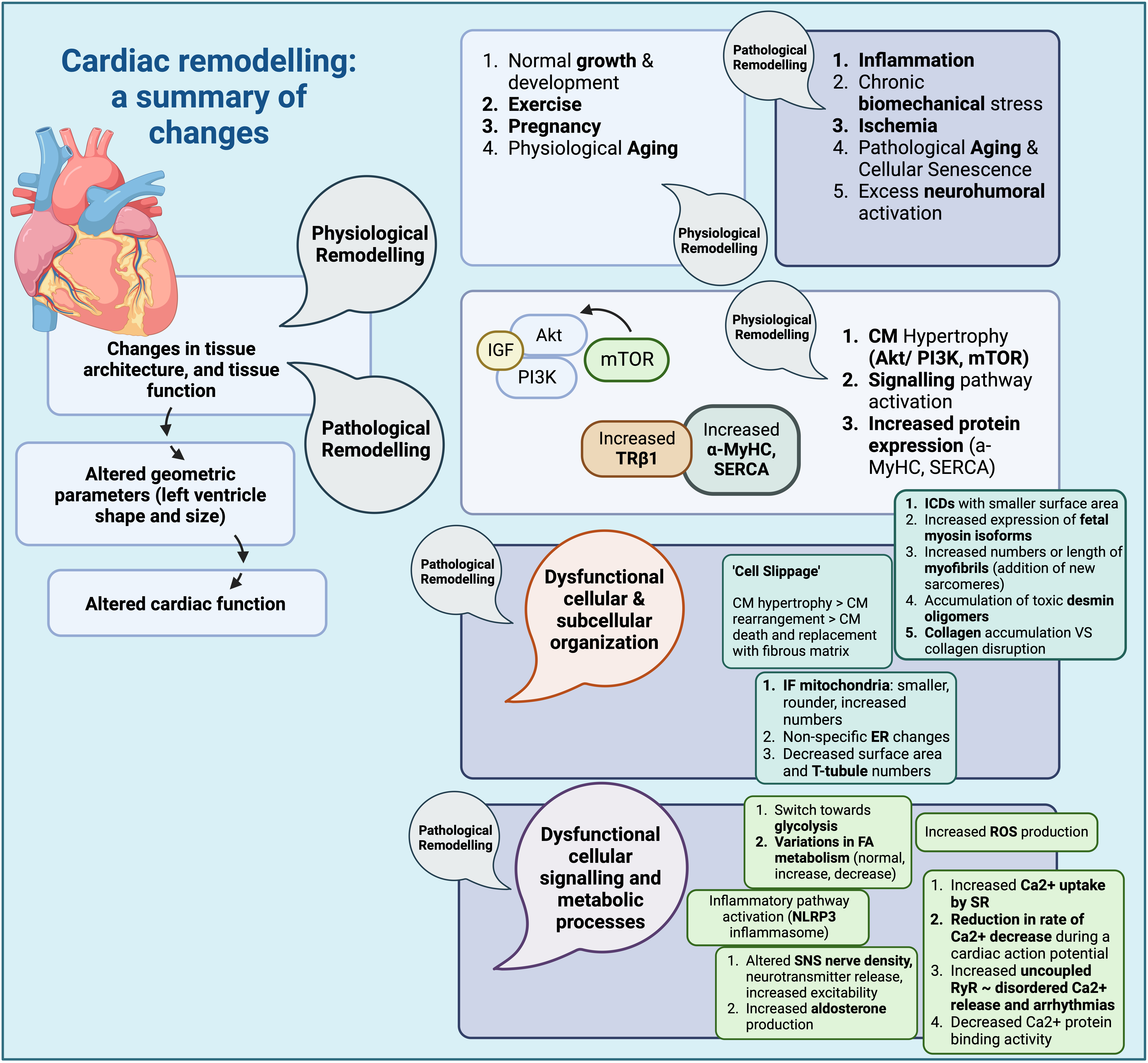
Fig. 1.

Cardiac remodelling: a summary of changes (Created with BioRender.com). Summary of structural changes and changes in signalling pathways, as well as other cellular processes during cardiac remodelling. CM, cardiomyocyte; IGF, insulin growth factor; PI3K, phosphoinositide 3 kinase; Akt, protein kinase B; mTOR, mammalian target of rapamycin; β-MyHC, β-Myosin heavy chain; α-MyHC, α-Myosin heavy chain; SERCA, sarcoendoplasmic reticulum calcium transport ATPase; TRβ1, thyroid hormone receptor β1; ICD, intercalated disks; ER, endoplasmic reticulum; FA, fatty acid; SNS, sympathetic nervous system; RyR, ryanodine receptor; SR, sarcoplasmic reticulum; ROS, reactive oxygen species; IF mitochondria, intrafibrillar mitochondria; NLRP3 inflammasome, nucleotide-binding domain, leucine-rich-containing family, pyrin domain-containing-3 inflammasome.

4. Cardiac Glucose Receptors and Remodelling
4.1 Endogenous Cardiac Glucose Receptors

Glucose transporters within cardiac tissue comprise both glucose transporters (GLUT) and sodium glucose cotransporters (SGLT) [119, 120, 121, 122, 123, 124, 125, 126]. GLUT1 and GLUT4 are the most commonly expressed forms, in fetal and adult hearts, respectively [127]. While both mediate glucose uptake in cardiomyocytes, GLUT1 is often described as insulin-independent, while GLUT4 is described as insulin-dependent [128, 129, 130, 131, 132]. Concerning SGLTs, studies that examine SGLT1 regulation within cardiac tissue have pointed to a possible role for leptin, which seems to increase cardiac SGLT1 expression. Furthermore, SGLT1 has been shown to colocalize with GLUT1 receptors and, at least in murine models, to increase with age. However, in terms of function, some studies do indicate a role for cardiac SGLT1 receptors in glucose uptake [132], while other differentiate between different cardiac SGLT1 variants, with some having no role in glucose uptake, due to the absence of glucose and Na+ binding domains [133]. On the other hand, so far no SGLT2 receptors have been identified in healthy heart tissue [134].

4.2 Cardiac Glucose Receptors in Cardiac Remodelling

As expected, the expression and activity of glucose receptors could be altered depending on the underlying states of the disease (summarized in Table 5 and Fig. 2), for example in the case of GLUT1, it appears to be overexpressed in chronic hypoxic states, possibly through HIF-1a binding. Furthermore, relevant studies have indicated a protective effect for increased GLUT1 expression in preventing cardiomyocyte death under low O2 tension [135]. Variable results have been observed with regard to GLUT1 receptors and cardiac remodelling, ranging from increased expression in post MI rat models [136], to decreased expression in the later stages of pathological remodelling in human samples of HF [137]. Furthermore, while GLUT1 expression appears to be increased in cases of hypertrophy, GLUT4 expression decreases [135], correlating with the general observation of down-regulation of an adult gene profile (including GLUT4) and subsequent predominance of a fetal gene expression profile, which includes GLUT1 [137]. GLUT4 is also downregulated in cases of diabetes, causing decreased cardiomyocyte glucose utilization, which can be reversed with insulin administration, or angiotensin-1 (AT-1) receptor blockade. The reversal observed with insulin could be attributed to any number of factors, including FA and ketone metabolism, which also affect GLUT4 expression [135]. Since local insulin resistance may be observed in hypertrophied cardiac tissue, it is thought that this might be attributed to glucose transporter derangements. Indeed, a decrease in the GLUT4/GLUT1 ratio is often identified [128].

Table 5.Glucose Receptors and their implications in cardiac remodelling.
Receptor Function
GLUT1 Overexpression in chronic hypoxic states (HIF-1a)-prevents cardiomyocyte death under chronic states of low oxygen tension
Increased expression, in post-MI remodelling (rat models)
Decreased expression, in late-stage heart failure (human samples)
Overexpressed in pathological cardiac hypertrophy
GLUT4 Downregulated in pathological cardiac hypertrophy
Downregulated in diabetes (impedes glucose utilization by cardiomyocytes) (reversal with insulin administration of AT1R blockade)
SGLT1 Downregulation in murine models of diabetes
Overexpression in murine models of ischemia by coronary artery ligation
Overexpression in human specimens of cardiomyopathy (diabetes, ischemia)
Upregulation during cardiac tissue injury
Role in glucose uptake contested with conflicting results from different studies

GLUT1, glucose tansporter 1; SGLT, sodium glucose cotransporter; HIF-1a, Hypoxia inducible factor-1a; MI, myocardial infarction; AT1R, angiotensin 1 receptor.
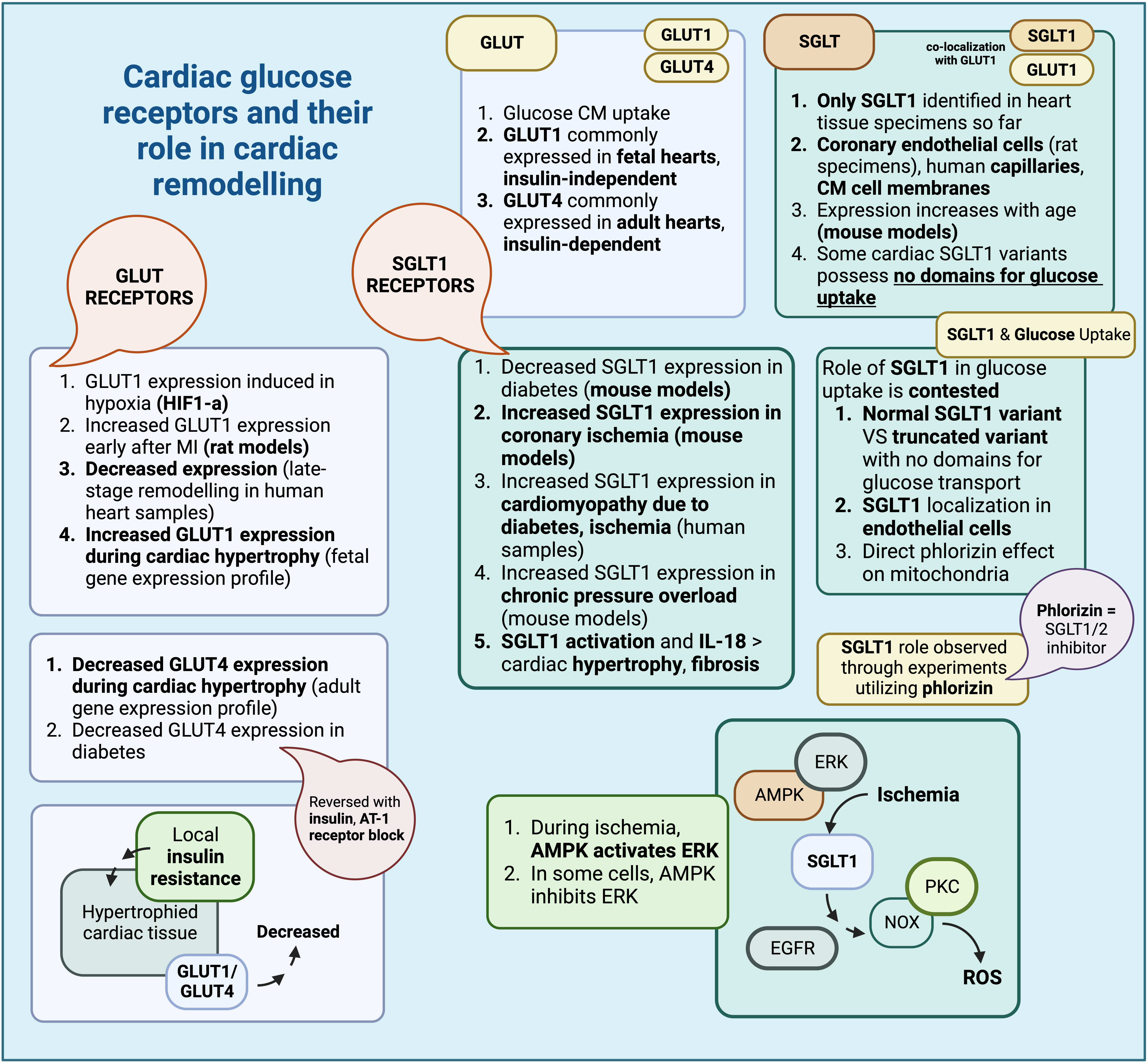
Fig. 2.

Cardiac glucose receptors and their role in cardiac remodelling (Created with BioRender.com). Summary of glucose receptor characteristics found within cardiac tissue and their respective roles in cardiac remodelling. GLUT, glucose transporter; SGLT, sodium glucose co-transporter; HIF-1a, hypoxia inducible factor-1 a; AT-1, angiotensin-1; AMPK, adenosine monophosphate-activated protein kinase; ERK, extracellular signal regulated kinase; CM, cardiomyocyte; IL-18, interleukin-18; PKC, protein kinase C; NOX, NADPH oxidase; EGFR, epidermal growth factor receptor; ROS, reactive oxygen species.

In the case of SGLT receptors, recent evidence appears to point to a role for them in the pathogenesis of cardiac remodelling; this is true for SGLT1 receptors, the only SGLT receptor type found within cardiac tissue so far. Oftentimes, SGLT1 derangements are associated with metabolic disturbances; indeed, SGLT1 expression appears to decrease in murine models of diabetes but increases in murine models of ischemia through coronary artery ligation. In human specimens, SGLT1 seems to increase in cases of cardiomyopathy due to diabetes and ischemia. Therefore, some authors theorize that upregulation of cardiac SGLT1 is an adaptation to tissue injury [132]; in fact, administration of phlorizin, an inhibitor of SGLT1, after restoration of coronary blood flow, did not lead to any restoration of ventricular function. ATP seems to be depleted earlier in these cases as well; thus, SGLT1, as observed in these experiments, might play a role in glucose uptake during injury [138]. However, conflicting reports do exist, which attribute no significant role for cardiac SGLT1s in glucose uptake whatsoever [133]; these different results could be perhaps attributed to various factors, including the presence of a normal and a truncated variant of SGLT1 [133], localization of SGLT1 receptors in endothelial cells [130], as well as the direct effect of phlorizin in mitochondria and mitochondrial ATPase [139].

Additional experiments evaluating the role of SGLT1 during ischemia and ischemia-reperfusion indicated that SGLT1 might be responsible for the observed myocardial injury, since SGLT1 knockdown was associated with milder issue injury. SGLT1 expression during ischemia is indirectly upregulated by adenosine monophosphate-activated protein kinase (AMPK) through extracellular signal regulated kinase (ERK) [140], although in other cells, AMPK appears to inhibit ERK instead [141]. SGLT1 causes PKC and NOX activation through EGFR, eventually contributing to oxidative stress and ROS species production [140]. There also appear to be conflicting results with respect to the association between cardiac injury and experimental SGLT1 inhibition, depending on whether the inhibition is carried out pharmacologically or through RNAi, with the resulting injury being exacerbated or ameliorated, respectively [140]. SGLT1 expression also appears to be upregulated in cases of chronic pressure overload, as shown in relevant murine models of transverse aortic constriction (TAC), and, along with IL-18, it might have a role in the development of subsequent cardiac hypertrophy and fibrosis [142]. In fact, transgenic expression of SGLT1 is associated with higher rates of tissue hypertrophy and cardiomyocyte size, along with signs of ventricular dysfunction ameliorated with transgenic SGLT1 suppression [143].

4.3 Effects of SGLT Inhibition on Cardiac Remodelling

With the emerging role of SGLT1 receptors during ischemic myocardial injury [140], their role in cardiomyocyte hypertrophy after injury [142], as well as results of preclinical studies of SGLT1 [138, 140], it seems that SGLT1 inhibition might be beneficial to the injury and possible remodelling observed after myocardial ischemia. Phlorizin, a non-selective SGLT inhibitor, has been used in preclinical studies for experimental inhibition of SGLT1 [138, 144], and although it can correct hyperglycemia, it has poor oral bioavailability. Its relevant analogues, including glucoside O and C, have been used to derive compounds such as empagliflozin and canagliflozin, approved drugs for the treatment of diabetes mellitus [145, 146].

SGLT1 inhibition can be achieved through selective SGLT1 inhibitors, or through substances that affect both SGLT1/SGLT2 receptors [145]. Some examples of SGLT1 inhibitors include KGA-2727, with promising results in mitigating ventricular remodelling and preventing increases in cardiomyocyte size, ANP, BNP, and IL-18, in murine models of myocardial infarction [147]. Mizagliflozin, which has completed phase II of clinical trials [145], appears to be useful in mitigating cardiomyocyte apoptosis, as well as the resulting cardiomyopathy observed in diabetes, through inhibition of the Jun N-terminal Kinase (JNK) and p38 pathways [148]. Sotagliflozin, a dual SGLT1/SGLT2 inhibitor, seems to alleviate some of the myocardial injury observed in pressure overload models, although this has been observed only in subjects with a normal diet [149]. Sotagliflozin is currently being used in Europe, approved for patients with type 1 diabetes and a body mass index (BMI) greater than 27 kg/m2, as an adjunct to insulin therapy. SGLT1 inhibition might be particularly useful in mitigating cardiac remodelling after injury in the setting of diabetes mellitus (DM), as well as in cases of cardiac injury independent of the presence of diabetes [145].

Relevant cardioprotective mechanisms induced by SGLT1 inhibition, summarised in Fig. 3, could include down-regulation of the JNK and p38 MAPK pathways, as well as prevention of cardiac fibrosis, which is also involved in the pathogenesis of diabetic cardiomyopathy (DCM), a diabetes-associated cardiomyopathy associated with increased cardiomyocyte size, cardiomyocyte apoptosis, and myocardial fibrosis, leading to ventricular dysfunction [148, 150, 151]. There are studies that point to a possible detrimental effect of diabetes on ventricular function and its contribution to pathological remodelling [152, 153], something that could be ameliorated with SGLT1 inhibition [145]. Another mechanism of action might include inhibition of the EGFR-ERK-PKC-Nox pathway (KGA-2727), particularly in the injured myocardium after myocardial infarction, affecting the subsequent remodelling [140, 147]. Finally, inhibition of SGLT1 might also prevent angiotensin II-mediated activation of SGLT1 receptors in vascular endothelial cells, in turn preventing ROS accumulation through NADPH activation, decreasing the incidence of endothelial senescence [154].
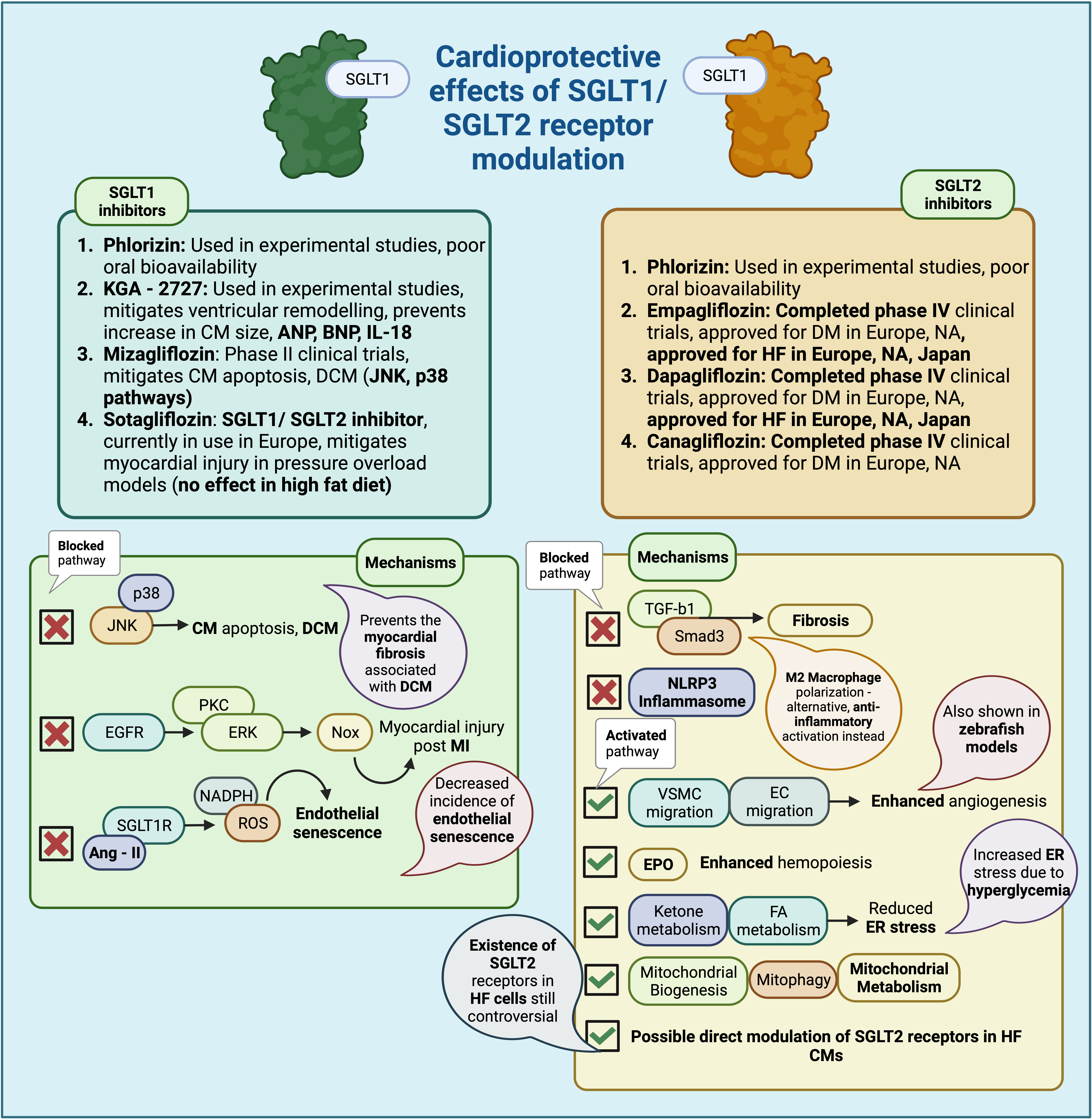
Fig. 3.

Cardioprotective effects of SGLT1/SGLT2 receptor modulation (Created with BioRender.com). Summary of the cardioprotective effects of SGLT1 and SGLT2 receptor inhibition, via inhibition or facilitation of specific biological pathways. SGLT, sodium glucose co-transporter; CM, cardiomyocyte; ANP, atrial natriuretic peptide; BNP, brain natriuteric peptide; IL-18, interleukin 18; DCM, diabetic cardiomyopathy; JNK, Jun N-terminal kinase; HF, heart failure; EGFR, epidermal growth factor receptor; PKC, protein kinase C; ERK, extracellular signal related kinase; NOX, NADPH oxidase; MI, myocardial infarction; Ang-II, angiotensin II; NADPH, Nicotinamide Adenine Dinucleotide Phosphate Hydrogen; ROS, reactive oxygen species; HF, heart failure; FA, fatty acid; ER, endoplasmic reticulum; TGF-β1, transforming growth factor-β1; Smad, Suppressor of mothers against decapentaplegic; NLRP3 inflammasome, nucleotide-binding domain, leucine- rich-containing family, pyrin domain-containing-3 inflammasome; VSMC, vascular smooth muscle cells; EC, endothelial cells; EPO, erythropoietin.

Although no SGLT2 receptors have been consistently found within cardiac tissue in relevant experiments, SGLT2 inhibition might also have cardioprotective effects through different mechanisms. The associated lack of hypoglycemia with SGLT2is, which could potentially exacerbate cardiac injury through increased sympathetic activation, also presents a favorable profile for use in cases of cardiovascular remodelling [37, 155]. Examples of SGLT2is include canagliflozin, empagliflozin, and dapagliflozin, all approved for the treatment of diabetes in both Europe and North America (NA); moreover, dapagliflozin and empagliflozin have also been approved for the treatment of HF and kidney disease, in Europe, NA and Japan [145].

Cardioprotective mechanisms of action for SGLT2is include weight loss, possibly related to inhibition of sodium and glucose reabsorption, reduction of plasma insulin and glucose levels, reduction in blood pressure, due to reduced sodium reabsorption and, over time, reduced RAAS activation, as well as increased erythropoietin (EPO) production which improves erythropoiesis, thus aiding in better tissue oxygenation [156]. Additional effects, more closely related to cardiovascular remodelling, include positive effects on inflammation, fibrosis-related signaling pathways, angiogenesis, cellular organelle function, and direct cardiomyocyte effects [157]. Mechanisms more closely related to the potential of SGLT2is to ameliorate or reverse the effects of cardiovascular remodelling include effects on inflammation, signalling pathways involved in cardiac fibrosis and inflammation, subcellular organelle function and ketone metabolism, angiogenesis, and perhaps possible direct effects on cardiomyocytes [156].

Anti-inflammatory effects of SGLT2is include inhibition of the NLRP3 inflammasome, through reduction of intracellular Ca2+ levels, and inhibition of NF-κβ-dependent release of pro-inflammatory cytokines [158, 159]. SGLT2is may also affect signaling pathways involved in cardiac fibrosis [160], a characteristic of many different pathologies, including ischemic cardiomyopathy, hypertrophic cardiomyopathy (HOCM), and hypertension. ‘Fibrotic remodelling’ is driven by macrophage and lymphocyte-induced myofibroblast activation. SGLT2is inhibit the TGF-b1/Smad3 pathway [161] and affect the polarization of M2 macrophages, halting progression of cardiac fibrosis [162]. One recently identified cardioprotective property of SGLTis includes their ability to modulate and possibly reduce the pro-inflammatory effects of epicardial fat in failing hearts; more specifically, it seems SGLT2is may reduce the amount of epicardial fat, thereby decreasing the amount of pro-inflammatory cytokines secreted, as has been observed in relevant experiments evaluating the effects of dapagliflozin [92]. Furthermore, in these experiments, this reduction in epicardial fat has exhibited a positive correlation with tumor necrosis factor-α (TNF-α) levels, overall ameliorating adverse cardiovascular events [163].

SGLT2is also enhance mitochondrial metabolism [160], facilitate the generation of new mitochondria (mitochondrial biogenesis) [164], and removal of senescent mitochondria (mitophagy) [165]. endoplasmic reticulum (ER) stress, often induced by high glucose concentrations, can also be alleviated by SGLT2is, through induction of enzymes involved in ketone and FA metabolism [166]. SGLT2is may also act upon and modulate ketone metabolism, an important aspect of their cardioprotective properties; it seems that SGLT2is can mildly increase levels of ketone compounds, levels of genes involved in ketolysis, as well as associated uptake of ketones, fatty-acids (FA) and branched chain amino acids (BCAA) [167]. This increase in ketone and FA uptake has been observed to occur in relevant swine intravascular balloon occlusion models, ameliorating the reduction in FA uptake occurring after the initial insult [58, 168].

SGLT2is have been further shown to act on pathways involved in angiogenesis, which, when disturbed, could contribute to adverse cardiac remodelling, especially after MI [169]. Pharmaceutical agents, such as sotagliflozin, have been used to enhance migration of vascular endothelial cells and smooth muscle cells [170], while others, such as empagliflozin and dapagliflozin, have been shown to enhance angiogenesis in an embryonic zebrafish model [171]. SGLT2is also affect hemopoiesis, by up-regulating EPO secretion by the kidney [5, 172].

Finally, SGLT2is may exert direct effects on cardiomyocytes, perhaps through the presence of SGLT2 receptors on cardiomyocytes themselves; more specifically, one relevant study detected the presence of SGLT2 receptors in end-stage HF cardiomyocytes, as well as transplanted cardiomyocytes within diabetic hosts, with overexpression of the SGLT2 receptor associated with cardiomyocyte metabolic derangements [173]. In addition, SGLT2is have recently been shown to affect ion transporters within cardiomyocytes, including Ca2+ and Na+ ion transporter proteins; more specifically, infusion of dapagliflozin has been shown to reduce systolic Ca2+ in murine ventricular cardiomyocytes [174], an effect also observed after infusion of empagliflozin, which has also been shown to influence intracellular Na+ concentration in studies involving rabbit cardiomyocytes [175]. These changes in turn have been shown to affect Ca2+ transients, as well as Na+ currents (INa), namely reducing late INa, with no effect on peak INa. This effect has been attributed to downregulation of various cardiomyocyte ion transporters, including the Na+/H+ exchanger (NHE), directly, or indirectly through AMPK phosphorylation, which may then affect the function of the Na+/Ca2+exchanger (NCX) [134, 176]. There also seem to be some direct effects on cardiomyocyte metabolism, namely upregulation of GLUT1, observed after empagliflozin infusion in human and murine cardiomyocytes [177], in turn leading to increased glucose uptake in these cells; however, SGLT2is did not seem to affect the activity of GLUT4 receptors [177].

5. Stem and Progenitor cells in Cardiac Remodelling

Stem cells are undifferentiated cells, with the ability for self-renewal and generation of differentiated progeny. There are many stem cell types, with variable potential for progeny generation, including mesenchymal stem cells (MSC), i.e., multipotent stem cells capable of generating tissues such as bone, muscle, and cartilage, and cardiac stem cells (CSC) [178, 179]. On the other hand, progenitor cells, are more lineage-specific and usually generate one or only a few lineage-specific cell types [180].

Local stem and progenitor cells have been associated with a number of pathological remodelling processes (Table 6, Ref. [181]), including the progressive change in cardiac tissue composition that is related to aging (mainly attributed to senescence of local progenitors [13]), and the contribution of local cardiac MSCs [182] to the pathological fibrosis observed during cardiac remodelling [12]. Some of these progenitors, including endothelial progenitors (EPC), are particularly important during cardiac tissue injury, as they contribute to local blood vessel generation, either de novo (vasculogenesis), or through existing vessels (angiogenesis) [183, 184]. Furthermore, with their secretomic profile, they contribute to multiple repair and regeneration processes [183], allow homing of other stem/progenitor cells, including cardiac progenitors [185], prevent MSC-derived deleterious effects such as fibrosis [186], as well as contribute to mitigation of inflammation and oxidative stress [187].

Table 6.Stem/Progenitor cell types implicated in the pathogenesis or progression of cardiac remodelling.
Cell type Alteration Effects/Additional information
MSC Pro-inflammatory, pro-fibrotic phenotype change PDGFRβ upregulation, interaction with local M1 macrophages and differentiation of cardiac MSCs into myofibroblasts.
HSPC Reduction in circulating HSPC populations ‘Three-Department’ model; defective mobilization from the bone marrow (BM)/damage to the niche itself, decreased survival of circulating HSPC, impaired trafficking of pro-vascular progenitors to target sites.
Increased HSPC retention in the bone marrow, increased HSPC differentiation towards myeloid lineages.
EPC Reduction in EPC populations, EPC senescence Continuous cardiovascular injury and EPC mobilization causing EPC depletion, increased oxidative stress, EPC senescence (increased SA β-gal).
Reduction in EPC levels (CD34+ KDR+EPC) increases risk of adverse cardiovascular events and restenosis after cardiovascular intervention.
CSC CSC senescence CSC senescence (p16, SA β-gal and γH2AX) impairs the potential for repair and regeneration after myocardial injury.
Elimination of senescent cells in mouse models improved cardiac function and remodelling after myocardial injury (Salerno et al., 2022 [181]).

PDGFRβ, Platelet derived growth factor receptor β; MSCs, mesenchymal stem cells; BM, bone marrow; HSPC, hematopoietic stem and progenitor cells; EPC, endothelial progenitor cells; SA β-gal, senescence associated β galactosidase; KDR, kinase insert domain receptor; γH2AX, γ phosphorylated form of the histone H2AX; CSC, cardiac stem cell.

Although MSCs have been known to contribute to tissue repair and mitigation of inflammatory responses [188, 189], some endogenous MSCs have been shown to exhibit a pro-inflammatory and pro-fibrotic phenotype, possibly contributing to the fibrotic response during pathological cardiac remodelling; this seems to occur either through upregulation of the of the PDGFRβ isoform, as opposed to the PDGFRα isoform normally expressed in cardiac MSCs, as well as through interaction with local M1 macrophages and eventual differentiation of cardiac MSCs into myofibroblasts [12].

Progenitor cells can be affected during pathological remodelling processes, usually due to chronic dysglycemia and diabetes; haematopoietic stem and progenitor cells (HSPC), for example, which can also contribute to the provascular cell pool [190], and more specifically, CD34+ HSPCs, have been found in reduced numbers in the peripheral circulation, in both T1DM and T2DM [191, 192]. The ‘three-department’ model has been developed to explain this defect in peripherally circulating HSPCs; defective mobilization from the bone marrow (BM) (stem cell niche) as well as damage to the niche itself, decreased survival of circulating HSPC in the periphery (something not yet proven in human specimens), and impaired trafficking of pro-vascular progenitors to target sites, are all believed to contribute to the apparent decrease in pro-vascular HSPC populations in diabetes. Furthermore, a shift in differentiation balance toward myeloid progenitors also appears to occur, possibly due to increased production of S100A8/9 proteins (alarmins) by pro- inflammatory cells. In the bone marrow, this leads to the release of oncostatin M (OSM) and CXCL12 production, which increases retention of HSPC, and promotes their differentiation toward a myeloid lineage, while in the periphery, this mainly promotes inflammation in target organs [193].

Endothelial progenitor cells (EPCs), derived from different sources, including the vascular wall [194], have a positive effect on post-injury cardiac remodelling, by aiding in myocardial repair [183]; decreased numbers of EPCs due to a number of factors, including continuous cardiovascular injury and mobilisation of EPCs with subsequent depletion in numbers, increased oxidative stress, and even EPC senescence (characterized by increased SA β-gal), have all been speculated to have an effect [195, 196]. More specifically, reduction in EPC levels (CD34 + KDR + EPC) increases the risk of adverse cardiovascular events, as shown by a meta-analysis of relevant studies examining the effect of circulating progenitor cells (CD34 + CD133 + CPC) and endothelial progenitors (EPC) on cardiovascular risk and restenosis after cardiovascular intervention [197].

Although the contribution of EPCs to the mitigation of injury has been shown, defining an EPC population has been a point of debate between different studies, since markers such as VEGFR-2 and CD34 can also be expressed by myeloid cell lineages; relevant studies differentiate between true EPCs, positive for CD34/VEGFR-2, and negative for CD45/CD14, that can generate both endothelial cells (EC) in vitro and integrate in growing vessels in vivo, and circulating angiogenic cells (CAC), expressing both endothelial (CD34/VEGFR-2) and hemopoietic lineage markers (CD45/CD14), and while capable of in vitro angiogenesis, they cannot integrate within growing vessels in vivo [198, 199]. Aldehyde dehydrogenase (ALDH) has been recently used to differentiate vascular progenitors from other hematopoietic stem and progenitor cell populations, with pro-vascular cells exhibiting high ALDH activity; this, along with other endothelial-specific markers, has been used to better identify vascular progenitor populations [198].

Finally, cardiac stem cell (CSC) senescence has also been shown to contribute to the development of varying disease phenotypes. A wide range of cardiac stem cell (CSC) populations have been identified, some existing in adult hearts and others in embryonic hearts [200]; CSC senescence, denoted by an increase in markers such as p16, SA β-gal and γH2AX, might contribute to tissue dysfunction and inflammation, as well as affect the potential for repair and regeneration after myocardial injury [201]. In fact, it appears that elimination of senescent cells in mouse models improved cardiac function and remodelling after myocardial injury [181].

6. SGLT Receptor Modulation and Effect on Stem and Progenitor Cells, in the Scope of Cardiovascular Remodelling

Recent research on the mechanisms with which stem and progenitor cells might contribute to the progression of pathological remodelling gave way to studies examining the effect of manipulation of the SGLT receptor on stem and progenitor cells in this context [12, 196, 202]. The need to answer this particular research question might be further explained by the effect of diabetes and dysglycemia on progenitors, as well as the associated cardiovascular risk [193], and the pathological remodelling that may occur in cases of diabetes [150]. Although it would be logical to assume that some effect on progenitor populations would be expected, given the beneficial effect of SGLTis on remodelling, so far studies have produced conflicting results; the primary setting for this proposed effect has mostly been investigated in the setting of diabetes mellitus, as SGLTis have been used primarily for their cardioprotective and antidiabetic effect.

SGLT inhibition and its effect on stem and progenitor cells in human patients was investigated for the first time in 2018, with a randomized clinical trial (RCT), by Bonora et al. [203]; dapagliflozin was evaluated in a total of 33 patients with T2DM for 12 weeks, although an extension period was added, up to 74 weeks [203]. Additionally, another 15 patients were added, which in this case received empagliflozin. The type of cells evaluated were circulating progenitors expressing CD34 antigen (CD3+-CPC) and endothelial progenitors, identified through their additional expression of KDR, a marker found primarily in cells of endothelial lineage (CD34+ KDR+-EPC). There was no direct significant effect on these cell populations in response to SGLT2i treatment, although there was some increase in progenitors after an extended period, which was attributed to better glycemic control by the researchers, correlated with HbA1c levels, rather than the SGLT2i treatment itself [203].

Another similar substudy followed in 2019, part of the EMPA-HEART Cardiolink-6 trial [204], by Hess et al. [205], in which researchers evaluated the role of empagliflozin in circulating vascular progenitors. Vascular progenitors were defined as a heterogeneous cellular group that includes bone marrow-derived EPCs (BM) (BM-EPC), angiogenic HSPCs, as well as monocyte-derived angiogenic macrophages [205]. Their levels were evaluated in patients, both at baseline and within 6 months after empagliflozin treatment. An additional marker was used to identify vascular progenitors, aldehyde dehydrogenase (ALDH), a detoxification enzyme found primarily within immature progenitors [206]. ALDH has been shown to better aid in identifying such cells, due to less variability in the expression of the ADLH gene, in contrast to other markers, such as CD133 and CD34, whose expression could vary depending on the stage of development [206]. Another cellular aspect that was quantified to better aid in cellular characterization was intracellular granular complexity, also termed side scatter property (SSC); thus, in this study, EPCs were defined, not only based on CD133 and CD34 expression, but also on SSC levels and ALDH expression [205]. High and middle complexity was used to denote pro-inflammatory granulocytes and monocytes, while low SSC was used to identify a heterogenous group of vascular progenitors with high expression of ALDH [190, 205]. Hence, vascular progenitors in this study were defined as a heterogeneous cellular group of ALDHhiSSClow CD133+ and ALDHhiSSClow CD34+ CD133; indeed, after 6 months of empagliflozin, the levels of these progenitor cell types increased, an event associated with stable or increased expression of antioxidant genes, including Nox1 and catalase, along with a decrease in pro-inflammatory granulocytes [205].

Some of the results of this study [205] seem to contradict some of the results of a previous study by Bonora et al. [203], while in the long term, even in the latter, cells staining positive for CD34 and VEGFRR2 also appeared to increase [203], even though a different SGLT2i was used each time. It seems that more detailed characterisation of vascular progenitor cells generated a different result in the study by Hess et al. [205]; the criteria used to characterise potential vascular progenitors generated further disagreement amongst research groups, since some authors noted that the populations identified through ALDH expression were heterogeneous, containing both vascular and hemopoietic progenitors [207]. However, the angiogenic potential of ALDHhiSSClow cells [208], as well as their ability to induce neoangiogenesis in immunodeficient mice [208] has been proven in relevant studies, despite the heterogeneity of the general population [209]. Therefore, it seems that ALDH activity is a useful indicator, in combination with other cellular markers, for identifying vascular progenitor populations [206], heterogeneous in origin though they may be [205, 207, 209].

Additional studies followed, examining the role of SGLT2 inhibitors, such as dapagliflozin, in the mobilization of hematopoietic cells from the bone marrow (BM); this was examined in mice models of T1DM and T2DM, as well as mouse models of carotid endothelial injury. While an increased ratio of granulocytes/lymphocytes was observed in subjects with diabetes, possibly due to the enhanced myelopoiesis associated with hyperglycemia, dapagliflozin did not appear to have any effect. An interesting finding in this study is the observation of CD49d+ granulocyte mobilization after dapagliflozin administration, which helped vascular repair within 3 days, in a murine model [210]. The role of SGLT2 inhibitors, and more specifically empagliflozin, on vascular progenitor populations was again, more recently examined in a substudy by Bakbak et al., 2023 [211], included within the EMPA-HEART 2 Cardiolink-7 trial [212]; in this case however, this effect was studied irrespective of T1DM or T2DM status, as only patients with cardiovascular risk factors were included. After 6 months of treatment, angiogenic progenitor cells as well as regenerative monocyte progenitor cells were found to be increased, while pro-inflammatory progenitors were found to be decreased. Thus, it seems that SGLT2 inhibitors can indeed exert a cardioprotective effect, through increasing angiogenic progenitors, even in the absence of diabetic status [211].

Finally, the administration of dapagliflozin also seemed to improve hematopoietic cell mobilization, as well as aid in the restoration of the vascular endothelium in areas of tissue damage, in relevant models of carotid injury; a mechanism for this was shown to be induced through the recruitment of BM-derived cells, through experimental inhibition of CXCR4 with the compound AMD3100, co-administered with dapagliflozin. Since SGLT2 expression has been detected at very low levels in the bone marrow, the authors concluded that a direct effect on the bone marrow by dapagliflozin seemed unlikely [210]. This mechanism could counteract the observed depletion of circulating angiogenic HSPCs in the diabetic state; furthermore, modulation of pro-inflammatory cell production from existing HSPC populations appears to mitigate vascular dysfunction, as well as endothelial damage [213].

In essence, the association between SGLT modulation and progenitor cells is relatively newfound, and therefore contradictory studies still exist (summarized in Table 7, Ref. [203, 204, 205, 208, 210, 211, 213]); if solidified by more studies, it could serve as additional clarification on the positive effects of SGLT2 inhibitors on the cardiovascular system [14].

Table 7.Recent studies on the association between SGLT receptor modulation and stem/progenitor cell activity, in the scope of cardiac remodelling and diabetes.
Publication SGLTi Cell Observations
Bonora et al., 2018 [203] Dapagliflozin, Empagliflozin (on additional 15 patients) CD34+-CPC No direct significant effect in response to SGLT2i treatment.
CD34+ KDR+-EPC Modest increase in progenitor numbers after an extended period, attributed to better glycemic control.
Verma et al., 2019 [204] Empagliflozin ALDHhiSSClow CD133+, ALDHhiSSClow CD34+ CD133+ cells Substudy of the EMPA-HEART Cardiolink-6 trial (Verma et al., 2019 [204]).
Hess et al., 2019 [205] Novel method to identify vascular progenitors, ALDH and SSC (intracellular complexity)-vascular progenitors from a variety of populations (BM-EPCs, angiogenic HSPCs, angiogenic monocytic-derived macrophages).
Bakbak et al., 2023 [211] Empagliflozin ALDHhiSSClowCD133+CD34+ (angiogenic progenitors), ALDHhiSSCmid CD163+ (regenerative monocyte progenitors), ALDHhiSSCmid CD86+CD163- (pro-inflammatory progenitors) Substudy of the EMPA-HEART Cardiolink-7 trial (Connelly et al., 2023).
Evaluation of 3 progenitor cell populations after 6 months of empagliflozin treatment vs placebo; angiogenic progenitors and regenerative monocyte progenitors were increased, while pro-inflammatory progenitors were decreased.
No T1DM, T2DM associations in this study-only patients with cardiovascular risk factors were included.
Cooper et al., 2018 [208] N/A ALDHhiSSClow ALDHhiSSClow cells have angiogenic potential, and can induce neo-angiogenesis in immunodeficient mice.
Albiero et al., 2021 [210] Dapagliflozin CD49d+ granulocytes T1DM, T2DM murine models, murine models of endothelial injury.
Dapagliflozin had no effect on the increased granulocyte/lymphocyte ratio observed in diabetic states.
Mobilization of CD49d+ granulocytes, after dapagliflozin administration, facilitating vascular repair within 3 days.
Hess et al., 2021 [213] Dapagliflozin HSPCs Improved HSPC mobilization, recruitment of BM-derived cells, modulation of pro-inflammatory cell production from existing HSPC populations tackles vascular dysfunction and endothelial damage in relevant models.

CPC, cardiac progenitor cells; KDR, kinase insert domain receptor; EPC, endothelial progenitor cells; SGLT2i, sodium glucose co-transporter 2 inhibitor; ALDH, aldehyde dehydrogenase; SSC, side scatter property; BM-EPC, Bone marrow endothelial progenitor cells; HSPC, hematopoietic stem and progenitor cells; T1DM, type 1 diabetes mellitus; T2DM, type 2 diabetes mellitus; BM, bone marrow.

7. Conclusions

Cardiac remodelling is a complex process that affects multiple subcellular structures and processes, from mitochondria and cell membranes, to gene expression and protein composition. Eventually, changes in tissue micro- and macroarchitecture can occur, impacting ventricular function and cardiac output. The mechanisms involved in cardiac remodelling have been extensively researched with new evidence constantly being discovered. Amongst the various pathophysiological mechanisms that can contribute to cardiac remodelling, various local stem and progenitors cell types seem to play a role. Cardiac MSCs contribute to the fibrotic reaction, while decreased populations of vascular progenitors and HSPCs, as well as senescence of local cardiac stem cells can alter the healing of local tissue injury and further contribute to the exacerbation of post-injury remodelling. Stem and progenitor cell populations have also been found to be affected during dysglycemic states and diabetes.

Glucose receptors, such as GLUT and SGLT, have been found at various locations, and though GLUT1 and GLUT4 exist within cardiac tissue, evidence pointing to the existence of cardiac SGLT1 and possibly SGLT2, is not yet well solidified. Notably, it has been proposed that the different locations of SGLT2 and SGLT1 could be related to different efficacy and safety characteristics between dual (i.e., sotagliflozin) and selective for SGLT2 inhibitors (i.e., empagliflozin) [214, 215, 216].

The association between SGLT inhibition and stem/progenitor cells has only recently emerged, with existing studies still producing conflicting results. Although vascular progenitors seem to be affected by SGLT2 modulation in some studies, others do not establish a significant connection. One of the reasons for these contradictory results seems to be the criteria used to identify endothelial progenitor populations, since different studies use different criteria. Therefore, more studies must be conducted so that universal criteria for defining each stem/progenitor cell population are defined and accepted, to better help interpret test results from different studies. Existing studies have explored the association between endothelial progenitors and SGLT2 modulation in the context of diabetes mellitus, as well as in patients with cardiovascular risk factors, without diabetic status; it might be interesting to further examine the effect of SGLT2 modulation on vascular progenitors in other disease entities that may lead to pathological cardiac remodelling as well, to further establish this connection between SGLT2 modulation and progenitor cells. Finally, EPCs are not the only types of progenitor cell involved in pathological remodelling; additional studies might be warranted that evaluate the effects of SGLT2is, if any, on dysfunctional cardiac MSCs, and whether the resulting fibrosis could be mitigated.

The cardioprotective effects of SGLT2 inhibitors are evident, with positive effects observed in people with or without diabetes; in addition, derangements in stem and progenitor cell numbers or function have been shown to contribute to the progression of pathological cardiac remodelling. Although the association between SGLT2is and progenitors is relatively new, if solidified, it could facilitate better management of pathological aspects of cardiac remodelling, further mitigating cardiac dysfunction.

Abbreviations

SLC5A1, solute carrier family 5 member 1; SLC5A2, solute carrier family 5 member 2; PCT, proximal convoluted tubule; SGLT2, sodium glucose co-transporter 2; SGLT1, sodium glucose co-transporter 1; MSC, mesenchymal stem cells; CSC, cardiac stem cells; EPCs, endothelial progenitor cells; HSPCs, hemopoietic stem progenitor cells; HF, heart failure; HFA, heart failure association; T1DM, Type 1 Diabetes Mellitus; T2DM, Type 2 Diabetes Mellitus; DM, diabetes mellitus; DCM, diabetic cardiomyopathy; HOCM, hypertrophic cardiomyopathy; LV, left ventricle; CO, cardiac output; BCAA, Branched chain amino acid; DHP, Dihydropyridine receptor; mTOR, mammalian target of rapamycin; PI3K, phosphoinositide 3 kinase; Rac1, Ras-related C3 botulinum toxin subtrate 1; Akt, protein kinase B; CM, cardiomyocyte; IGF, insulin-like growth factor; SIRT6, sirtuin 6; SR, sarcoendoplasmic reticulum; β-MyHC, β-Myosin heavy chain; α-MyHC, α-Myosin heavy chain; SERCA, sarcoendoplasmic reticulum calcium transport ATPase; TH, thyroid hormone; TR, thyroid hormone receptor; TRα, thyroid hormone receptor a; TRβ, thyroid hormone receptor b; ROS, reactive oxygen species; ICD, intercalated disk; ECM, extracellular matrix; SMAD3, Suppressor of Mothers against Decapentaplegic 3; TGF-β1, transforming growth factor-β1; Stat3, signal transducer and activator of transcription 3; EphrinB2, Eph family receptor interacting protein B2; ATP, adenosine triphosphate; HIF-1a, hypoxia inducible factor-1a; FA, Fatty acid; EF, ejection fraction; NADPH, Nicotinamide Adenine Dinucleotide Phosphate; NOX, NADPH Oxidase; NOS, nitric oxide synthase; GHSPx, gluthathione peroxidase; SOD, superoxide dismutase; MI, myocardial infarction; ETC, electron transport chain; NF-κβ, nuclear factor kappa-light-chain-enhancer of activated B cells; TNF-α, tumor necrosis factor-alpha; Bax, Bcl2-associated X protein; IHD, ischemic heart disease; NLRP3, NLR family pyrin domain containing 3; IL-Ib, interleukin Ib; IL-6, interleukin- 6; IL-18, interleukin-18; LTCC, L-type Calcium Channel; RyR2, Ryanodine receptor 2; MCU, mitochondrial calcium uniporter; NCX, Sodium Calcium exchanger (Na+/Ca2+ exchanger); PMCA, plasma membrane Ca2+ ATPase; TATS, cardiac transverse-axial tubular system; NKA, Na+/K+ ATPase; NHE, Na+/H+ Exchanger; RAAS, renin angiotensin aldosterone system; NA, noradrenaline; AT1R, angiotensin-1 receptor; miRNA, micro ribonucleic acid; LVWT, left ventricular diameter systole; LVDD, left ventricular diastole diastole; EF, ejection fraction; GLUT, glucose transporter; AT-1, angiotensin-1; ATPase, adenosine-5-triphosphatase; AMPK, adenosine monophosphate-activated protein kinase; ERK, extracellular signal-regulated kinase; PKC, protein kinase C; EGFR, epidermal growth factor receptor; RNAi, ribonucleic acid interference; TAC, transverse aortic constriction; ANP, atrial natriuretic peptide; BNP, brain natriuretic peptide; JNK, Jun N-terminal Kinase; BMI, body mass index; DM, diabetes mellitus; MAPK, mitogen activated protein kinases; PKA, Protein kinase A; PKC, Protein kinase C; RAAS, renin angiotensin aldosterone system; ER, endoplasmic reticulum; MI, myocardial infarction; EPO, erythropoietin; PDGFR, platelet growth factor receptor; BM, bone marrow; OSM, oncostatin M; CPCs, cardiac progenitor cells; VEGFR-2, vascular endothelial growth factor receptor 2; SA β-gal, senescence associated beta galactosidase; CXCL12, C-X-C motif chemokine ligand 12; KDR, kinase insert domain receptor; VSMC, Vascular smooth muscle cell; EC, endothelial cells; CAC, circulating angiogenic cells; ALDH, aldehyde dehydrogenase; CSC, cardiac stem cells; γH2AX, γ phosphorylated form of the histone H2AX; BM, bone marrow; BM-EPC, bone marrow endothelial progenitor cells; SSC, side scatter property; NFAT, Nuclear factor of activated T-cells; miR, micro ribonucleic acid; FoxO3, Forkhead transcription factor O subfamily member 3a; RA, Rheumatoid Arthritis; SLE, Systemic lupus erythematosus; PM, polymyositis; DM, dermatomyositis; pSS, primary Sjögren’s Syndrome; SSc, Systemic sclerosis; p53, Tumor protein p53; MMP14, Matrix metalloproteinase 14; AMPK, 5’ AMP (Adenosine monophosphate)-activated protein kinase; GSSG, Oxidized glutathione; GSH, Reduced glutathione.

Author Contributions

TMS and KCC made substantial contributions to conception and design of the manuscript, carried out the literature search, drafted and wrote the manuscript. TK assisted in literature research, DM, FM, CDM and DK reviewed the literature and edited the manuscript. All authors contributed to editorial changes in the manuscript. All authors read and approved the final manuscript. All authors have participated sufficiently in the work and agreed to be accountable for all aspects of the work.

Ethics Approval and Consent to Participate

Not applicable.

Acknowledgment

Figures were created with BioRender.com.

Funding

This research received no external funding.

Conflict of Interest

TK has received honoraria for lectures from AstraZeneca, Boehringer Ingelheim, Pharmaserve Lilly, and Novo Nordisk, for advisory boards from Novo Nordisk and Boehringer Ingelheim, and has participated in sponsored studies by Eli-Lilly and Novo Nordisk. C. David Mazer reports advisory board honoraria and/or consulting fees from Amgen, AstraZeneca, BioAge, Boehringer Ingelheim, Cardior, PhaseBio and Sandoz; and reports stipends for serving on the Data and Safety Monitoring Board of Beth Israel Deaconess Medical Center, Cerus, and Takeda; Dr. Mazer is supported by a merit award from the University of Toronto Department of Anesthesiology, and holds the Cara Phelan Chair in Critical Care at St. Michael’s Hospital. TMS, KCC, FM, DM, DK have no conflicts of interest to report.

References
[1]
Azevedo PS, Polegato BF, Minicucci MF, Paiva SAR, Zornoff LAM. Cardiac Remodeling: Concepts, Clinical Impact, Pathophysiological Mechanisms and Pharmacologic Treatment. Arquivos Brasileiros De Cardiologia. 2016; 106: 62–69.
[2]
Song P, Zhao Q, Zou MH. Targeting senescent cells to attenuate cardiovascular disease progression. Ageing Research Reviews. 2020; 60: 101072.
[3]
Bozkurt B, Coats AJS, Tsutsui H, Abdelhamid CM, Adamopoulos S, Albert N, et al. Universal definition and classification of heart failure: a report of the Heart Failure Society of America, Heart Failure Association of the European Society of Cardiology, Japanese Heart Failure Society and Writing Committee of the Universal Definition of Heart Failure: Endorsed by the Canadian Heart Failure Society, Heart Failure Association of India, Cardiac Society of Australia and New Zealand, and Chinese Heart Failure Association. European Journal of Heart Failure. 2021; 23: 352–380.
[4]
Savarese G, Becher PM, Lund LH, Seferovic P, Rosano GMC, Coats AJS. Global burden of heart failure: a comprehensive and updated review of epidemiology. Cardiovascular Research. 2023; 118: 3272–3287.
[5]
Lopaschuk GD, Verma S. Mechanisms of Cardiovascular Benefits of Sodium Glucose Co-Transporter 2 (SGLT2) Inhibitors: A State-of-the-Art Review. JACC. Basic to Translational Science. 2020; 5: 632–644.
[6]
Dunlay SM, Givertz MM, Aguilar D, Allen LA, Chan M, Desai AS, et al. Type 2 Diabetes Mellitus and Heart Failure: A Scientific Statement From the American Heart Association and the Heart Failure Society of America: This statement does not represent an update of the 2017 ACC/AHA/HFSA heart failure guideline update. Circulation. 2019; 140: e294–e324.
[7]
Sano R, Shinozaki Y, Ohta T. Sodium-glucose cotransporters: Functional properties and pharmaceutical potential. Journal of Diabetes Investigation. 2020; 11: 770–782.
[8]
Adachi T, Yasuda K, Okamoto Y, Shihara N, Oku A, Ueta K, et al. T-1095, a renal Na+-glucose transporter inhibitor, improves hyperglycemia in streptozotocin-induced diabetic rats. Metabolism: Clinical and Experimental. 2000; 49: 990–995.
[9]
Martins CNG, Bau AA, Silva LM da, Filho ORC. Possible Mechanisms of Action of SGLT2 Inhibitors in Heart Failure. ABC Heart Fail Cardiomyop. 2021; 1: 33–43.
[10]
Moura FA, Berg DD, Bellavia A, Dwyer JP, Mosenzon O, Scirica BM, et al. Risk Assessment of Kidney Disease Progression and Efficacy of SGLT2 Inhibition in Patients With Type 2 Diabetes. Diabetes Care. 2023; 46: 1807–1815.
[11]
Karangelis D, Mazer CD, Stakos D, Tzifa A, Loggos S, Verma S, et al. Cardio-Protective Effects of Sodium-Glucose Co-Transporter 2 Inhibitors: Focus on Heart Failure. Current Pharmaceutical Design. 2021; 27: 1051–1060.
[12]
Hamid T, Xu Y, Ismahil MA, Rokosh G, Jinno M, Zhou G, et al. Cardiac Mesenchymal Stem Cells Promote Fibrosis and Remodeling in Heart Failure: Role of PDGF Signaling. JACC. Basic to Translational Science. 2022; 7: 465–483.
[13]
Cianflone E, Torella M, Biamonte F, De Angelis A, Urbanek K, Costanzo FS, et al. Targeting Cardiac Stem Cell Senescence to Treat Cardiac Aging and Disease. Cells. 2020; 9: 1558.
[14]
Li J, Zhou L, Gong H. New insights and advances of sodium-glucose cotransporter 2 inhibitors in heart failure. Frontiers in Cardiovascular Medicine. 2022; 9: 903902.
[15]
Wright EM, Loo DDF, Hirayama BA. Biology of human sodium glucose transporters. Physiological Reviews. 2011; 91: 733–794.
[16]
Gyimesi G, Pujol-Giménez J, Kanai Y, Hediger MA. Sodium-coupled glucose transport, the SLC5 family, and therapeutically relevant inhibitors: from molecular discovery to clinical application. Pflugers Archiv: European Journal of Physiology. 2020; 472: 1177–1206.
[17]
Ghezzi C, Loo DDF, Wright EM. Physiology of renal glucose handling via SGLT1, SGLT2 and GLUT2. Diabetologia. 2018; 61: 2087–2097.
[18]
Tan Y, Zhang Z, Zheng C, Wintergerst KA, Keller BB, Cai L. Mechanisms of diabetic cardiomyopathy and potential therapeutic strategies: preclinical and clinical evidence. Nature Reviews. Cardiology. 2020; 17: 585–607.
[19]
Tracy E, Rowe G, LeBlanc AJ. Cardiac tissue remodeling in healthy aging: the road to pathology. American Journal of Physiology. Cell Physiology. 2020; 319: C166–C182.
[20]
Cohn JN, Ferrari R, Sharpe N. Cardiac remodeling–concepts and clinical implications: a consensus paper from an international forum on cardiac remodeling. Behalf of an International Forum on Cardiac Remodeling. Journal of the American College of Cardiology. 2000; 35: 569–582.
[21]
Sayin BY, Oto A. Left Ventricular Hypertrophy: Etiology-Based Therapeutic Options. Cardiology and Therapy. 2022; 11: 203–230.
[22]
Seward JB, Casaclang-Verzosa G. Infiltrative cardiovascular diseases: cardiomyopathies that look alike. Journal of the American College of Cardiology. 2010; 55: 1769–1779.
[23]
Pan SY, Tian HM, Zhu Y, Gu WJ, Zou H, Wu XQ, et al. Cardiac damage in autoimmune diseases: Target organ involvement that cannot be ignored. Frontiers in Immunology. 2022; 13: 1056400.
[24]
Lisco G, Giagulli VA, Iovino M, Zupo R, Guastamacchia E, De Pergola G, et al. Endocrine system dysfunction and chronic heart failure: a clinical perspective. Endocrine. 2022; 75: 360–376.
[25]
Rhee SS, Pearce EN. Update: Systemic Diseases and the Cardiovascular System (II). The endocrine system and the heart: a review. Revista Espanola De Cardiologia. 2011; 64: 220–231.
[26]
Mladěnka P, Applová L, Patočka J, Costa VM, Remiao F, Pourová J, et al. Comprehensive review of cardiovascular toxicity of drugs and related agents. Medicinal Research Reviews. 2018; 38: 1332–1403.
[27]
Wu QQ, Xiao Y, Yuan Y, Ma ZG, Liao HH, Liu C, et al. Mechanisms contributing to cardiac remodelling. Clinical Science (London, England: 1979). 2017; 131: 2319–2345.
[28]
Vega RB, Konhilas JP, Kelly DP, Leinwand LA. Molecular Mechanisms Underlying Cardiac Adaptation to Exercise. Cell Metabolism. 2017; 25: 1012–1026.
[29]
Shioi T, Kang PM, Douglas PS, Hampe J, Yballe CM, Lawitts J, et al. The conserved phosphoinositide 3-kinase pathway determines heart size in mice. The EMBO Journal. 2000; 19: 2537–2548.
[30]
DeBosch B, Treskov I, Lupu TS, Weinheimer C, Kovacs A, Courtois M, et al. Akt1 is required for physiological cardiac growth. Circulation. 2006; 113: 2097–2104.
[31]
Sundaresan NR, Vasudevan P, Zhong L, Kim G, Samant S, Parekh V, et al. The sirtuin SIRT6 blocks IGF-Akt signaling and development of cardiac hypertrophy by targeting c-Jun. Nature Medicine. 2012; 18: 1643–1650.
[32]
Gao XM, Wong G, Wang B, Kiriazis H, Moore XL, Su YD, et al. Inhibition of mTOR reduces chronic pressure-overload cardiac hypertrophy and fibrosis. Journal of Hypertension. 2006; 24: 1663–1670.
[33]
Cox EJ, Marsh SA. A systematic review of fetal genes as biomarkers of cardiac hypertrophy in rodent models of diabetes. PLoS ONE. 2014; 9: e92903.
[34]
Hong JH, Zhang HG. Transcription Factors Involved in the Development and Prognosis of Cardiac Remodeling. Frontiers in Pharmacology. 2022; 13: 828549.
[35]
Ucar A, Gupta SK, Fiedler J, Erikci E, Kardasinski M, Batkai S, et al. The miRNA-212/132 family regulates both cardiac hypertrophy and cardiomyocyte autophagy. Nature Communications. 2012; 3: 1078.
[36]
Kinugawa K, Yonekura K, Ribeiro RC, Eto Y, Aoyagi T, Baxter JD, et al. Regulation of thyroid hormone receptor isoforms in physiological and pathological cardiac hypertrophy. Circulation Research. 2001; 89: 591–598.
[37]
Dhingra NK, Mistry N, Puar P, Verma R, Anker S, Mazer CD, et al. SGLT2 inhibitors and cardiac remodelling: a systematic review and meta-analysis of randomized cardiac magnetic resonance imaging trials. ESC Heart Failure. 2021; 8: 4693–4700.
[38]
Carreño JE, Apablaza F, Ocaranza MP, Jalil JE. Cardiac hypertrophy: molecular and cellular events. Revista Espanola De Cardiologia. 2006; 59: 473–486.
[39]
Dorn GW, 2nd. Apoptotic and non-apoptotic programmed cardiomyocyte death in ventricular remodelling. Cardiovascular Research. 2009; 81: 465–473.
[40]
Canty JM, Jr. Myocardial injury, troponin release, and cardiomyocyte death in brief ischemia, failure, and ventricular remodeling. American Journal of Physiology. Heart and Circulatory Physiology. 2022; 323: H1–H15.
[41]
Chai R, Xue W, Shi S, Zhou Y, Du Y, Li Y, et al. Cardiac Remodeling in Heart Failure: Role of Pyroptosis and Its Therapeutic Implications. Frontiers in Cardiovascular Medicine. 2022; 9: 870924.
[42]
Ghardashi Afousi A, Gaeini A, Rakhshan K, Naderi N, Darbandi Azar A, Aboutaleb N. Targeting necroptotic cell death pathway by high-intensity interval training (HIIT) decreases development of post-ischemic adverse remodelling after myocardial ischemia / reperfusion injury. Journal of Cell Communication and Signaling. 2019; 13: 255–267.
[43]
Voglhuber J, Holzer M, Radulović S, Thai PN, Djalinac N, Matzer I, et al. Functional remodelling of perinuclear mitochondria alters nucleoplasmic Ca2+ signalling in heart failure. Philosophical Transactions of the Royal Society of London. Series B, Biological Sciences. 2022; 377: 20210320.
[44]
Adams RA, Liu Z, Hsieh C, Marko M, Lederer WJ, Jafri MS, et al. Structural Analysis of Mitochondria in Cardiomyocytes: Insights into Bioenergetics and Membrane Remodeling. Current Issues in Molecular Biology. 2023; 45: 6097–6115.
[45]
Pinali C, Bennett H, Davenport JB, Trafford AW, Kitmitto A. Three-dimensional reconstruction of cardiac sarcoplasmic reticulum reveals a continuous network linking transverse-tubules: this organization is perturbed in heart failure. Circulation Research. 2013; 113: 1219–1230.
[46]
Guo A, Zhang C, Wei S, Chen B, Song LS. Emerging mechanisms of T-tubule remodelling in heart failure. Cardiovascular Research. 2013; 98: 204–215.
[47]
Wu HD, Xu M, Li RC, Guo L, Lai YS, Xu SM, et al. Ultrastructural remodelling of Ca(2+) signalling apparatus in failing heart cells. Cardiovascular Research. 2012; 95: 430–438.
[48]
Ibrahim M, Siedlecka U, Buyandelger B, Harada M, Rao C, Moshkov A, et al. A critical role for Telethonin in regulating t-tubule structure and function in the mammalian heart. Human Molecular Genetics. 2013; 22: 372–383.
[49]
Yang H, Schmidt LP, Wang Z, Yang X, Shao Y, Borg TK, et al. Dynamic Myofibrillar Remodeling in Live Cardiomyocytes under Static Stretch. Scientific Reports. 2016; 6: 20674.
[50]
Dhalla NS, Saini-Chohan HK, Rodriguez-Leyva D, Elimban V, Dent MR, Tappia PS. Subcellular remodelling may induce cardiac dysfunction in congestive heart failure. Cardiovascular Research. 2009; 81: 429–438.
[51]
Bonda TA, Szynaka B, Sokołowska M, Dziemidowicz M, Winnicka MM, Chyczewski L, et al. Remodeling of the intercalated disc related to aging in the mouse heart. Journal of Cardiology. 2016; 68: 261–268.
[52]
Pinali C, Bennett HJ, Davenport JB, Caldwell JL, Starborg T, Trafford AW, et al. Three-dimensional structure of the intercalated disc reveals plicate domain and gap junction remodeling in heart failure. Biophysical Journal. 2015; 108: 498–507.
[53]
Agnetti G, Halperin VL, Kirk JA, Chakir K, Guo Y, Lund L, et al. Desmin modifications associate with amyloid-like oligomers deposition in heart failure. Cardiovascular Research. 2014; 102: 24–34.
[54]
Silva AC, Pereira C, Fonseca ACRG, Pinto-do-Ó P, Nascimento DS. Bearing My Heart: The Role of Extracellular Matrix on Cardiac Development, Homeostasis, and Injury Response. Frontiers in Cell and Developmental Biology. 2021; 8: 621644.
[55]
Huang XR, Chung ACK, Yang F, Yue W, Deng C, Lau CP, et al. Smad3 mediates cardiac inflammation and fibrosis in angiotensin II-induced hypertensive cardiac remodeling. Hypertension (Dallas, Tex.: 1979). 2010; 55: 1165–1171.
[56]
Su SA, Yang D, Wu Y, Xie Y, Zhu W, Cai Z, et al. EphrinB2 Regulates Cardiac Fibrosis Through Modulating the Interaction of Stat3 and TGF-β/Smad3 Signaling. Circulation Research. 2017; 121: 617–627.
[57]
Aksentijević D, Shattock MJ. With a grain of salt: Sodium elevation and metabolic remodelling in heart failure. Journal of Molecular and Cellular Cardiology. 2021; 161: 106–115.
[58]
Matsuura TR, Puchalska P, Crawford PA, Kelly DP. Ketones and the Heart: Metabolic Principles and Therapeutic Implications. Circulation Research. 2023; 132: 882–898.
[59]
Gibb AA, Hill BG. Metabolic Coordination of Physiological and Pathological Cardiac Remodeling. Circulation Research. 2018; 123: 107–128.
[60]
Ritterhoff J, Young S, Villet O, Shao D, Neto FC, Bettcher LF, et al. Metabolic Remodeling Promotes Cardiac Hypertrophy by Directing Glucose to Aspartate Biosynthesis. Circulation Research. 2020; 126: 182–196.
[61]
Sant’Ana PG, Tomasi LCD, Murata GM, Vileigas DF, Mota GAF, Souza SLBD, et al. Hypoxia-Inducible Factor 1-Alpha and Glucose Metabolism during Cardiac Remodeling Progression from Hypertrophy to Heart Failure. International Journal of Molecular Sciences. 2023; 24: 6201.
[62]
Chandler MP, Kerner J, Huang H, Vazquez E, Reszko A, Martini WZ, et al. Moderate severity heart failure does not involve a downregulation of myocardial fatty acid oxidation. American Journal of Physiology. Heart and Circulatory Physiology. 2004; 287: H1538–H1543.
[63]
Heather LC, Cole MA, Lygate CA, Evans RD, Stuckey DJ, Murray AJ, et al. Fatty acid transporter levels and palmitate oxidation rate correlate with ejection fraction in the infarcted rat heart. Cardiovascular Research. 2006; 72: 430–437.
[64]
Kimura I, Inoue D, Maeda T, Hara T, Ichimura A, Miyauchi S, et al. Short-chain fatty acids and ketones directly regulate sympathetic nervous system via G protein-coupled receptor 41 (GPR41). Proceedings of the National Academy of Sciences of the United States of America. 2011; 108: 8030–8035.
[65]
Lukasova M, Malaval C, Gille A, Kero J, Offermanns S. Nicotinic acid inhibits progression of atherosclerosis in mice through its receptor GPR109A expressed by immune cells. The Journal of Clinical Investigation. 2011; 121: 1163–1173.
[66]
Shimazu T, Hirschey MD, Newman J, He W, Shirakawa K, Le Moan N, et al. Suppression of oxidative stress by β-hydroxybutyrate, an endogenous histone deacetylase inhibitor. Science (New York, N.Y.). 2013; 339: 211–214.
[67]
Aubert G, Martin OJ, Horton JL, Lai L, Vega RB, Leone TC, et al. The Failing Heart Relies on Ketone Bodies as a Fuel. Circulation. 2016; 133: 698–705.
[68]
Griendling KK, Touyz RM, Zweier JL, Dikalov S, Chilian W, Chen YR, et al. Measurement of Reactive Oxygen Species, Reactive Nitrogen Species, and Redox-Dependent Signaling in the Cardiovascular System: A Scientific Statement From the American Heart Association. Circulation Research. 2016; 119: e39–e75.
[69]
Woo SH, Kim JC, Eslenur N, Trinh TN, Do LNH. Modulations of Cardiac Functions and Pathogenesis by Reactive Oxygen Species and Natural Antioxidants. Antioxidants (Basel, Switzerland). 2021; 10: 760.
[70]
Mongirdienė A, Skrodenis L, Varoneckaitė L, Mierkytė G, Gerulis J. Reactive Oxygen Species Induced Pathways in Heart Failure Pathogenesis and Potential Therapeutic Strategies. Biomedicines. 2022; 10: 602.
[71]
Krijnen PAJ, Meischl C, Hack CE, Meijer CJLM, Visser CA, Roos D, et al. Increased Nox2 expression in human cardiomyocytes after acute myocardial infarction. Journal of Clinical Pathology. 2003; 56: 194–199.
[72]
Chen YR, Zweier JL. Cardiac mitochondria and reactive oxygen species generation. Circulation Research. 2014; 114: 524–537.
[73]
Kohlhaas M, Liu T, Knopp A, Zeller T, Ong MF, Böhm M, et al. Elevated cytosolic Na+ increases mitochondrial formation of reactive oxygen species in failing cardiac myocytes. Circulation. 2010; 121: 1606–1613.
[74]
Zhao X, He G, Chen YR, Pandian RP, Kuppusamy P, Zweier JL. Endothelium-derived nitric oxide regulates postischemic myocardial oxygenation and oxygen consumption by modulation of mitochondrial electron transport. Circulation. 2005; 111: 2966–2972.
[75]
Ieda M, Tsuchihashi T, Ivey KN, Ross RS, Hong TT, Shaw RM, et al. Cardiac fibroblasts regulate myocardial proliferation through beta1 integrin signaling. Developmental Cell. 2009; 16: 233–244.
[76]
Ramachandra CJA, Cong S, Chan X, Yap EP, Yu F, Hausenloy DJ. Oxidative stress in cardiac hypertrophy: From molecular mechanisms to novel therapeutic targets. Free Radical Biology & Medicine. 2021; 166: 297–312.
[77]
Shah AK, Bhullar SK, Elimban V, Dhalla NS. Oxidative Stress as A Mechanism for Functional Alterations in Cardiac Hypertrophy and Heart Failure. Antioxidants (Basel, Switzerland). 2021; 10: 931.
[78]
Liu RM, Desai LP. Reciprocal regulation of TGF-β and reactive oxygen species: A perverse cycle for fibrosis. Redox Biology. 2015; 6: 565–577.
[79]
Purnomo Y, Piccart Y, Coenen T, Prihadi JS, Lijnen PJ. Oxidative stress and transforming growth factor-β1-induced cardiac fibrosis. Cardiovascular & Hematological Disorders Drug Targets. 2013; 13: 165–172.
[80]
Clempus RE, Griendling KK. Reactive oxygen species signaling in vascular smooth muscle cells. Cardiovascular Research. 2006; 71: 216–225.
[81]
Incalza MA, D’Oria R, Natalicchio A, Perrini S, Laviola L, Giorgino F. Oxidative stress and reactive oxygen species in endothelial dysfunction associated with cardiovascular and metabolic diseases. Vascular Pharmacology. 2018; 100: 1–19.
[82]
Chen PY, Qin L, Li G, Wang Z, Dahlman JE, Malagon-Lopez J, et al. Endothelial TGF-β signalling drives vascular inflammation and atherosclerosis. Nature Metabolism. 2019; 1: 912–926.
[83]
Goumans MJ, Liu Z, ten Dijke P. TGF-beta signaling in vascular biology and dysfunction. Cell Research. 2009; 19: 116–127.
[84]
Tan Y, Cheong MS, Cheang WS. Roles of Reactive Oxygen Species in Vascular Complications of Diabetes: Therapeutic Properties of Medicinal Plants and Food. Oxygen. 2022; 2: 246–268.
[85]
Low EL, Baker AH, Bradshaw AC. TGFβ, smooth muscle cells and coronary artery disease: a review. Cellular Signalling. 2019; 53: 90–101.
[86]
Fonseca FA, Izar MC. Role of Inflammation in Cardiac Remodeling After Acute Myocardial Infarction. Frontiers in Physiology. 2022; 13: 927163.
[87]
Shen S, Wang Z, Sun H, Ma L. Role of NLRP3 Inflammasome in Myocardial Ischemia-Reperfusion Injury and Ventricular Remodeling. Medical Science Monitor: International Medical Journal of Experimental and Clinical Research. 2022; 28: e934255.
[88]
Sokolova M, Sjaastad I, Louwe MC, Alfsnes K, Aronsen JM, Zhang L, et al. NLRP3 Inflammasome Promotes Myocardial Remodeling During Diet-Induced Obesity. Frontiers in Immunology. 2019; 10: 1621.
[89]
Ridker PM, Rifai N, Stampfer MJ, Hennekens CH. Plasma concentration of interleukin-6 and the risk of future myocardial infarction among apparently healthy men. Circulation. 2000; 101: 1767–1772.
[90]
Gao R, Shi H, Chang S, Gao Y, Li X, Lv C, et al. The selective NLRP3-inflammasome inhibitor MCC950 reduces myocardial fibrosis and improves cardiac remodeling in a mouse model of myocardial infarction. International Immunopharmacology. 2019; 74: 105575.
[91]
Fan J, Ren M, Adhikari BK, Wang H, He Y. The NLRP3 Inflammasome as a Novel Therapeutic Target for Cardiac Fibrosis. Journal of Inflammation Research. 2022; 15: 3847–3858.
[92]
Salvatore T, Galiero R, Caturano A, Vetrano E, Rinaldi L, Coviello F, et al. Dysregulated Epicardial Adipose Tissue as a Risk Factor and Potential Therapeutic Target of Heart Failure with Preserved Ejection Fraction in Diabetes. Biomolecules. 2022; 12: 176.
[93]
Iacobellis G. Epicardial adipose tissue in contemporary cardiology. Nature Reviews. Cardiology. 2022; 19: 593–606.
[94]
Kho C, Lee A, Hajjar RJ. Altered sarcoplasmic reticulum calcium cycling–targets for heart failure therapy. Nature Reviews. Cardiology. 2012; 9: 717–733.
[95]
Beutner G, Sharma VK, Giovannucci DR, Yule DI, Sheu SS. Identification of a ryanodine receptor in rat heart mitochondria. The Journal of Biological Chemistry. 2001; 276: 21482–21488.
[96]
Njegic A, Wilson C, Cartwright EJ. Targeting Ca2+ Handling Proteins for the Treatment of Heart Failure and Arrhythmias. Frontiers in Physiology. 2020; 11: 1068.
[97]
Sutanto H, Lyon A, Lumens J, Schotten U, Dobrev D, Heijman J. Cardiomyocyte calcium handling in health and disease: Insights from in vitro and in silico studies. Progress in Biophysics and Molecular Biology. 2020; 157: 54–75.
[98]
Eisner DA, Caldwell JL, Kistamás K, Trafford AW. Calcium and Excitation-Contraction Coupling in the Heart. Circulation Research. 2017; 121: 181–195.
[99]
Zaglia T, Ceriotti P, Campo A, Borile G, Armani A, Carullo P, et al. Content of mitochondrial calcium uniporter (MCU) in cardiomyocytes is regulated by microRNA-1 in physiologic and pathologic hypertrophy. Proceedings of the National Academy of Sciences of the United States of America. 2017; 114: E9006–E9015.
[100]
Piacentino V, 3rd, Weber CR, Chen X, Weisser-Thomas J, Margulies KB, Bers DM, et al. Cellular basis of abnormal calcium transients of failing human ventricular myocytes. Circulation Research. 2003; 92: 651–658.
[101]
Aksentijevic D, O’Brien BA, Eykyn TR, Shattock MJ. Is there a causal link between intracellular Na elevation and metabolic remodelling in cardiac hypertrophy? Biochemical Society Transactions. 2018; 46: 817–827.
[102]
Ferrantini C, Crocini C, Coppini R, Vanzi F, Tesi C, Cerbai E, et al. The transverse-axial tubular system of cardiomyocytes. Cellular and Molecular Life Sciences: CMLS. 2013; 70: 4695–4710.
[103]
Dries E, Santiago DJ, Gilbert G, Lenaerts I, Vandenberk B, Nagaraju CK, et al. Hyperactive ryanodine receptors in human heart failure and ischaemic cardiomyopathy reside outside of couplons. Cardiovascular Research. 2018; 114: 1512–1524.
[104]
Lyu Y, Verma VK, Lee Y, Taleb I, Badolia R, Shankar TS, et al. Remodeling of t-system and proteins underlying excitation-contraction coupling in aging versus failing human heart. NPJ Aging and Mechanisms of Disease. 2021; 7: 16.
[105]
Peterzan MA, Lygate CA, Neubauer S, Rider OJ. Metabolic remodeling in hypertrophied and failing myocardium: a review. American Journal of Physiology. Heart and Circulatory Physiology. 2017; 313: H597–H616.
[106]
Iwai T, Tanonaka K, Inoue R, Kasahara S, Motegi K, Nagaya S, et al. Sodium accumulation during ischemia induces mitochondrial damage in perfused rat hearts. Cardiovascular Research. 2002; 55: 141–149.
[107]
Maack C, Cortassa S, Aon MA, Ganesan AN, Liu T, O’Rourke B. Elevated cytosolic Na+ decreases mitochondrial Ca2+ uptake during excitation-contraction coupling and impairs energetic adaptation in cardiac myocytes. Circulation Research. 2006; 99: 172–182.
[108]
Chun YS, Hyun JY, Kwak YG, Kim IS, Kim CH, Choi E, et al. Hypoxic activation of the atrial natriuretic peptide gene promoter through direct and indirect actions of hypoxia-inducible factor-1. The Biochemical Journal. 2003; 370: 149–157.
[109]
Weidemann A, Klanke B, Wagner M, Volk T, Willam C, Wiesener MS, et al. Hypoxia, via stabilization of the hypoxia-inducible factor HIF-1alpha, is a direct and sufficient stimulus for brain-type natriuretic peptide induction. The Biochemical Journal. 2008; 409: 233–242.
[110]
Althaus M, Pichl A, Clauss WG, Seeger W, Fronius M, Morty RE. Nitric oxide inhibits highly selective sodium channels and the Na+/K+-ATPase in H441 cells. American Journal of Respiratory Cell and Molecular Biology. 2011; 44: 53–65.
[111]
Baloglu E. Hypoxic Stress-Dependent Regulation of Na,K-ATPase in Ischemic Heart Disease. International Journal of Molecular Sciences. 2023; 24: 7855.
[112]
Gordan R, Gwathmey JK, Xie LH. Autonomic and endocrine control of cardiovascular function. World Journal of Cardiology. 2015; 7: 204–214.
[113]
Gardner RT, Ripplinger CM, Myles RC, Habecker BA. Molecular Mechanisms of Sympathetic Remodeling and Arrhythmias. Circulation. Arrhythmia and Electrophysiology. 2016; 9: e001359.
[114]
Wang Y, Jiang W, Chen H, Zhou H, Liu Z, Liu Z, et al. Sympathetic Nervous System Mediates Cardiac Remodeling After Myocardial Infarction in a Circadian Disruption Model. Frontiers in Cardiovascular Medicine. 2021; 8: 668387.
[115]
Delcayre C, Silvestre JS, Garnier A, Oubenaissa A, Cailmail S, Tatara E, et al. Cardiac aldosterone production and ventricular remodeling. Kidney International. 2000; 57: 1346–1351.
[116]
Adamcova M, Kawano I, Simko F. The Impact of microRNAs in Renin-Angiotensin-System-Induced Cardiac Remodelling. International Journal of Molecular Sciences. 2021; 22: 4762.
[117]
Aimo A, Vergaro G, González A, Barison A, Lupón J, Delgado V, et al. Cardiac remodelling - Part 2: Clinical, imaging and laboratory findings. A review from the Study Group on Biomarkers of the Heart Failure Association of the European Society of Cardiology. European Journal of Heart Failure. 2022; 24: 944–958.
[118]
Fan G, Guo DL. The effect of sodium-glucose cotransporter-2 inhibitors on cardiac structure remodeling and function: A meta-analysis of randomized controlled trials. European Journal of Internal Medicine. 2023; 114: 49–57.
[119]
Fajardo VM, Feng I, Chen BY, Perez-Ramirez CA, Shi B, Clark P, et al. GLUT1 overexpression enhances glucose metabolism and promotes neonatal heart regeneration. Scientific Reports. 2021; 11: 8669.
[120]
Doege H, Bocianski A, Scheepers A, Axer H, Eckel J, Joost HG, et al. Characterization of human glucose transporter (GLUT) 11 (encoded by SLC2A11), a novel sugar-transport facilitator specifically expressed in heart and skeletal muscle. The Biochemical Journal. 2001; 359: 443–449.
[121]
Grover-McKay M, Walsh SA, Thompson SA. Glucose transporter 3 (GLUT3) protein is present in human myocardium. Biochimica et Biophysica Acta. 1999; 1416: 145–154.
[122]
Koepsell H. Glucose transporters in brain in health and disease. Pflugers Archiv: European Journal of Physiology. 2020; 472: 1299–1343.
[123]
Mora S, Pessin J. Glucose/Sugar Transport in Mammals. In Lennarz WJ, Lane MD (eds.) Encyclopedia of Biological Chemistry (pp. 391–394.). Academic Press: Waltham. 2013.
[124]
Waller AP, George M, Kalyanasundaram A, Kang C, Periasamy M, Hu K, et al. GLUT12 functions as a basal and insulin-independent glucose transporter in the heart. Biochimica et Biophysica Acta. 2013; 1832: 121–127.
[125]
Wende AR, Kim J, Holland WL, Wayment BE, O’Neill BT, Tuinei J, et al. Glucose transporter 4-deficient hearts develop maladaptive hypertrophy in response to physiological or pathological stresses. American Journal of Physiology. Heart and Circulatory Physiology. 2017; 313: H1098–H1108.
[126]
Willaert A, Khatri S, Callewaert BL, Coucke PJ, Crosby SD, Lee JGH, et al. GLUT10 is required for the development of the cardiovascular system and the notochord and connects mitochondrial function to TGFβ signaling. Human Molecular Genetics. 2012; 21: 1248–1259.
[127]
Abel ED. Glucose transport in the heart. Frontiers in Bioscience: a Journal and Virtual Library. 2004; 9: 201–215.
[128]
Paternostro G, Pagano D, Gnecchi-Ruscone T, Bonser RS, Camici PG. Insulin resistance in patients with cardiac hypertrophy. Cardiovascular Research. 1999; 42: 246–253.
[129]
Zhou L, Cryan EV, D’Andrea MR, Belkowski S, Conway BR, Demarest KT. Human cardiomyocytes express high level of Na+/glucose cotransporter 1 (SGLT1). Journal of Cellular Biochemistry. 2003; 90: 339–346.
[130]
Elfeber K, Stümpel F, Gorboulev V, Mattig S, Deussen A, Kaissling B, et al. Na(+)-D-glucose cotransporter in muscle capillaries increases glucose permeability. Biochemical and Biophysical Research Communications. 2004; 314: 301–305.
[131]
Vrhovac I, Balen Eror D, Klessen D, Burger C, Breljak D, Kraus O, et al. Localizations of Na(+)-D-glucose cotransporters SGLT1 and SGLT2 in human kidney and of SGLT1 in human small intestine, liver, lung, and heart. Pflugers Archiv: European Journal of Physiology. 2015; 467: 1881–1898.
[132]
Banerjee SK, McGaffin KR, Pastor-Soler NM, Ahmad F. SGLT1 is a novel cardiac glucose transporter that is perturbed in disease states. Cardiovascular Research. 2009; 84: 111–118.
[133]
Ferté L, Marino A, Battault S, Bultot L, Van Steenbergen A, Bol A, et al. New insight in understanding the contribution of SGLT1 in cardiac glucose uptake: evidence for a truncated form in mice and humans. American Journal of Physiology. Heart and Circulatory Physiology. 2021; 320: H838–H853.
[134]
Chen S, Coronel R, Hollmann MW, Weber NC, Zuurbier CJ. Direct cardiac effects of SGLT2 inhibitors. Cardiovascular Diabetology. 2022; 21: 45.
[135]
Shao D, Tian R. Glucose Transporters in Cardiac Metabolism and Hypertrophy. Comprehensive Physiology. 2015; 6: 331–351.
[136]
Rosenblatt-Velin N, Montessuit C, Papageorgiou I, Terrand J, Lerch R. Postinfarction heart failure in rats is associated with upregulation of GLUT-1 and downregulation of genes of fatty acid metabolism. Cardiovascular Research. 2001; 52: 407–416.
[137]
Razeghi P, Young ME, Alcorn JL, Moravec CS, Frazier OH, Taegtmeyer H. Metabolic gene expression in fetal and failing human heart. Circulation. 2001; 104: 2923–2931.
[138]
Kashiwagi Y, Nagoshi T, Yoshino T, Tanaka TD, Ito K, Harada T, et al. Expression of SGLT1 in Human Hearts and Impairment of Cardiac Glucose Uptake by Phlorizin during Ischemia-Reperfusion Injury in Mice. PLoS ONE. 2015; 10: e0130605.
[139]
Hawley SA, Ford RJ, Smith BK, Gowans GJ, Mancini SJ, Pitt RD, et al. The Na+/Glucose Cotransporter Inhibitor Canagliflozin Activates AMPK by Inhibiting Mitochondrial Function and Increasing Cellular AMP Levels. Diabetes. 2016; 65: 2784–2794.
[140]
Li Z, Agrawal V, Ramratnam M, Sharma RK, D’Auria S, Sincoular A, et al. Cardiac sodium-dependent glucose cotransporter 1 is a novel mediator of ischaemia/reperfusion injury. Cardiovascular Research. 2019; 115: 1646–1658.
[141]
Du J, Guan T, Zhang H, Xia Y, Liu F, Zhang Y. Inhibitory crosstalk between ERK and AMPK in the growth and proliferation of cardiac fibroblasts. Biochemical and Biophysical Research Communications. 2008; 368: 402–407.
[142]
Matsushita N, Ishida N, Ibi M, Saito M, Sanbe A, Shimojo H, et al. Chronic Pressure Overload Induces Cardiac Hypertrophy and Fibrosis via Increases in SGLT1 and IL-18 Gene Expression in Mice. International Heart Journal. 2018; 59: 1123–1133.
[143]
Ramratnam M, Sharma RK, D’Auria S, Lee SJ, Wang D, Huang XYN, et al. Transgenic knockdown of cardiac sodium/glucose cotransporter 1 (SGLT1) attenuates PRKAG2 cardiomyopathy, whereas transgenic overexpression of cardiac SGLT1 causes pathologic hypertrophy and dysfunction in mice. Journal of the American Heart Association. 2014; 3: e000899.
[144]
Numata S, McDermott JP, Sanchez G, Mitra A, Blanco G. The sodium-glucose cotransporter isoform 1 (SGLT-1) is important for sperm energetics, motility, and fertility†. Biology of Reproduction. 2022; 106: 1206–1217.
[145]
Zhao M, Li N, Zhou H. SGLT1: A Potential Drug Target for Cardiovascular Disease. Drug Design, Development and Therapy. 2023; 17: 2011–2023.
[146]
Nakagaito M, Joho S, Ushijima R, Nakamura M, Kinugawa K. Comparison of Canagliflozin, Dapagliflozin and Empagliflozin Added to Heart Failure Treatment in Decompensated Heart Failure Patients With Type 2 Diabetes Mellitus. Circulation Reports. 2019; 1: 405–413.
[147]
Sawa Y, Saito M, Ishida N, Ibi M, Matsushita N, Morino Y, et al. Pretreatment with KGA-2727, a selective SGLT1 inhibitor, is protective against myocardial infarction-induced ventricular remodeling and heart failure in mice. Journal of Pharmacological Sciences. 2020; 142: 16–25.
[148]
Lin N, Lin H, Yang Q, Lu W, Sun Z, Sun S, et al. SGLT1 Inhibition Attenuates Apoptosis in Diabetic Cardiomyopathy via the JNK and p38 Pathway. Frontiers in Pharmacology. 2021; 11: 598353.
[149]
Young SL, Ryan L, Mullins TP, Flint M, Steane SE, Walton SL, et al. Sotagliflozin, a Dual SGLT1/2 Inhibitor, Improves Cardiac Outcomes in a Normoglycemic Mouse Model of Cardiac Pressure Overload. Frontiers in Physiology. 2021; 12: 738594.
[150]
Murtaza G, Virk HUH, Khalid M, Lavie CJ, Ventura H, Mukherjee D, et al. Diabetic cardiomyopathy - A comprehensive updated review. Progress in Cardiovascular Diseases. 2019; 62: 315–326.
[151]
Romero-Becerra R, Santamans AM, Folgueira C, Sabio G. p38 MAPK Pathway in the Heart: New Insights in Health and Disease. International Journal of Molecular Sciences. 2020; 21: 7412.
[152]
Yap J, Tay WT, Teng THK, Anand I, Richards AM, Ling LH, et al. Association of Diabetes Mellitus on Cardiac Remodeling, Quality of Life, and Clinical Outcomes in Heart Failure With Reduced and Preserved Ejection Fraction. Journal of the American Heart Association. 2019; 8: e013114.
[153]
Zhang G, Shi K, Yan WF, Li XM, Li Y, Guo YK, et al. Effects of diabetes mellitus on left ventricular function and remodeling in hypertensive patients with heart failure with reduced ejection fraction: assessment with 3.0 T MRI feature tracking. Cardiovascular Diabetology. 2022; 21: 69.
[154]
Park SH, Belcastro E, Hasan H, Matsushita K, Marchandot B, Abbas M, et al. Angiotensin II-induced upregulation of SGLT1 and 2 contributes to human microparticle-stimulated endothelial senescence and dysfunction: protective effect of gliflozins. Cardiovascular Diabetology. 2021; 20: 65.
[155]
Khunti K, Davies M, Majeed A, Thorsted BL, Wolden ML, Paul SK. Hypoglycemia and risk of cardiovascular disease and all-cause mortality in insulin-treated people with type 1 and type 2 diabetes: a cohort study. Diabetes Care. 2015; 38: 316–322.
[156]
Chen Y, Peng D. New insights into the molecular mechanisms of SGLT2 inhibitors on ventricular remodeling. International Immunopharmacology. 2023; 118: 110072.
[157]
Mason T, Coelho-Filho OR, Verma S, Chowdhury B, Zuo F, Quan A, et al. Empagliflozin Reduces Myocardial Extracellular Volume in Patients With Type 2 Diabetes and Coronary Artery Disease. JACC. Cardiovascular Imaging. 2021; 14: 1164–1173.
[158]
Elrakaybi A, Laubner K, Zhou Q, Hug MJ, Seufert J. Cardiovascular protection by SGLT2 inhibitors - Do anti-inflammatory mechanisms play a role? Molecular Metabolism. 2022; 64: 101549.
[159]
Kim SR, Lee SG, Kim SH, Kim JH, Choi E, Cho W, et al. SGLT2 inhibition modulates NLRP3 inflammasome activity via ketones and insulin in diabetes with cardiovascular disease. Nature Communications. 2020; 11: 2127.
[160]
Tan Y, Yu K, Liang L, Liu Y, Song F, Ge Q, et al. Sodium-Glucose Co-Transporter 2 Inhibition With Empagliflozin Improves Cardiac Function After Cardiac Arrest in Rats by Enhancing Mitochondrial Energy Metabolism. Frontiers in Pharmacology. 2021; 12: 758080.
[161]
Daud E, Ertracht O, Bandel N, Moady G, Shehadeh M, Reuveni T, et al. The impact of empagliflozin on cardiac physiology and fibrosis early after myocardial infarction in non-diabetic rats. Cardiovascular Diabetology. 2021; 20: 132.
[162]
Lee TM, Chang NC, Lin SZ. Dapagliflozin, a selective SGLT2 Inhibitor, attenuated cardiac fibrosis by regulating the macrophage polarization via STAT3 signaling in infarcted rat hearts. Free Radical Biology & Medicine. 2017; 104: 298–310.
[163]
Sato T, Aizawa Y, Yuasa S, Kishi S, Fuse K, Fujita S, et al. The effect of dapagliflozin treatment on epicardial adipose tissue volume. Cardiovascular Diabetology. 2018; 17: 6.
[164]
Yurista SR, Silljé HHW, Oberdorf-Maass SU, Schouten EM, Pavez Giani MG, Hillebrands JL, et al. Sodium-glucose co-transporter 2 inhibition with empagliflozin improves cardiac function in non-diabetic rats with left ventricular dysfunction after myocardial infarction. European Journal of Heart Failure. 2019; 21: 862–873.
[165]
Cai C, Guo Z, Chang X, Li Z, Wu F, He J, et al. Empagliflozin attenuates cardiac microvascular ischemia/reperfusion through activating the AMPKα1/ULK1/FUNDC1/mitophagy pathway. Redox Biology. 2022; 52: 102288.
[166]
Moellmann J, Mann PA, Kappel BA, Kahles F, Klinkhammer BM, Boor P, et al. The sodium-glucose co-transporter-2 inhibitor ertugliflozin modifies the signature of cardiac substrate metabolism and reduces cardiac mTOR signalling, endoplasmic reticulum stress and apoptosis. Diabetes, Obesity & Metabolism. 2022; 24: 2263–2272.
[167]
Selvaraj S, Fu Z, Jones P, Kwee LC, Windsor SL, Ilkayeva O, et al. Metabolomic Profiling of the Effects of Dapagliflozin in Heart Failure With Reduced Ejection Fraction: DEFINE-HF. Circulation. 2022; 146: 808–818.
[168]
Santos-Gallego CG, Requena-Ibanez JA, San Antonio R, Ishikawa K, Watanabe S, Picatoste B, et al. Empagliflozin Ameliorates Adverse Left Ventricular Remodeling in Nondiabetic Heart Failure by Enhancing Myocardial Energetics. Journal of the American College of Cardiology. 2019; 73: 1931–1944.
[169]
Oduk Y, Zhu W, Kannappan R, Zhao M, Borovjagin AV, Oparil S, et al. VEGF nanoparticles repair the heart after myocardial infarction. American Journal of Physiology. Heart and Circulatory Physiology. 2018; 314: H278–H284.
[170]
Luo LL, Han JX, Wu SR, Kasim V. Intramuscular injection of sotagliflozin promotes neovascularization in diabetic mice through enhancing skeletal muscle cells paracrine function. Acta Pharmacologica Sinica. 2022; 43: 2636–2650.
[171]
Huttunen R, Sainio A, Hjelt A, Haapanen-Saaristo AM, Määttä J, Rummukainen P, et al. Distinctive effects of SGLT2 inhibitors on angiogenesis in zebrafish embryos. Biomedicine & Pharmacotherapy = Biomedecine & Pharmacotherapie. 2022; 156: 113882.
[172]
Mazer CD, Hare GMT, Connelly PW, Gilbert RE, Shehata N, Quan A, et al. Effect of Empagliflozin on Erythropoietin Levels, Iron Stores, and Red Blood Cell Morphology in Patients With Type 2 Diabetes Mellitus and Coronary Artery Disease. Circulation. 2020; 141: 704–707.
[173]
Marfella R, Scisciola L, D’Onofrio N, Maiello C, Trotta MC, Sardu C, et al. Sodium-glucose cotransporter-2 (SGLT2) expression in diabetic and non-diabetic failing human cardiomyocytes. Pharmacological Research. 2022; 184: 106448.
[174]
Hamouda NN, Sydorenko V, Qureshi MA, Alkaabi JM, Oz M, Howarth FC. Dapagliflozin reduces the amplitude of shortening and Ca(2+) transient in ventricular myocytes from streptozotocin-induced diabetic rats. Molecular and Cellular Biochemistry. 2015; 400: 57–68.
[175]
Baartscheer A, Schumacher CA, Wüst RCI, Fiolet JWT, Stienen GJM, Coronel R, et al. Empagliflozin decreases myocardial cytoplasmic Na+ through inhibition of the cardiac Na+/H+ exchanger in rats and rabbits. Diabetologia. 2017; 60: 568–573.
[176]
Gao J, Xue G, Zhan G, Wang X, Li J, Yang X, et al. Benefits of SGLT2 inhibitors in arrhythmias. Frontiers in Cardiovascular Medicine. 2022; 9: 1011429.
[177]
Mustroph J, Lücht CM, Wagemann O, Sowa T, Hammer KP, Sag CM, et al. Empagliflozin enhances human and murine cardiomyocyte glucose uptake by increased expression of GLUT1. Diabetologia. 2019; 62: 726–729.
[178]
Ding DC, Shyu WC, Lin SZ. Mesenchymal stem cells. Cell Transplantation. 2011; 20: 5–14.
[179]
Lin SC, Talbot P. Stem Cells. In Wexler P (ed.) Encyclopedia of Toxicology (pp. 390–394). Third edn. Academic Press: Oxford. 2014.
[180]
Tesche LJ, Gerber DA. Tissue-derived stem and progenitor cells. Stem Cells International. 2010; 2010: 824876.
[181]
Salerno N, Marino F, Scalise M, Salerno L, Molinaro C, Filardo A, et al. Pharmacological clearance of senescent cells improves cardiac remodeling and function after myocardial infarction in female aged mice. Mechanisms of Ageing and Development. 2022; 208: 111740.
[182]
Chong JJH, Chandrakanthan V, Xaymardan M, Asli NS, Li J, Ahmed I, et al. Adult cardiac-resident MSC-like stem cells with a proepicardial origin. Cell Stem Cell. 2011; 9: 527–540.
[183]
Huang H, Huang W. Regulation of Endothelial Progenitor Cell Functions in Ischemic Heart Disease: New Therapeutic Targets for Cardiac Remodeling and Repair. Frontiers in Cardiovascular Medicine. 2022; 9: 896782.
[184]
Madeddu P. Therapeutic angiogenesis and vasculogenesis for tissue regeneration. Experimental Physiology. 2005; 90: 315–326.
[185]
Urbich C, Aicher A, Heeschen C, Dernbach E, Hofmann WK, Zeiher AM, et al. Soluble factors released by endothelial progenitor cells promote migration of endothelial cells and cardiac resident progenitor cells. Journal of Molecular and Cellular Cardiology. 2005; 39: 733–742.
[186]
Lin F, Zeng Z, Song Y, Li L, Wu Z, Zhang X, et al. YBX-1 mediated sorting of miR-133 into hypoxia/reoxygenation-induced EPC-derived exosomes to increase fibroblast angiogenesis and MEndoT. Stem Cell Research & Therapy. 2019; 10: 263.
[187]
Yang Z, von Ballmoos MW, Faessler D, Voelzmann J, Ortmann J, Diehm N, et al. Paracrine factors secreted by endothelial progenitor cells prevent oxidative stress-induced apoptosis of mature endothelial cells. Atherosclerosis. 2010; 211: 103–109.
[188]
Bagno L, Hatzistergos KE, Balkan W, Hare JM. Mesenchymal Stem Cell-Based Therapy for Cardiovascular Disease: Progress and Challenges. Molecular Therapy: the Journal of the American Society of Gene Therapy. 2018; 26: 1610–1623.
[189]
Bolli RAR, Arshia A, Hassan SA, Dasari C, Nong Y, Guo Y, et al. Cardiac Mesenchymal Cells Cultured at Physiologic Oxygen Tension Have Superior Therapeutic Efficacy in Heart Failure Caused by Myocardial Infarction. Frontiers in Cell and Developmental Biology. 2021; 9: 662415.
[190]
Terenzi DC, Al-Omran M, Quan A, Teoh H, Verma S, Hess DA. Circulating Pro-Vascular Progenitor Cell Depletion During Type 2 Diabetes: Translational Insights Into the Prevention of Ischemic Complications in Diabetes. JACC. Basic to Translational Science. 2018; 4: 98–112.
[191]
Boscari F, D’Anna M, Bonora BM, Tresso S, Cappellari R, Avogaro A, et al. Effects of glucose variability on hematopoietic stem/progenitor cells in patients with type 1 diabetes. Journal of Endocrinological Investigation. 2021; 44: 119–126.
[192]
Fadini GP, Boscaro E, de Kreutzenberg S, Agostini C, Seeger F, Dimmeler S, et al. Time course and mechanisms of circulating progenitor cell reduction in the natural history of type 2 diabetes. Diabetes Care. 2010; 33: 1097–1102.
[193]
Fadini GP, Albiero M. Impaired Hematopoietic Stem/Progenitor Cell Traffic and Multi-organ Damage in Diabetes. Stem Cells (Dayton, Ohio). 2022; 40: 716–723.
[194]
Ingram DA, Mead LE, Moore DB, Woodard W, Fenoglio A, Yoder MC. Vessel wall-derived endothelial cells rapidly proliferate because they contain a complete hierarchy of endothelial progenitor cells. Blood. 2005; 105: 2783–2786.
[195]
Fadini GP, Agostini C, Avogaro A. Endothelial progenitor cells and vascular biology in diabetes mellitus: current knowledge and future perspectives. Current Diabetes Reviews. 2005; 1: 41–58.
[196]
Hill JM, Zalos G, Halcox JPJ, Schenke WH, Waclawiw MA, Quyyumi AA, et al. Circulating endothelial progenitor cells, vascular function, and cardiovascular risk. The New England Journal of Medicine. 2003; 348: 593–600.
[197]
Rigato M, Avogaro A, Fadini GP. Levels of Circulating Progenitor Cells, Cardiovascular Outcomes and Death: A Meta-Analysis of Prospective Observational Studies. Circulation Research. 2016; 118: 1930–1939.
[198]
Hess DA, Verma S, Bhatt D, Bakbak E, Terenzi DC, Puar P, et al. Vascular repair and regeneration in cardiometabolic diseases. European Heart Journal. 2022; 43: 450–459.
[199]
Urbich C, Heeschen C, Aicher A, Dernbach E, Zeiher AM, Dimmeler S. Relevance of monocytic features for neovascularization capacity of circulating endothelial progenitor cells. Circulation. 2003; 108: 2511–2516.
[200]
Mehanna RA, Essawy MM, Barkat MA, Awaad AK, Thabet EH, Hamed HA, et al. Cardiac stem cells: Current knowledge and future prospects. World Journal of Stem Cells. 2022; 14: 1–40.
[201]
Lewis-McDougall FC, Ruchaya PJ, Domenjo-Vila E, Shin Teoh T, Prata L, Cottle BJ, et al. Aged-senescent cells contribute to impaired heart regeneration. Aging Cell. 2019; 18: e12931.
[202]
Yan C, Xu Z, Huang W. Cellular Senescence Affects Cardiac Regeneration and Repair in Ischemic Heart Disease. Aging and Disease. 2021; 12: 552–569.
[203]
Bonora BM, Cappellari R, Albiero M, Avogaro A, Fadini GP. Effects of SGLT2 Inhibitors on Circulating Stem and Progenitor Cells in Patients With Type 2 Diabetes. The Journal of Clinical Endocrinology and Metabolism. 2018; 103: 3773–3782.
[204]
Verma S, Mazer CD, Yan AT, Mason T, Garg V, Teoh H, et al. Effect of Empagliflozin on Left Ventricular Mass in Patients With Type 2 Diabetes Mellitus and Coronary Artery Disease: The EMPA-HEART CardioLink-6 Randomized Clinical Trial. Circulation. 2019; 140: 1693–1702.
[205]
Hess DA, Terenzi DC, Trac JZ, Quan A, Mason T, Al-Omran M, et al. SGLT2 Inhibition with Empagliflozin Increases Circulating Provascular Progenitor Cells in People with Type 2 Diabetes Mellitus. Cell Metabolism. 2019; 30: 609–613.
[206]
Terenzi DC, Bakbak E, Trac JZ, Al-Omran M, Quan A, Teoh H, et al. Isolation and characterization of circulating pro-vascular progenitor cell subsets from human whole blood samples. STAR Protocols. 2021; 2: 100311.
[207]
Fadini GP. SGLT-2 Inhibitors and Circulating Progenitor Cells in Diabetes. Cell Metabolism. 2020; 31: 883.
[208]
Cooper TT, Sherman SE, Kuljanin M, Bell GI, Lajoie GA, Hess DA. Inhibition of Aldehyde Dehydrogenase-Activity Expands Multipotent Myeloid Progenitor Cells with Vascular Regenerative Function. Stem Cells (Dayton, Ohio). 2018; 36: 723–736.
[209]
Hess DA, Terenzi DC, Verma S. SGLT-2 Inhibitors and Regenerative Cell Exhaustion. Cell Metabolism. 2020; 31: 884–885.
[210]
Albiero M, Tedesco S, Amendolagine FI, D’Anna M, Migliozzi L, Zuccolotto G, et al. Inhibition of SGLT2 Rescues Bone Marrow Cell Traffic for Vascular Repair: Role of Glucose Control and Ketogenesis. Diabetes. 2021; 70: 1767–1779.
[211]
Bakbak E, Verma S, Krishnaraj A, Quan A, Wang CH, Pan Y, et al. Empagliflozin improves circulating vascular regenerative cell content in people without diabetes with risk factors for adverse cardiac remodeling. American Journal of Physiology. Heart and Circulatory Physiology. 2023; 325: H1210–H1222.
[212]
Connelly KA, Mazer CD, Puar P, Teoh H, Wang CH, Mason T, et al. Empagliflozin and Left Ventricular Remodeling in People Without Diabetes: Primary Results of the EMPA-HEART 2 CardioLink-7 Randomized Clinical Trial. Circulation. 2023; 147: 284–295.
[213]
Hess DA, Terenzi DC, Verma S. Heal Thyself: SGLT2 Inhibition Limits Regenerative Cell Exhaustion and Heals Damaged Vessels. Diabetes. 2021; 70: 1620–1622.
[214]
Koufakis T, Doumas M, Zebekakis P, Kotsa K. Dual sodium-glucose cotransporter (SGLT) 1/2 versus pure SGLT2 inhibitors: two distinct drug categories or one class with multiple faces? Expert Opinion on Pharmacotherapy. 2022; 23: 1497–1502.
[215]
Koufakis T, Mustafa OG, Tsimihodimos V, Ajjan RA, Kotsa K. Insights Into the Results of Sotagliflozin Cardiovascular Outcome Trials: Is Dual Inhibition the Cherry on the Cake of Cardiorenal Protection? Drugs. 2021; 81: 1365–1371.
[216]
Koufakis T, Papanas N, Dimitriadis G, Zebekakis P, Kotsa K. Interpreting the results of the VERTIS-CV trial: Is this the end of the “class effect” perspective? Journal of Diabetes. 2020; 12: 942–945.

Publisher’s Note: IMR Press stays neutral with regard to jurisdictional claims in published maps and institutional affiliations.

Share
Back to top