IMR Press / FBS / Volume 16 / Issue 1 / DOI: 10.31083/j.fbs1601006
Open Access Short Communication
The Relationship between Mitochondrial Genome Mutations in Monocytes and the Development of Obesity and Coronary Heart Disease
Show Less
1 Laboratory of Cellular and Molecular Pathology of Cardiovascular System, Petrovsky National Research Centre of Surgery, 119435 Moscow, Russia
2 Medical Institute, Рeoples’ Friendship University of Russia Named after Patrice Lumumba (RUDN University), 117198 Moscow, Russia
*Correspondence: taya0077@mail.ru (Taisiya V. Tolstik)
Front. Biosci. (Schol Ed) 2024, 16(1), 6; https://doi.org/10.31083/j.fbs1601006
Submitted: 10 January 2024 | Revised: 20 February 2024 | Accepted: 23 February 2024 | Published: 13 March 2024
Copyright: © 2024 The Author(s). Published by IMR Press.
This is an open access article under the CC BY 4.0 license.
Abstract

Background: Metabolic disorders, including obesity, are often accompanied by an increased risk of cardiovascular complications. Monocytes are the common link between obesity and cardiovascular diseases (CVDs). The bias of innate cellular immunity towards pro-inflammatory activation stimulates the development of diseases associated with chronic inflammation, in particular metabolic disorders, including obesity, as well as CVDs. Disorders in the functional state of monocytes and activation of inflammation may be associated with mitochondrial dysfunction. Mutations accumulating in mitochondrial DNA with age may lead to mitochondrial dysfunction and may be considered a potential marker for developing chronic inflammatory diseases. Methods: The present study aimed to study the relationship between mitochondrial heteroplasmy in CD14+ monocytes and cardiovascular risk factors in 22 patients with obesity and coronary heart disease (CHD) by comparing them to 22 healthy subjects. Results: It was found that single-nucleotide variations (SNV) A11467G have a negative correlation with total cholesterol (r = –0.82, p < 0.05), low density lipoproteins (LDL) (r = –0.82, p < 0.05), with age (r = –0.57, p < 0.05) and with mean carotid intima-media thickness (cIMT) (r = –0.43, p < 0.05) and a positive correlation with HDL level (r = 0.71, p < 0.05). SNV 576insC positively correlated with body mass index (BMI) (r = 0.60, p < 0.001) and LDL level (r = 0.43, p < 0.05). SNV A1811G positively correlated with mean cIMT (r = 0.60, p < 0.05). Conclusions: It was revealed that some variants of mitochondrial DNA (mtDNA) heteroplasmy are associated with CVD risk factors. The results demonstrate the potential for using these molecular genetic markers to develop personalized CVD and metabolic disorder treatments.

Keywords
mtDNA mutations
monocytes
inflammation
mitochondria
cardiovascular diseases
1. Introduction

Cardiovascular diseases (CVDs) remain the leading cause of disability and mortality worldwide [1]. Cardiovascular events are complications in many pathological conditions, including metabolic disorders [2]. The impaired lipid and carbohydrate metabolisms in obesity lead to hyperinsulinemia, insulin resistance, increased cholesterol levels, hormonal regulation disorders, and chronic systemic inflammation, which are important pathogenetic mechanisms in developing diabetes mellitus and atherosclerosis underlying the development of CVD [3]. In the development of obesity and CVD, an important role is assigned to cells of the immune system, particularly mononuclear phagocytes, including monocytes, which are key cells in the development of inflammatory processes associated with metabolic and cardiovascular disorders [4]. Circulating blood monocytes are transformed into tissue macrophages that secrete pro-inflammatory molecules and phagocytize lipids, forming foam cells, an important stage in chronic inflammation and the formation of atherosclerotic lesions in the vascular wall [5].

Currently, mitochondrial dysfunction is considered a possible factor in the disturbance of the functional status of monocytes, contributing to the pro-inflammatory activation of monocytes and the development of metabolic disorders, particularly obesity and CVD [6]. Mitochondria are involved in reactive oxygen species (ROS) production, glucose-derived pyruvate degradation, fatty acid oxidation, signaling pathway regulation, and immune cell differentiation, thus regulating various biological processes [7, 8]. Numerous studies have demonstrated the role mitochondrial DNA (mtDNA) mutations play in the development of mitochondrial dysfunction [6]. mtDNA polymorphisms are associated with certain metabolic diseases. For example, mitochondrial variants MT-16320 are associated with blood glucose levels, MT-8706 and MT-8898 with waist–hip ratio, and MT-8414 and MT-16189 polymorphisms may increase the risk of type 2 diabetes [9]. As a result of the conducted studies on the complete mtDNA genome of patients with CVD, 11 heterogeneous “hot spots” were identified, which cover the main genes, such as nicotinamide adenine dinucleotide (NADH) dehydrogenase (ND) subunits 1, 4, 5, and 6, cytochrome C oxidase I (CoI), 16S rRNA, and the D-loop [10]. Mutations in the ND and CoI genes directly affect adenosine triphosphate (ATP) production. In addition, mtDNA mutations can affect the processes of mitochondrial autophagy, leading to unstable mitochondrial division and an increased risk of myocardial infarction [11].

Previous studies have demonstrated variants of mtDNA heteroplasmy associated with atherosclerosis and CVD, particularly mitochondrial mutations A11467G, 576insC, and 10958ins [12, 13]. At the same time, the single-nucleotide variation (SNV) A1811G was identified as anti-atherogenic since the A1811G heteroplasmy level was higher in the control group than in patients with atherosclerosis [14]. This study aimed to investigate the level of heteroplasmic mtDNA mutations in CD14+ peripheral blood monocytes in patients with coronary heart disease (CHD) and obesity compared to healthy participants in the control group, as well as to study the relationship between the studied variants of mtDNA heteroplasmy and traditional cardiovascular risk factors.

2. Materials and Methods
2.1 Design of the Study

The present study included 22 patients (9 men and 13 women) aged 50 to 70 years with a high body mass index (BMI) >30 kg/m2 who had previously been diagnosed with CHD based on the results of the investigation, including computed tomography angiography, 12-lead electrocardiogram, and echocardiogram. Additionally, 22 participants with normal body weight and without clinical signs of CHD manifestations were included. The exclusion criteria were the presence of severe chronic diseases that could affect the results of chronic infectious and oncological diseases. The study was conducted in accordance with the Helsinki Declaration of 1975 (revised version of 2013) and was approved by the Local Ethics Committee of the Petrovsky National Research Centre of Surgery (protocol # 3, December 13, 2022). All participants signed an informed consent form to participate in the study. The study participants underwent a clinical and laboratory examination, which included the determination of traditional cardiovascular risk factors, namely, body mass index (BMI), the presence of arterial hypertension, and biochemical blood analysis, including the level of glycemia and lipid profile parameters, as well as measuring the thickness of the intima–medial layer of the carotid arteries (cIMT). Arterial hypertension was diagnosed based on patients’ medical history; data were obtained from outpatient records. Glycemia and lipid profile analyses were performed after an overnight fasting of 8 to 14 hours using standard laboratory methods. The ultrasound investigation of the carotid arteries was performed using high-resolution ultrasound with the fixation of images of the distal segment of the left and right common carotid arteries in three projections [15]. The intima–media thickness measurements were performed using dedicated software (M’Ath, Metris SRL, Argenteuil, France) [16]. Calculating the mean cIMT value was based on the results of six measurements. Table 1 shows the main clinical and laboratory indicators of the study participants.

Table 1.Characteristics of study participants.
Control Group of patients with obesity and CHD Significance, p
Age, years 58.1 (3.5) 61.2 (4.6) >0.05
Gender, w/m 10/12 9/13 >0.05
BMI, kg/m2 23.3 (4.1) 39.6 (5.4) <0.001
Arterial hypertension, % 23% 91% <0.05
Statin administration, % 32% 86% <0.05
Glucose, mmol/L 5.0 (0.6) 5.1 (0.5) >0.05
Total cholesterol, mmol/L 5.2 (0.6) 5.8 (0.8) <0.05
HDL, mmol/L 1.5 (0.8) 1.34 (0.4) >0.05
LDL, mmol/L 2.3 (1.5) 2.5 (1.3) >0.05
Triglycerides, mmol/L 3.2 (1.8) 2.4 (1.0) <0.05
cIMT, mm 0.7 (0.2) 0.8 (0.1) >0.05

CHD, coronary heart disease; BMI, body mass index; HDL, high-density lipoprotein cholesterol; LDL, low-density lipoprotein cholesterol; cIMT, thickness of the intima–medial layer of the common carotid artery.

2.2 Isolation of CD14+ Monocytes

A total of 9 mL of whole blood was collected in a vacutainer containing K2-EDTA as an anticoagulant, and the CD14+ monocytes were isolated using Ficoll gradient centrifugation. Next, immunomagnetic separation was conducted using LS Columns (Miltenyi Biotec Inc., Santa Barbara, CA, USA) and nanoparticles (Miltenyi Biotec, Santa Barbara, CA, USA) to isolate the CD14+ cells. Subsequently, at least 1 million CD14+ cells were obtained. The purity of the isolated cell population was evaluated by flow cytometry, which revealed at least 95% CD14+ cells. Isolated monocytes tested negative for mycoplasma. The CD14+ monocytes were resuspended in a phosphate-salt buffer (Sigma-Aldrich, St. Louis, MO, USA) and stored at –70 °C for subsequent DNA isolation.

2.3 DNA Isolation

Total DNA was isolated according to a standard protocol using phenol–chloroform extraction [17]. The concentration and quality of the obtained DNA from CD14+ monocytes were determined using a spectrophotometer (NanoPhotometer, Implen GmbH, München, Germany). The working DNA concentration in the samples used to study the amount of mitochondrial DNA heteroplasmy by polymerase chain reaction (PCR) was 6 ng/µL.

2.4 PCR Analysis

The following mitochondrial genome mutations associated with mitochondrial dysfunction were selected for PCR analysis (Table 2). These mutations were analyzed by digital PCR. To measure the level of relative DNA expression, primers specific to the transcripts of the corresponding genes were selected using Primer-BLAST (National Center for Biotechnology Information, NIH, Bethesda, MD, USA). Primers were ordered from Evrogen (Evrogen, Moscow, Russia). Currently, the primer sequences are at the patent stage, meaning they cannot be published in Open Access. The level of mtDNA heteroplasmy for the selected mutations was determined by digital PCR (QIAcuity Eight, Qiagen, Hilden, Germany). The QuantiFast SYBR® Green RT-PCR kit (Qiagen, Hilden, Germany) was used to amplify the reaction mixture. The reaction was carried out according to the manufacturer’s protocol. The level of mtDNA heteroplasmy was calculated as a percentage of the mutant mitochondrial DNA templates relative to the total amount of mitochondrial DNA templates [18, 19].

Table 2.Characteristics of mutations used in the study.
Mutation Localization Mutation characteristics Exerted effect
576insC Region D-loops Cytosine insertion (C) Reading frame shift
A1811G rRNA–16S Change adenine (A) to guanine (G) Impaired translation process affects protein synthesis cell function
10958ins ND4—NADH dehydrogenase subunit 4 Insertion of adenine–thymine–cytosine–adenine (ATCA) Impaired respiratory chain complex I function
A11467G ND4—NADH dehydrogenase subunit 4 Change adenine (A) to guanine (G) Impaired respiratory chain complex I function
2.5 Statistical Analysis

Statistical analysis was performed using the R project software (version 2023.03.1+446, R Foundation for Statistical Computing, Vienna, Austria) for statistical computing. Cochran’s equation was used to estimate sample size. The Shapiro–Wilk criterion was applied to check the normality of the distribution of quantitative features. Methods of variational statistics, such as the mean (M) and standard deviation (SD), were used for the analysis. The difference between groups was assessed using the Mann–Whitney analysis. Differences were considered significant at p < 0.05. Correlation analysis of quantitative values was carried out using Spearman’s correlation coefficient. To assess the strength of the correlation relationship, the following correlation coefficient values were used: very weak coupling at r = 0–0.2, weak coupling at r = 0.2–0.29, moderate coupling at r = 0.3–0.49, medium coupling at r = 0.5–0.69, and high coupling at r = 0.7–1.

3. Results

The level of the studied variants of mtDNA heteroplasmy in the monocytes of study participants is presented in Table 3. The level of the SNV A11467G was significantly higher in the control group than in the study participants with obesity and CHD (p < 0.05), and the level of the 576insC mtDNA variant was significantly higher in the groups with obesity and CHD compared with the control (p < 0.001). The other studied variants of mtDNA heteroplasmy, namely, A1811G and 10958ins, did not differ significantly in the studied groups.

Table 3.The level of mitochondrial DNA (mtDNA) heteroplasmic variants studied in the study participants.
mtDNA heteroplasmy, % Control Group of patients with obesity and CHD Significance, p
A11467G 53 (8) 44 (14) <0.05*
576insC 37 (6) 78 (23) <0.001**
A1811G 40 (22) 48 (18) >0.05
10958ins 52 (17) 59 (25) >0.05

*statistically significant differences compared to the control group p < 0.05; ** statistically significant differences compared to the control group p < 0.001.
The data are presented as mean and standard deviation (mean (SD)).

A statistical analysis of the relationship between the studied variants of mtDNA heteroplasmy and traditional CVD risk factors, in particular, age, BMI, blood lipid profile indicators, and cIMT, was carried out. Fig. 1 demonstrates the result of the relationship using Spearman’s rank correlation coefficient between the levels of heteroplasmic mtDNA variants and the age, BMI, total cholesterol, HDL, LDL, and mean cIMT of the study participants.
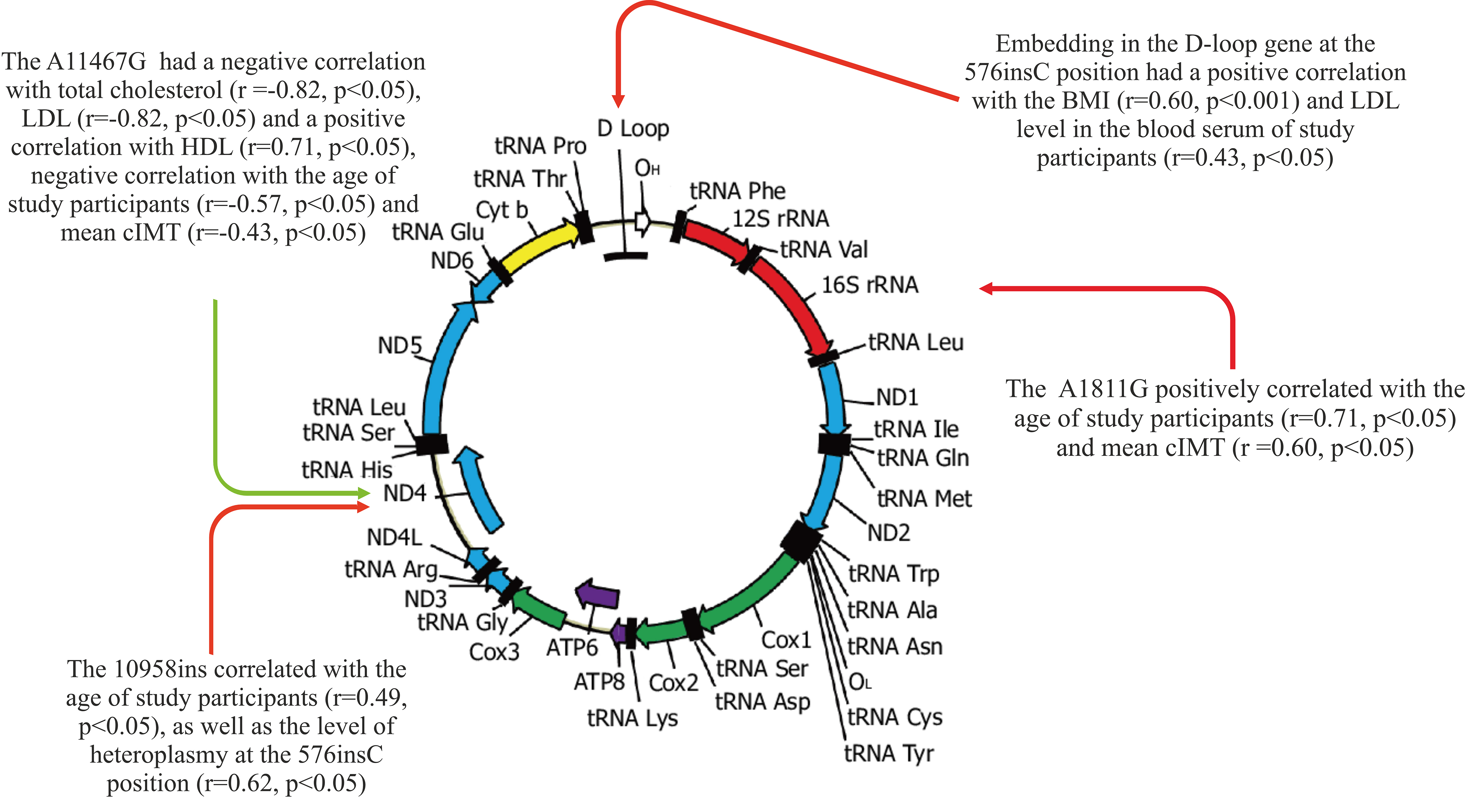
Fig. 1.

The relationship between the studied mtDNA mutations and traditional CVD risk factors. CVD, cardiovascular disease.

The results of the correlation analysis showed that the A11467G mutation in the ND4 gene had a negative correlation with total cholesterol (r = –0.82, p < 0.05), LDL (r = –0.82, p < 0.05) and a positive correlation with HDL (r = 0.71, p < 0.05), as well as a negative correlation with age (r = –0.57, p < 0.05) and mean cIMT (r = –0.43, p < 0.05) of the study participants.

Embedding in the D-loop gene at the 576insC position had a positive correlation with the BMI (r = 0.60, p < 0.001) and LDL level in the blood serum of the study participants (r = 0.43, p < 0.05). The SNV A1811G in the ribosomal RNA rRNA–16S gene positively correlated with the age (r = 0.71, p < 0.05) and mean cIMT (r = 0.60, p < 0.05) of the study participants.

A significant relationship was revealed between the mitochondrial mutation 10958ins, which is located in the ND4 gene, and the age of the study participants (r = 0.49, p < 0.05), as well as the level of heteroplasmy at the 576insC position (r = 0.62, p < 0.05).

Multivariable logistic regression analysis was conducted to reveal the potential contribution of independent variables in the development of obesity and CHD. It was demonstrated that the heteroplasmy level of the mitochondrial genome variant 576insC is a significant predictor of CHD (odds ratio: 1.93; 95% confidence interval: 1.50–2.62).

4. Discussion

In this study, mtDNA heteroplasmic variants associated with CHD and obesity were identified for the first time in the CD14+ monocyte population. A high mutational level of 576insC was detected in obesity and CHD. In contrast, the level of SNV A11467G was significantly lower in patients with CHD and obesity compared to participants in the control group. The A11467G mutation refers to haplogroup U and its descendants; it is a synonymous change, which means that it does not lead to a change in the amino acid encoded by this gene and, therefore, can have a positive effect [20]. The results of the correlation analysis demonstrate that a high level of A11467G is associated with an improvement in cardiovascular risk factors, namely lower levels of total cholesterol, low-density lipoproteins, higher levels of high-density lipoproteins, as well as lower values of cIMT, concurrently, the level of A11467G heteroplasmy decreases with age. The protective effect of mtDNA mutations may be because different mtDNA variants have different effectiveness in the process of energy metabolism and detoxification in cells. Some mtDNA variants may be more resistant to oxidative stress and other negative influences, which helps cells cope with metabolic changes more effectively [21]. The other variants of mitochondrial heteroplasmy studied in this project were positively associated with traditional CVD risk factors, including age, BMI, total cholesterol, and cIMT. Among the studied variants of mitochondrial heteroplasmy, only the 576insC mtDNA mutation was elevated in patients with CHD and obesity and positively correlated with BMI, which indicates the important role of this mtDNA mutation in CD14+ monocytes in the development of metabolic disorders; however, this mutation was identified for the first time; hence, requires further study.

Since mtDNA encodes several proteins involved in the functioning of the respiratory chain, mutations in mtDNA can disrupt the functioning of the respiratory chain, which is the process of energy production in mitochondria [22]. This can lead to an increase in the formation of ROS and a change in the metabolism of glucose and lipids. Changes in metabolism can contribute to the development of atherosclerosis, as elevated lipid levels and oxidative stress lead to cell damage, inflammation, and the formation of atherosclerotic plaques [23]. For example, it was previously found that mitochondrial mutations A1811G and G9477A correlate with the degree of pro-inflammatory activation of monocytes [24]. Activation of monocytes may be associated with various immunological responses and inflammatory processes. This study showed that the A1811G mutation in the ribosomal RNA gene rRNA–16S positively correlates with the age of study participants and cIMT, confirming the important role of immuno-inflammatory mechanisms in atherogenesis.

Previous studies have identified many mtDNA mutations associated with the development of CHD [25]. Some studies confirm the possible involvement of mtDNA mutations in the development of atherosclerosis [26]. These mutations can be classified into several main categories based on different mechanisms of action. The first category includes mutations in transport RNAs (tRNAs) that lead to the destabilization of base pairing and a change in the secondary structure of tRNAs, which can lead to accelerated degradation of tRNAs and a subsequent decrease in the level of mitochondrial proteins [27]. The second category includes mutations in the components of phosphorylating oxidation (OXPHOS), resulting in a reduction in ATP synthesis and an increase in the formation of ROS, which in turn contributes to the dysfunction of energy metabolism and mitochondrial damage. The third category involves mutations in the mtDNA D-loop, which can disrupt the normal replication process and decrease the number of mtDNA copies [28]. However, some studies have shown that mtDNA heteroplasmy can have a protective effect since various mutations in mtDNA can lead to intergenomic competition and intergenomic complementary interaction. Most of the detected mtDNA mutations are non-pathogenic. However, the combination of various factors, including genetic and environmental, can contribute to the development of clinical manifestations of the disease [25].

5. Conclusions

The mtDNA heteroplasmic variants identified in this study, associated with traditional CVD risk factors in CHD and obesity, can be used to develop personalized therapeutic strategies in patients with high cardiovascular risk against the background of metabolic disorders. Mitochondrial genome editing may be considered a possible therapeutic approach for atherosclerosis and CVD in the future [29]. Moreover, the purpose of such an intervention may be both the removal of genetic variants in the mitochondrial genome associated with pathology and the introduction of potentially beneficial changes in the functioning of mitochondria. Thus, the SNV A11467G in the ND4 gene can be considered a potential target for editing the mitochondrial genome in the treatment of CVD since an increase in the level of A11467G heteroplasmy is associated with positive changes in a number of traditional CVD risk factors. The study of the main molecular mechanisms in each specific mtDNA mutation will help to develop more accessible diagnostic tools for the timely detection of patients with high risk of atherosclerosis and the prevention of metabolic disorders and concomitant CVD. However, the widespread and successful use of mtDNA decoding methods in clinical practice requires a standardized approach to interpreting mtDNA variability and the level of heteroplasmy, which is the subject of further research.

Availability of Data and Materials

Data are available from the corresponding author upon request.

Author Contributions

AMM and VAK designed the research study. AIB, TVT performed the research. TVK and YVM analyzed the data. TVT and TVK wrote the manuscript. TVK and VAK revised the manuscript. All authors contributed to editorial changes in the manuscript. All authors read and approved the final manuscript. All authors have participated sufficiently in the work and agreed to be accountable for all aspects of the work.

Ethics Approval and Consent to Participate

This work was performed in accordance with the Declaration of Helsinki of 1975 and its revised version of 2013. The study was approved by the Local Ethics Committee of the Petrovsky National Research Centre of Surgery at December, 13, 2022 (approval number # 3).

Acknowledgment

Not applicable.

Funding

This work was supported by the Ministry of Science and Higher Education of the Russian Federation—the state task of Petrovsky National Research Centre of Surgery (Project # 123030700026-8).

Conflict of Interest

The authors declare no conflict of interest.

References
[1]
Cardiovascular Diseases. Available at: https://www.who.int/health-topics/cardiovascular-diseases#tab=tab_1 (Accessed: 10 January 2024).
[2]
Nedosugova LV, Markina YV, Bochkareva LA, Kuzina IA, Petunina NA, Yudina IY, et al. Inflammatory Mechanisms of Diabetes and Its Vascular Complications. Biomedicines. 2022; 10: 1168.
[3]
Mamatov AU, Orozmatov TT, Madaminov ZhB, Abdymanap Kyzy A, Chuvasheva ND. Obesity and the risk of developing cardiovascular diseases: a look at the modern problem. Scientific Heritage. 2021; 64-2: 35–42.
[4]
Stunault MI, Bories G, Guinamard RR, Ivanov S. Metabolism Plays a Key Role during Macrophage Activation. Mediators of Inflammation. 2018; 2018: 2426138.
[5]
Kirichenko TV, Markina YV, Bogatyreva AI, Tolstik TV, Varaeva YR, Starodubova AV. The Role of Adipokines in Inflammatory Mechanisms of Obesity. International Journal of Molecular Sciences. 2022; 23: 14982.
[6]
Rambold AS, Pearce EL. Mitochondrial Dynamics at the Interface of Immune Cell Metabolism and Function. Trends in Immunology. 2018; 39: 6–18.
[7]
Dai W, Jiang L. Dysregulated Mitochondrial Dynamics and Metabolism in Obesity, Diabetes, and Cancer. Frontiers in Endocrinology. 2019; 10: 570.
[8]
Todosenko N, Khaziakhmatova O, Malashchenko V, Yurova K, Bograya M, Beletskaya M, et al. Mitochondrial Dysfunction Associated with mtDNA in Metabolic Syndrome and Obesity. International Journal of Molecular Sciences. 2023; 24: 12012.
[9]
Merry TL, Chan A, Woodhead JST, Reynolds JC, Kumagai H, Kim SJ, et al. Mitochondrial-derived peptides in energy metabolism. American Journal of Physiology. Endocrinology and Metabolism. 2020; 319: E659–E666.
[10]
Liu C, Fetterman JL, Liu P, Luo Y, Larson MG, Vasan RS, et al. Deep sequencing of the mitochondrial genome reveals common heteroplasmic sites in NADH dehydrogenase genes. Human Genetics. 2018; 137: 203–213.
[11]
Lampert MA, Orogo AM, Najor RH, Hammerling BC, Leon LJ, Wang BJ, et al. BNIP3L/NIX and FUNDC1-mediated mitophagy is required for mitochondrial network remodeling during cardiac progenitor cell differentiation. Autophagy. 2019; 15: 1182–1198.
[12]
Khotina VA, Bagheri Ekta M, Baig MS, Wu WK, Grechko AV, Sukhorukov VN. Challenges of mitochondrial DNA editing in mammalian cells: focus on treatment of cardiovascular disease. Vessel Plus. 2022; 6: 65.
[13]
Hagen CM, Aidt FH, Hedley PL, Jensen MK, Havndrup O, Kanters JK, et al. Mitochondrial haplogroups modify the risk of developing hypertrophic cardiomyopathy in a Danish population. PLoS ONE. 2013; 8: e71904.
[14]
Sazonova MA, Zhelankin AV, Barinova VA, Sinyov VV, Khasanova ZB, Postnov AY, et al. Mutations of mitochondrial genome in carotid atherosclerosis. Frontiers in Genetics. 2015; 6: 111.
[15]
Amato M, Veglia F, de Faire U, Giral P, Rauramaa R, Smit AJ, et al. Carotid plaque-thickness and common carotid IMT show additive value in cardiovascular risk prediction and reclassification. Atherosclerosis. 2017; 263: 412–419.
[16]
Beux F, Carmassi S, Salvetti MV, Ghiadoni L, Huang Y, Taddei S, et al. Automatic evaluation of arterial diameter variation from vascular echographic images. Ultrasound in Medicine & Biology. 2001; 27: 1621–1629.
[17]
Köchl S, Niederstätter H, Parson W. DNA extraction and quantitation of forensic samples using the phenol-chloroform method and real-time PCR. Methods in Molecular Biology (Clifton, N.J.). 2005; 297: 13–30.
[18]
Xie F, Guo W, Wang X, Zhou K, Guo S, Liu Y, et al. Mutational profiling of mitochondrial DNA reveals an epithelial ovarian cancer-specific evolutionary pattern contributing to high oxidative metabolism. Clinical and Translational Medicine. 2024; 14: e1523.
[19]
Kirichenko TV, Ryzhkova AI, Sinyov VV, Sazonova MD, Orekhova VA, Karagodin VP, et al. Impact of Mitochondrial DNA Mutations on Carotid Intima-Media Thickness in the Novosibirsk Region. Life (Basel, Switzerland). 2020; 10: 160.
[20]
Divne AM, Råsten-Almqvist P, Rajs J, Gyllensten U, Allen M. Analysis of the mitochondrial genome in sudden infant death syndrome. Acta Paediatrica (Oslo, Norway: 1992). 2003; 92: 386–388.
[21]
Schaefer PM, Scherer Alves L, Lvova M, Huang J, Rathi K, Janssen K, et al. Combination of common mtDNA variants results in mitochondrial dysfunction and a connective tissue dysregulation. Proceedings of the National Academy of Sciences of the United States of America. 2022; 119: e2212417119.
[22]
Liu Y, Huang Y, Xu C, An P, Luo Y, Jiao L, et al. Mitochondrial Dysfunction and Therapeutic Perspectives in Cardiovascular Diseases. International Journal of Molecular Sciences. 2022; 23: 16053.
[23]
Dabravolski SA, Nikiforov NG, Zhuravlev AD, Orekhov NA, Grechko AV, Orekhov AN. Role of the mtDNA Mutations and Mitophagy in Inflammaging. International Journal of Molecular Sciences. 2022; 23: 1323.
[24]
Orekhov AN, Poznyak AV, Sobenin IA, Nikifirov NN, Ivanova EA. Mitochondrion as a Selective Target for the Treatment of Atherosclerosis: Role of Mitochondrial DNA Mutations and Defective Mitophagy in the Pathogenesis of Atherosclerosis and Chronic Inflammation. Current Neuropharmacology. 2020; 18: 1064–1075.
[25]
Matam K, Shaik NA, Aggarwal S, Diwale S, Banaganapalli B, Al-Aama JY, et al. Evidence for the presence of somatic mitochondrial DNA mutations in right atrial appendage tissues of coronary artery disease patients. Molecular Genetics and Genomics: MGG. 2014; 289: 533–540.
[26]
Orekhov AN, Ivanova EA, Markin AM, Nikiforov NG, Sobenin IA. Genetics of Arterial-Wall-Specific Mechanisms in Atherosclerosis: Focus on Mitochondrial Mutations. Current Atherosclerosis Reports. 2020; 22: 54.
[27]
Zhang Z, Liu M, He J, Zhang X, Chen Y, Li H. Maternally inherited coronary heart disease is associated with a novel mitochondrial tRNA mutation. BMC Cardiovascular Disorders. 2019; 19: 293.
[28]
Liu H, Liu X, Zhou J, Li T. Mitochondrial DNA Is a Vital Driving Force in Ischemia-Reperfusion Injury in Cardiovascular Diseases. Oxidative Medicine and Cellular Longevity. 2022; 2022: 6235747.
[29]
Khotina VA, Vinokurov AY, Bagheri Ekta M, Sukhorukov VN, Orekhov AN. Creation of Mitochondrial Disease Models Using Mitochondrial DNA Editing. Biomedicines. 2023; 11: 532.

Publisher’s Note: IMR Press stays neutral with regard to jurisdictional claims in published maps and institutional affiliations.

Share
Back to top