- Academic Editors
-
-
-
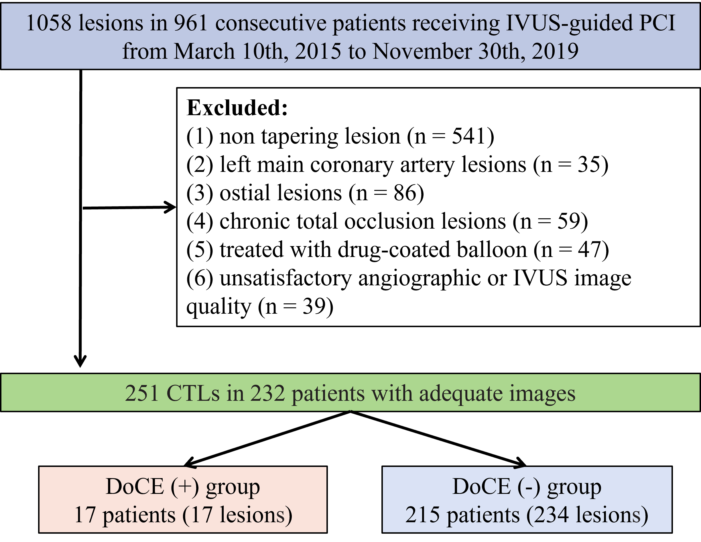
Background: This study aimed to assess the clinical significance of generating a volumetric stent expansion index for tapering lesions through intravascular ultrasound (IVUS). Previous IVUS studies have used minimal stent area (MSA) to predict adverse outcomes. Methods: A total of 251 tapering lesions were treated in this study via IVUS guidance in 232 patients. Eight stent expansion indices were evaluated to determine the association of these indices with device-oriented clinical endpoints (DoCEs) after two-year follow-ups. These were the ILUMIEN III and IV standards, the ULTIMATE (Intravascular Ultrasound Guided Drug Eluting Stents Implantation in “All-Comers” Coronary Lesions) standard, the IVUS-XPL (Impact of Intravascular Ultrasound Guidance on the Outcomes of Xience Prime Stents in Long Lesions) standard, the minimal volumetric expansion index (MVEI) using the Huo-Kassab or linear model, the MSA/vessel area at the MSA cross-section, the traditional stent expansion (MSA/mean proximal and distal reference lumen cross-sectional area), and MSA. Results: The MVEI was the only stent expansion index that correlated significantly with the two-year DoCEs (hazard ratio [HR], 1.91; 95% confidence interval [CI]: 1.16–3.96; p = 0.028). In the ROC analysis, the area under the curve for the MVEI was 0.71 (p = 0.002), with an optimal cut-off value of 62.2 for predicting the DoCEs. Conclusions: This is the first study to use IVUS for tapering lesions and demonstrate that the MVEI is an independent predictor of two-year DoCEs.