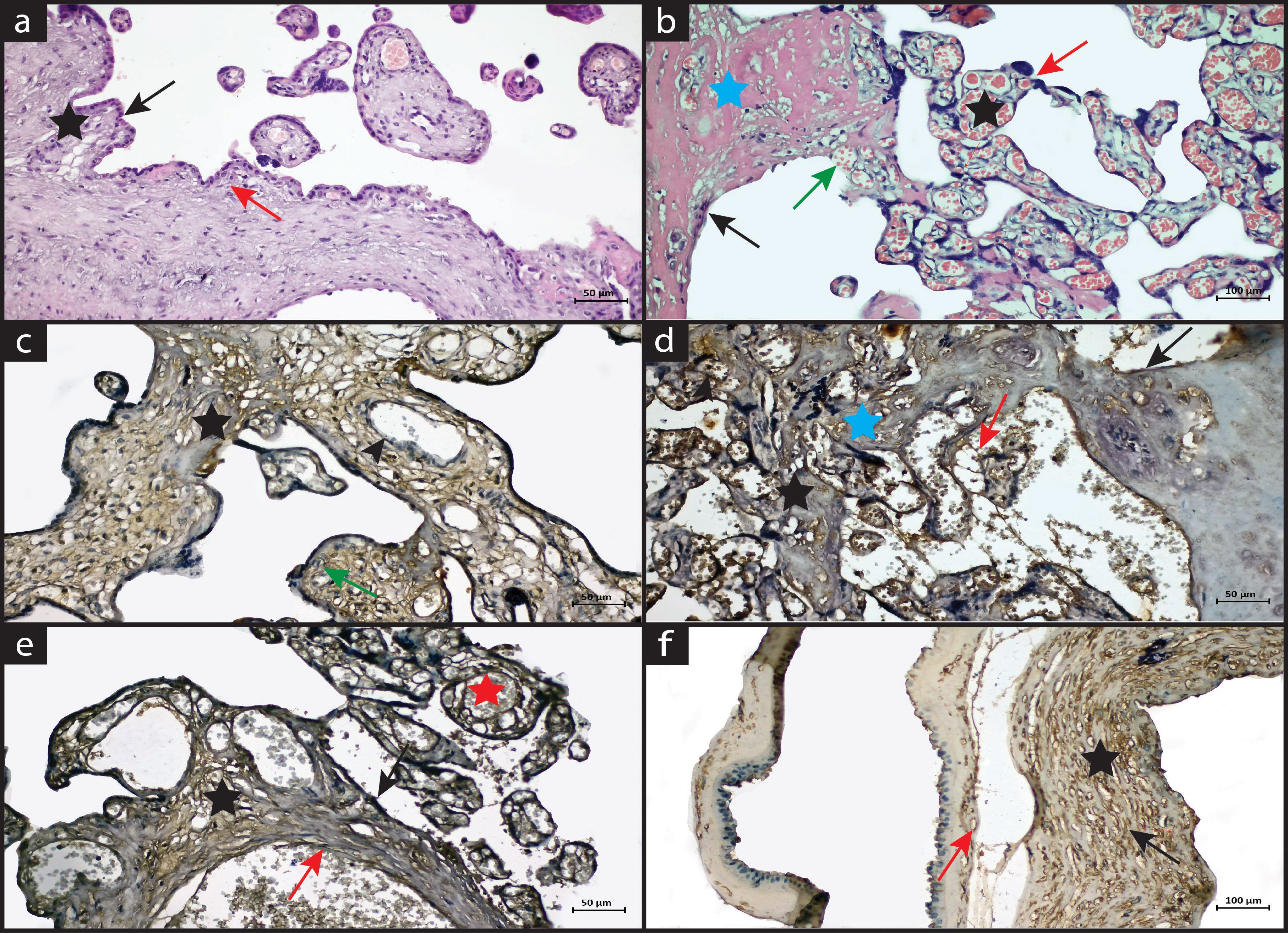
IMR Press / CEOG / Volume 50 / Issue 8 / DOI: 10.31083/j.ceog5008173
Open Access Original Research
The Investigation of Caspase-3 and Tumor Necrosis Factor-Alpha Expression in Placentas of Patients with Preterm Premature Rupture of Membranes
Show Less
1 Department of Gynecology and Obstetrics, Harran University Faculty of Medicine, 63300 Şanlıurfa, Turkey
2 Department of Histology and Embryology, Dicle University Faculty of Medicine, 21280 Diyarbakır, Turkey
3 Department of Gynecology and Obstetrics, Health Sciences University, Gazi Yaşargil Training and Research Hospital, 21070 Diyarbakır, Turkey
*Correspondence: sampson_21@hotmail.com (Süleyman Cemil Oğlak)
Clin. Exp. Obstet. Gynecol. 2023, 50(8), 173; https://doi.org/10.31083/j.ceog5008173
Submitted: 25 May 2023 | Revised: 17 June 2023 | Accepted: 30 June 2023 | Published: 16 August 2023
Copyright: © 2023 The Author(s). Published by IMR Press.
This is an open access article under the CC BY 4.0 license.
Abstract

Background: Caspase-3 is involved in the execution of apoptosis and is widely used as an apoptotic marker. Tumor necrosis factor-α (TNF-α) released from activated macrophages has various functions such as modulation of cell growth and differentiation, immunoregulation, coagulation, and regulation of endothelial cell function. This study investigated the immunohistochemical staining of caspase-3 and TNF-α expression in the placentas of pregnant women with preterm premature rupture of membranes (PPROM). Methods: Placentas of 25 healthy, and 25 women with PPROM were processed for routine histological tissue processing. Placentas were stained with hematoxylin-eosin, caspase-3, and TNF-α immunostaining. Results: Normal placental histology was observed in the control group. Amniotic epithelium, vascular structures, and fibrinoid accumulation were histologically normal. Leukocyte infiltration, thinned vessel walls with dilatation and congestion, syncytial nodes, and fibrinoid accumulation were increased in the PPROM group. The immune activity of caspase-3 expression was mainly negative in placental components such as syncytial nodes, vascular endothelium, fibrinoid accumulation, and macrophages in the control group. In the PPROM group, caspase-3 positive reaction was increased in the amniotic membrane and epithelium, endothelial cells, fibrinoid accumulation, and areas of inflammatory cell infiltration. In the control group, negative TNF-α expression was observed in the placental membranes and structures. In the PPROM group, TNF-α expression was increased in inflammatory cells, endothelial cells, and syncytial nodes. Conclusions: Placentas of patients with PPROM showed loss and weakened membranes with increased placental pathology, and increased expression of caspase-3 and TNF-α. We suggest that caspase-3 and TNF-α signaling pathways can be used as a marker in the progression of PPROM.

Keywords
caspase-3
tumor necrosis factor-alpha
placenta
premature rupture of membranes
Figures
Fig. 1.
Share
Back to top