- Academic Editor
-
-
-
†These authors contributed equally.
Background: To explore the feasibility of radiomic models using
different magnetic resonance imaging (MRI) sequences combined with clinical
information in evaluating the status of lymphovascular space invasion (LVSI) in
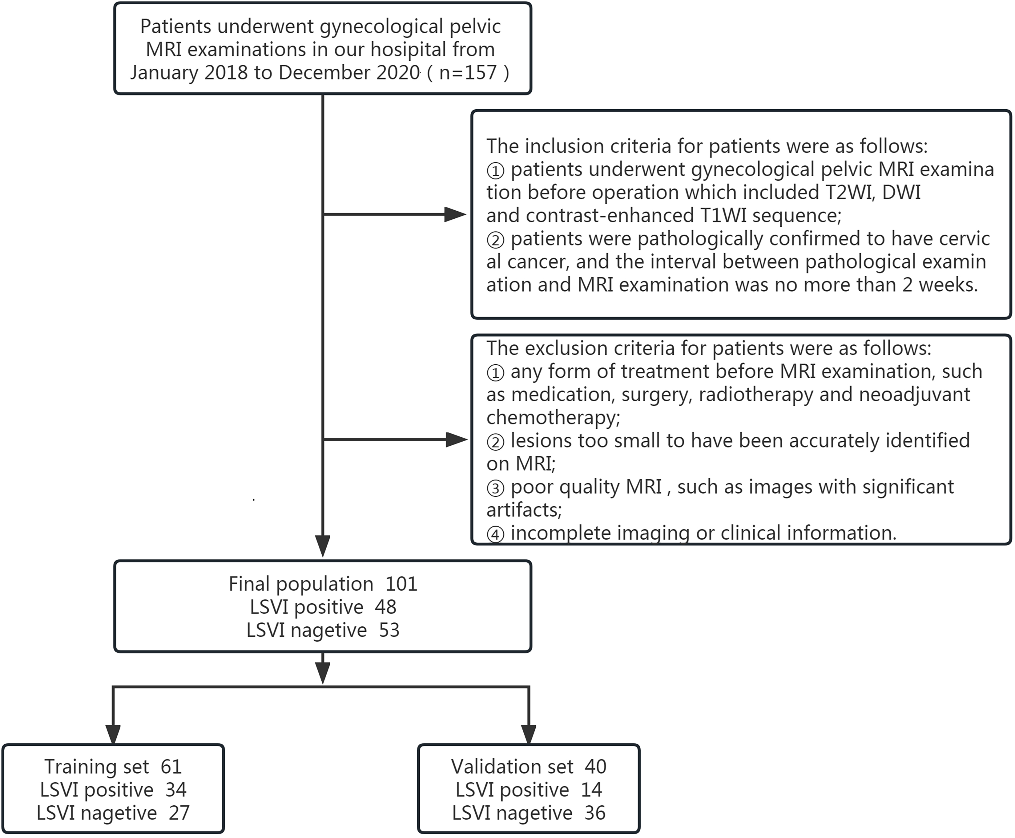
cervical cancer. Methods: One hundred one cervical cancer patients were
included from January 2018 to December 2020. All patients underwent 3.0T MRI
examination including T2 weighted imaging (T2WI), diffusion weighted imaging
(DWI) and contrast-enhanced T1 weighted imaging (T1WI + C) enhanced sequences.
Age, preoperative squamous cell carcinoma (SCC) associated antigen value and the
depth of muscular invasion were collected. The 101 patients were divided into
training set and validation set. Three different models were developed using
T2WI, DWI and T1WI + C parameters respectively. One model was developed combining
the three different sequences. The diagnostic performance of each model was
compared via receiver operating characteristic curve analysis. Results:
Forty-eight cases were pathologically confirmed with lymphovascular space
invasion. The average SCC value of the LVSI positive group (10.82