Background: Tanshinone IIA (TSIIA) is an element of the effective
ingredients of Salvia miltiorrhiza Bunge (Labiatae), exhibits a
significant therapeutic effect in brain neuroprotection. The focus of this study
was the examination of synaptic plasticity of in Mg-free-induced epileptic
hippocampus neurons and how TSIIA protects against it. Methods: The
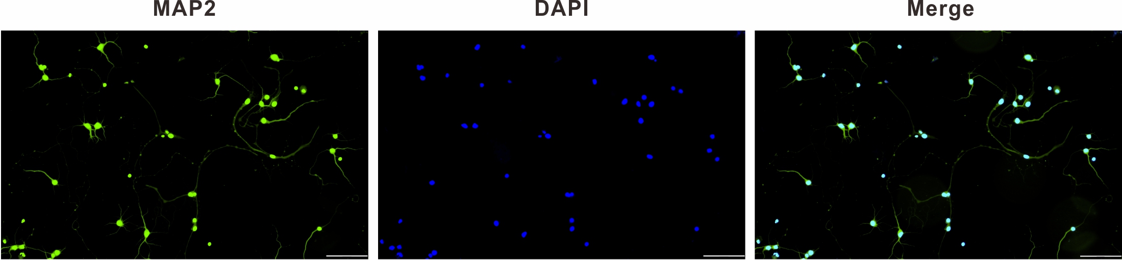
purity of the primary hippocampal neurons extracted from Sprague Dawley rats was
assessed within 24 hours by microtubule-associated protein (MAP2)
immunofluorescence staining. A hippocampal neuron model for
Mg-free-induced spontaneous recurrent epileptiform discharge was
developed, five experimental groups were then randomized: blank (Blank), model
(Model), TSIIA (TSIIA, 20 µM), LY294002 (LY294002, 25 µM), and
TSIIA+LY294002 (TSIIA+LY294002, 20 µM+25 µM). FIJI software was used
to examine variations of neurite complexity, total length of hippocampal neurons,
number of primary dendrites and density of dendritic spines. Developmental
regulation brain protein (Drebrin) and brain-derived neurotrophic factor (BDNF)
expression was evaluated using immunofluorescence staining and the relative
expression of phospho-protein kinase B (p-Akt)/Akt, BDNF, synaptophysin (SYN) and postsynaptic density 95
(PSD-95) determined by Western blot.
Results: In contrast to the model group, TSIIA drastically reduced
damage to synaptic plasticity of hippocampal neurons caused by epilepsy
(p 0.05). The TSIIA group showed a significant increase in the
relative expression of PSD-95, SYN, BDNF, and p-Akt/Akt (p 0.01).
Conclusions: TSIIA was effective in reducing harm to the synaptic
plasticity of hippocampal neurons induced by persistent status epilepticus, with
the possible mechanism being regulation of the phosphatidylinositol 3-kinase
56 (PI3K)/Akt signaling pathway.