- Academic Editor
-
-
-
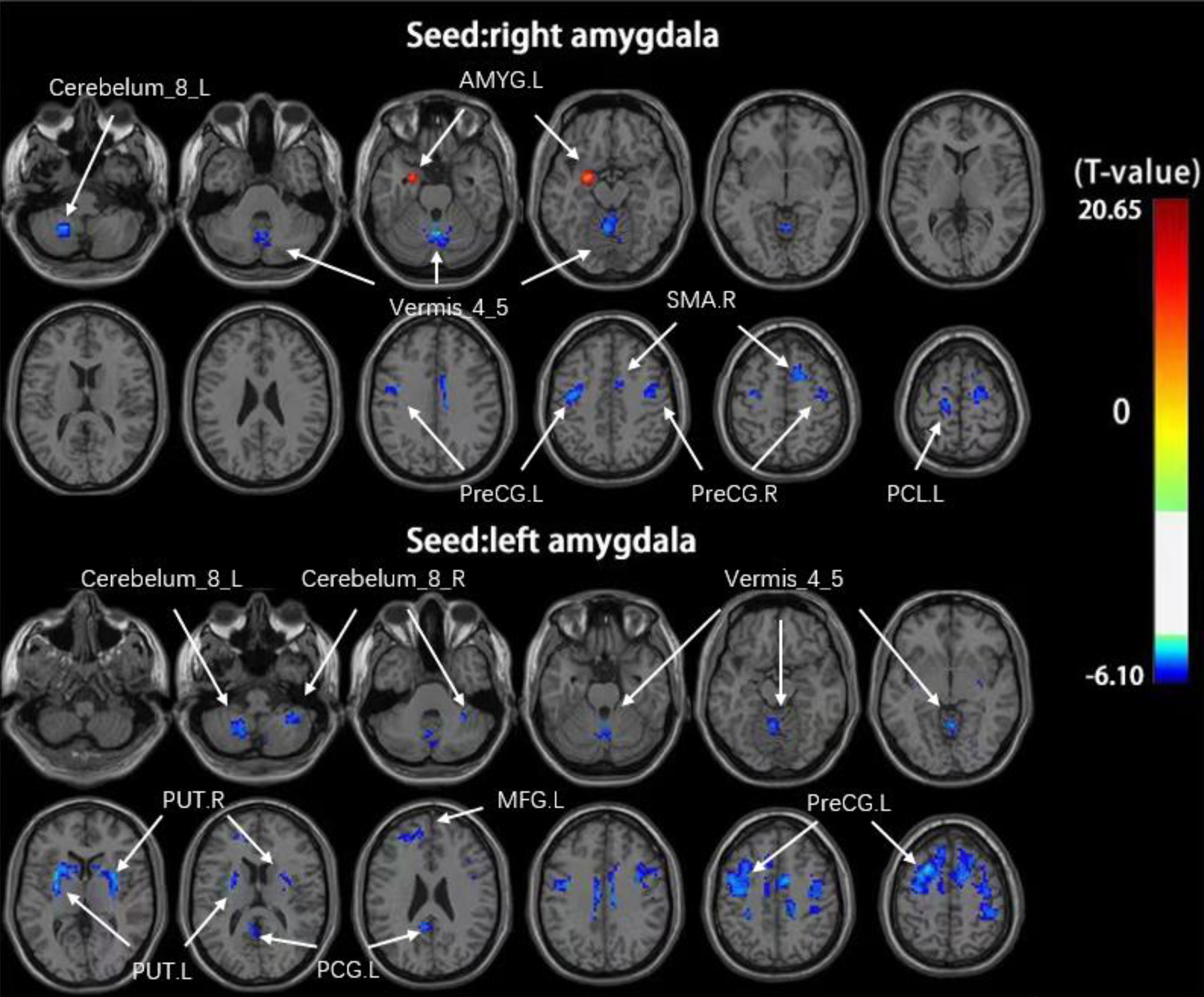
Background: Glaucoma patients frequently present with depressive symptoms, the development of which is closely associated with amygdalar activity. However, no studies to date have documented glaucoma-related changes in the functional connectivity (FC) of the amygdala. Accordingly, resting-state functional magnetic resonance imaging (rs-fMRI) analyses were herein used to evaluate changes in amygdalar FC in primary angle-closure glaucoma (PACG) patients. Methods: In total, this study enrolled 36 PACG patients and 33 healthy controls (HCs). Complete eye exams were conducted for all PACG patients. After the preprocessing of magnetic resonance imaging (MRI) data, the bilateral amygdala was selected as a seed point, followed by the comparison of resting-state FC between the PACG and HC groups. Then, those brain regions exhibiting significant differences between these groups were identified, and relationships between the FC coefficient values for these regions and clinical variables of interest were assessed. Results: These analyses revealed that as compared to HC individuals, PACG patients exhibited reductions in FC between the amygdala and the cerebellum_8, vermis_4_5, anterior central gyrus, supplementary motor area, paracentral lobule, putamen, middle frontal gyrus, and posterior cingulate gyrus, while enhanced FC was detected between the right and left amygdala. No significant correlations between these changes in amygdalar any any disease-related clinical parameters or disease duration were noted. Conclusions: Patients with PACG exhibit extensive resting state abnormalities with respect to the FC between the amygdala and other regions of the brain, suggesting that dysregulated amygdalar FC may play a role in the pathophysiology of PACG.