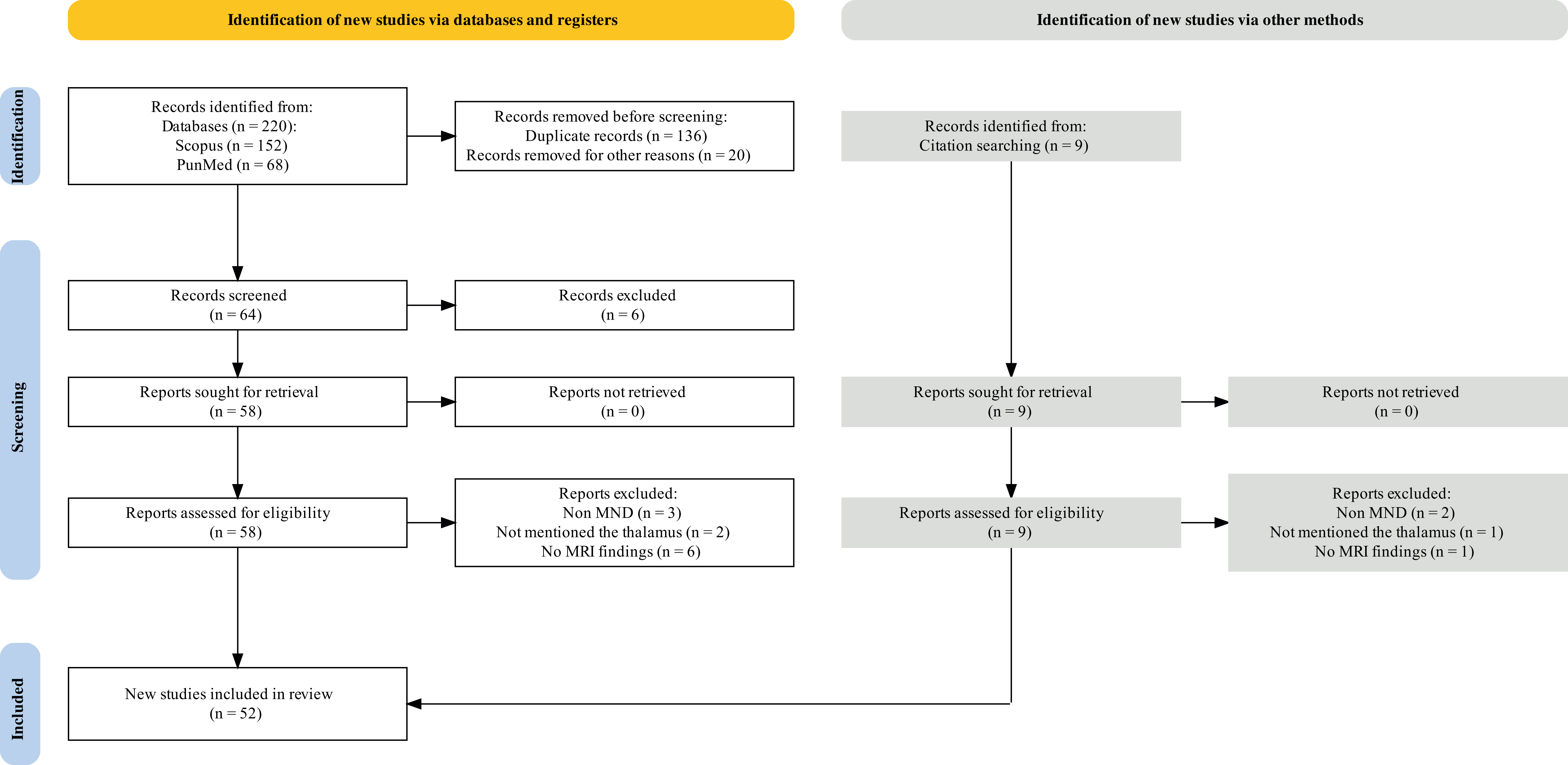
Background: Motor neuron diseases (MNDs) are progressive neurodegenerative disorders characterized by motor impairment and non-motor symptoms. The involvement of the thalamus in MNDs, especially in conditions such as amyotrophic lateral sclerosis (ALS), and its interaction with frontotemporal dementia (FTD), has garnered increasing research interest. This systematic review analyzed magnetic resonance imaging (MRI) studies that focused on thalamic alterations in MNDs to understand the significance of these changes and their correlation with clinical outcomes. Methods: Following PRISMA 2020 guidelines, the PubMed and Scopus databases were searched from inception to June 2023 for studies related to MRI findings in the thalamus of patients with MNDs. Eligible studies included adult patients diagnosed with ALS or other forms of MND who underwent brain MRI, with outcomes related to thalamic alterations. Studies were evaluated for risk of bias using the Newcastle-Ottawa scale. Results: A total of 52 studies (including 3009 MND patients and 2181 healthy controls) used various MRI techniques, including volumetric analysis, diffusion tensor imaging, and functional MRI, to measure thalamic volume, connectivity, and other alterations. This review confirmed significant thalamic changes in MNDs, such as atrophy and microstructural degradation, which are associated with disease severity, progression, and functional disability. Thalamic involvement varies across different MND subtypes and is influenced by the presence of cognitive impairment and mutations in genes including chromosome 9 open reading frame 72 (C9orf72). The synthesis of findings across studies indicates that thalamic pathology is a prevalent early biomarker of MNDs that contributes to motor and cognitive deficits. The thalamus is a promising target for monitoring as its dysfunction underpins a variety of clinical symptoms in MNDs. Conclusions: Thalamic alterations provide valuable insights into the pathophysiology and progression of MNDs. Multimodal MRI techniques are potent tools for detecting dynamic thalamic changes, indicating structural integrity, connectivity disruption, and metabolic activity.