IMR Press / JIN / Volume 23 / Issue 3 / DOI: 10.31083/j.jin2303056
Open Access Original Research
Association of Retinal Nerve Fiber Layer Thickness with Brain Microstructural Changes in Participants with White Matter Hyperintensities
Show Less
1 Department of Neurology, The Second Affiliated Hospital and Yuying Children's Hospital of Wenzhou Medical University, 325027 Wenzhou, Zhejiang, China
2 Critical Care Medicine, The First Affiliated Hospital, Zhejiang University School of Medicine, 310003 Hangzhou, Zhejiang, China
3 Department of Radiology, The Second Affiliated Hospital and Yuying Children's Hospital of Wenzhou Medical University, 325027 Wenzhou, Zhejiang, China
4 China-USA Neuroimaging Research Institute, Department of Radiology of the Second Affiliated Hospital and Yuying Children’s Hospital, Wenzhou Medical University, 325027 Wenzhou, Zhejiang, China
*Correspondence: caoyungang2003@163.com (Yungang Cao); wzhanzhao@aliyun.com (Zhao Han)
These authors contributed equally.
J. Integr. Neurosci. 2024, 23(3), 56; https://doi.org/10.31083/j.jin2303056
Submitted: 19 August 2023 | Revised: 20 October 2023 | Accepted: 25 October 2023 | Published: 11 March 2024
Copyright: © 2024 The Author(s). Published by IMR Press.
This is an open access article under the CC BY 4.0 license.
Abstract

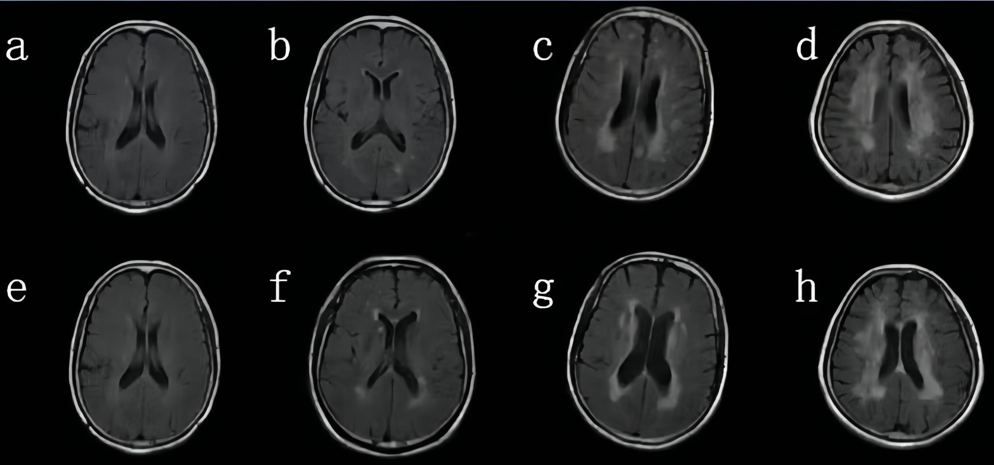
Purpose: White matter hyperintensity (WMH) is suggested to cause stroke and dementia in older adults. Retinal structural thicknesses revealed by optical coherence tomography (OCT) are associated with structural changes in the brain. We aimed to explore the association between the peripapillary retinal nerve fiber layer (RNFL) and cerebral microstructural changes in participants with white matter hyperintensities (WMH). Methods: Seventy-four participants (37 controls, healthy control (HC), and 37 older adults with WMH) underwent retinal and brain imaging using OCT and magnetic resonance imaging (MRI) respectively. Peripapillary RNFL thickness was assessed by the OCT. Gray matter volume (GMV) was assessed from a T1-weighted MRI. White matter integrity was assessed with diffusion tensor imaging (DTI) while WMH severity was assessed with the Fazekas scale. All participants underwent a neuropsychological examination (Mini-Mental State Examination, MMSE). Results: Older adults with WMH showed thinner peripapillary RNFL (p = 0.004) thickness when compared with the control group after adjusting for age, hypertension and gender. In our older adults with WMH, RNFL thickness correlated with fractional anisotropy (FA) in the superior longitudinal fasciculus (SLF) (Rho = –0.331, p < 0.001). In older adults with WMH, RNFL was significantly associated with MMSE scores (Rho = 0.422, p < 0.001) and Fazekas scores (Rho = –0.381, p = 0.022) respectively. Conclusions: We suggest neurodegeneration of peripapillary RNFL in older adults with WMH was associated with cerebral microstructural volume, impaired cerebral axonal damage, and cognitive performances. OCT metrics may provide evidence of neurodegeneration that may underpin WMH and cerebral microstructural changes in the brain. Clinical Trial Registration: This study was registered online at the China Clinical Trial Registration Center (registration number: ChiCTR-ROC-17011819).

Keywords
white matter hyperintensity
optical coherence tomography
peripapillary retinal nerve fiber layer
diffusion tensor imaging
cognition
Funding
SAHoWMU-CR2017-01-212/Clinical Scientific Research Fund of the Second Affiliated Hospital of Wenzhou Medical University
82271344/National Natural Science Foundation of China
81771267/National Natural Science Foundation of China
Figures
Fig. 1.
Share
Back to top